
Abstract
Objective
This study was to investigate the expression and clinical significance of RRBP1 in esophageal carcinoma.
Materials and methods
RRBP1 expression was detected in 120 esophageal carcinoma and matched adjacent normal tissues, and the relationship of RRBP1 with clinicopathological characteristics and prognosis was analyzed.
Results
RRBP1 was highly expressed in esophageal carcinoma tissues compared with matched adjacent normal tissues (P<0.05). Moreover, RRBP1 expression was associated with T stage, lymph node metastasis, and TNM stage in esophageal carcinoma (P<0.05). Survival analysis revealed that RRBP1, T stage, lymph node metastasis, and TNM stage were significantly associated with patients’ prognosis.
Conclusion
RRBP1 is highly expressed in esophageal carcinoma and can serve as a potential biomarker to predict patients’ prognosis.
Introduction
Esophageal carcinoma is one of the most common malignant tumors in China, which accounts for the sixth most common cause of cancer-related death in the world.Citation1,Citation2 Surgical resection is the main treatment for esophageal cancer patients; however, the 5-year survival rate of esophageal cancer patients after surgery is still less than 25%.Citation3,Citation4 Currently, the incidence of esophageal carcinoma is still increasing in China.Citation1,Citation5,Citation6 The early diagnosis of esophageal carcinoma is a tough challenge.Citation7,Citation8 Thus, it would be meaningful to explore novel molecular biomarkers associated with the early diagnosis and prognosis of esophageal cancer.
RRBP1 is an endoplasmic reticulum membrane protein, which plays a critical role in the transportation and secretion of nascent proteins.Citation9 Recently, RRBP1 over-expression has been frequently observed in lung cancer, breast cancer, and colorectal cancer.Citation10–Citation12 Moreover, RRBP1 correlates with shorter survival and can serve as a valuable prognostic factor in Her-2-positive breast cancer patients.Citation13 RRBP1 over-expression contributes to the progression of colorectal cancer and is useful for predicting patients’ prognosis.Citation14 Thus, this evidence suggests that RRBP1 may be a key oncogene involved in tumor formation and progression. However, the expression and clinical significance of RRBP1 have never been reported in esophageal carcinoma.
In this study, we detected the expression of RRBP1 in 120 cases of esophageal carcinoma and matched adjacent normal tissues, and analyzed the correlation between RRBP1 expression and clinicopathological features. Moreover, whether RRBP1 could be a potential prognostic biomarker in patients with esophageal carcinoma was further assessed.
Materials and methods
Patients and samples
One hundred and twenty esophageal carcinoma (without chemotherapy and radiotherapy before surgery) specimens were collected from patients presenting to Cangzhou Central Hospital during 2010–2014. Matched adjacent normal tissues were collected 3 cm from esophageal carcinoma tissue. Patients included 57 males and 63 females with a mean age of 58 years (range, 32–74 years). Clinical pathological characteristics including age, gender, history of smoking, tumor location, T stage, lymph node metastasis, and TNM stage were obtained from hospital records. Follow-up time was from the day of surgery. No patient was lost during follow-up and the follow-up duration ranged from 1 to 65 months (mean, 38.1 months). All the samples were diagnosed as squamous cell carcinoma. The pathological diagnosis was confirmed by two pathologists in Cangzhou Central Hospital.
Quantitative real-time polymerase chain reaction (q-RT-PCR)
All tissues were frozen in liquid nitrogen. RNA was extracted by RNAisoTM PLUS (Thermo Fisher Scientific, Waltham, MA, USA) and reverse transcribed into cDNA by cDNA Synthesis Kit (TaKaRa Corp, Dalian, China). Quantitative analysis of RRBP1 was performed using 7500 SYBR Green Fast Real-Time PCR System (Thermo Fisher Scientific,). The reaction conditions were 95°C for 10 min, followed 95° for 15 s for 40 cycles and 60°C for 60 s. The primer sequences of RRBP1 were 5′-TGAATCCTCCAAAGACCACA-3′ and 5′-CTTTCCCTCTCGCGTCTCT-3′. The primer sequences of GAPDH were 5′-CTGAACGGGAAGCTCACTGG-3′ and 5′-TGAGGTCCACCACCCTGTTG-3′. The experiments were repeated three times under the same conditions.
Western blot analysis
All tissues were frozen in liquid nitrogen. Proteins were extracted by protease inhibitors and quantified by the Pierce BCA Protein Assay Kit (Thermo Fisher Scientific). An amount of 50 μg per sample was resolved on 5% sodium dodecyl sulfate polyacrylamide gel electrophoresis and transferred onto polyvinylidene fluoride membranes. After blocking in 5% fat-free milk at room temperature, membranes were incubated with RRBP1 (Epitomics, Inc., Burlingame, CA, USA) (diluted 1:1000) and GAPDH (Zhongshan Corp, Beijing, China) (diluted 1:1000) antibodies overnight at 4°C. Then, membranes were incubated with horseradish peroxidase-conjugated secondary antibodies for 1 h. The signals were measured by enhanced chemiluminescence detection reagents.
Immunohistochemical (IHC) staining
Sections (2 μm thick) were deparaffinized with xylene and rehydrated in graded ethanol. Endogenous peroxidase was wiped off with 3% hydrogen peroxide and antigenicity was repaired by 0.01 mol/L sodium citrate buffer (pH 6.0). All sections were incubated with rabbit monoclonal RRBP1 antibody (Epitomics, Inc.) (diluted 1:200) at room temperature for 2 h. After incubation with secondary biotinylated antibody, sections were stained with diaminobenzidine (DAB) and hematoxylin.
The staining of RRBP1 was analyzed by semi-quantitative method. The staining intensity was scored as blank (0), weak (1), moderate (2), and strong (3). The percentage of positive cells was scored as <5% (0), ≥5% –<25% (1), 25% –50% (2), and >50% (3). The scores were calculated by multiplying these two values (ranging from 0 to 9). These scores (≥4) were defined as RRBP1 high-expression, and others were defined as RRBP1 low-expression (<4). All IHC scores were assessed by two pathologists independently without the clinical information.
Statistical analysis
All data were analyzed with SPSS software (version 19.0; IBM Corporation, Armonk, NY, USA). IHC results were analyzed by chi-square test. Survival analysis was performed by the Kaplan–Meier method and log-rank test. Multivariate analysis was assessed by Cox’s proportional hazards model. The comparison of two-sample mean was evaluated using independent samples t-test. P-value of <0.05 was defined as statistically significant.
Ethics statement
This study was approved by the Cangzhou Central Hospital Ethnics Committee. All patients signed informed consent and agreed to the use of their tissue samples in this study.
Results
RRBP1 is highly expressed in esophageal carcinoma
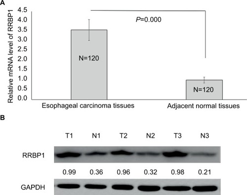
First, we detected the expression of RRBP1 in 120 esophageal carcinoma specimens and matched adjacent normal tissues by qRT-PCR and Western blot assays. qRT-PCR results indicated that RRBP1 mRNA level was significantly higher in esophageal carcinoma tissues compared with matched adjacent normal tissues (, P=0.000). Meanwhile, Western blot results revealed that RRBP1 protein was highly expressed in esophageal carcinoma tissues compared with matched adjacent normal tissues (, P=0.000). These data indicated that RRBP1 was highly expressed in esophageal carcinoma.
Figure 1 RRBP1 expression.
Notes: The expression of RRBP was detected in esophageal carcinoma and matched adjacent normal tissues by qRT-PCR (A) and Western blot (B). T, esophageal carcinoma tissue; N, matched adjacent normal esophageal tissue.
RRBP1 expression correlates with clinical pathological characteristics in esophageal carcinoma
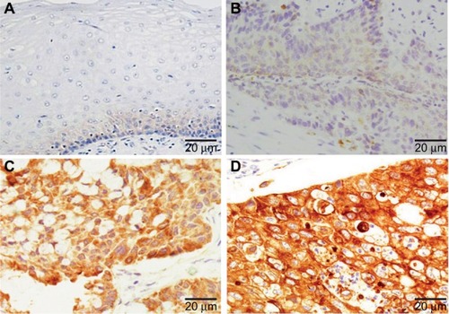
Subsequently, we detected the expression of RRBP1 in 120 esophageal carcinoma specimens and matched adjacent normal tissues by IHC. As shown in , positive expression of RRBP1 was located in cell cytoplasm and easily observed in esophageal carcinoma tissues, but was hardly detected in normal esophageal tissues. The high-expression rates of RRBP1 in esophageal carcinoma and normal esophageal tissues were 59.2% and 11.7%, respectively, and the difference was statistically significant (, P=0.000). Moreover, RRBP1 expression was associated with T stage, lymph node metastasis, and TNM stage in esophageal carcinoma (, P<0.05), but was not associated with age, gender, history of smoking, and tumor location (, P>0.05).
Table 1 RRBP1 expression in esophageal carcinoma and normal esophageal tissues by immunohistochemical staining
Table 2 RRBP1 expression correlation with clinicopathological characteristics in esophageal carcinoma
Figure 2 RRBP1 expression was detected in esophageal carcinoma and matched adjacent normal tissues by immunohistochemical staining.
Notes: (A) Adjacent normal tissues; (B) weak staining of RRBP1 in esophageal carcinoma; (C) moderate staining of RRBP1 in esophageal carcinoma; (D) strong staining of RRBP1 in esophageal carcinoma.
High-expression of RRBP1 predicts an unfavorable survival rate in esophageal carcinoma patients
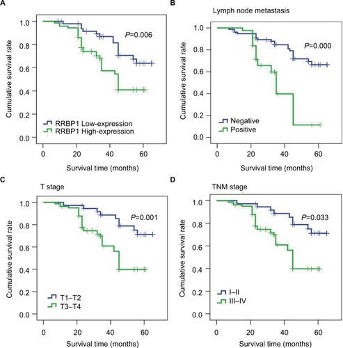
Then, we further analyzed the correlation between RRBP1 expression and patients’ survival by Kaplan–Meier method and Cox’s proportional hazards model. Kaplan–Meier analysis revealed that the median survival time of patients with RRBP1 high-expression was 43 months, which was significantly shorter compared with those with RRBP1 low-expression (56 months) (, , P=0.006). Moreover, T stage, lymph node metastasis, and TNM stage rather than age, gender, and history of smoking were confirmed to be associated with patients’ survival (, , P<0.05). Furthermore, multivariate Cox regression analysis showed RRBP1 high-expression was significantly associated with unfavorable survival rate in esophageal carcinoma. Except for age, gender, history of smoking and tumor location, T stage, lymph node metastasis and TNM stage were also confirmed to be correlated with patients’ survival (, P<0.05).
Table 3 Patient survival: Kaplan–Meier survival analysis
Table 4 Patients’ survival evaluation by multivariate Cox regression analysis
Figure 3 Kaplan–Meier survival analysis.
Notes: Results indicated that RRBP1 expression (A), lymph node metastasis (B), T stage (C), and TNM (D) stage were associated with patients’ prognosis.
Discussion
RRBP1, an endoplasmic reticulum membrane protein, is mainly located on the endoplasmic reticulum membrane and plays an important role in the transportation and secretion of nascent proteins.Citation9,Citation11,Citation15 Moreover, RRBP1 is crucial for the terminal differentiation of secretory tissues and the procollagen biosynthesis of secretory tissues.Citation16–Citation19 Recently, RRBP1 has been reported to be connected to the regulation of unfolded protein response signaling molecules and the accumulation of perinuclear autophagosomes of cancer cells.Citation10,Citation20,Citation21 In addition, RRBP1 was confirmed as an oncogene highly expressed in lung cancer, breast cancer, and colorectal cancer.Citation10–Citation12 RRBP1 over-expression predicts unfavorable survival rates in colorectal cancer patients.Citation14 However, the expression and clinical significance of RRBP1 have never been reported in esophageal carcinoma.
In this study, in order to investigate the clinical significance of RRBP1 in esophageal carcinoma, we detected the expression of RRBP1 in 120 cases of esophageal carcinoma and matched adjacent normal tissues by qRT-PCR, Western blot, and IHC assays. qRT-PCR and Western blot results both showed that RRBP1 was highly expressed in esophageal carcinoma tissues compared to matched adjacent normal tissues, suggesting that RRBP1 high-expression might contribute to the occurrence of esophageal carcinoma. Meanwhile, IHC results showed that RRBP1 high-expression was observed in 59.2% esophageal carcinoma, but only in 11.7% matched adjacent normal tissues. Thus, IHC results were consistent with qRT-PCR and Western blot results, which further supported that RRBP1 high-expression was correlated with the occurrence of esophageal carcinoma. In addition, our data revealed that RRBP1 expression was associated with T stage, lymph node metastasis, and TNM stage in esophageal carcinoma, which suggested that RRBP1 expression might be connected to the progression of esophageal carcinoma. Survival analysis showed that patients with RRBP1 high-expression presented shorter survival rates compared with those with RRBP1 low-expression, indicating that RRBP1 might serve as a prognostic biomarker in esophageal carcinoma. It is well-known that T stage, lymph node metastasis, and TNM stage are key factors associated with the progression of esophageal carcinoma and patients’ survival.Citation22–Citation28 In the present study, our data also indicated that T stage, lymph node metastasis, and TNM stage were independent prognostic factors in esophageal carcinoma. Thus, our data suggested that RRBP1 high-expression might contribute to the progression of esophageal carcinoma, which results in a poorer prognosis. In addition, Liang et al reported that RRBP1 was a valuable prognostic factor in Her-2-positive breast cancer patients.Citation13 Pan et al reported that RRBP1 promoted the progression of colorectal cancer and predicted prognosis.Citation14
Conclusion
This paper is the first to report that RRBP1 is an oncogene highly expressed in esophageal carcinoma. Additionally, our data indicate that RRBP1 may be connected with the occurrence and progression of esophageal carcinoma, and serve as an independent prognostic factor to predict patients’ prognosis. Of course, further investigations are needed to validate our findings.
Acknowledgments
Thanks to all patients who agreed to participate in this study.
Author contributions
All authors contributed toward data analysis, drafting and revising the paper and agree to be accountable for all aspects of the work.
Disclosure
The authors report no conflicts of interest in this work.
References
- HanTShuTDongSChemokine-like factor-like MARVEL transmembrane domain-containing 3 expression is associated with a favorable prognosis in esophageal squamous cell carcinomaOncol Lett20171352982298828521405
- JemalABrayFCenterMMFerlayJWardEFormanDGlobal cancer statisticsCA Cancer J Clin2011612699021296855
- HuJMLiuKLiuJHCD163 as a marker of M2 macrophage, contribute to predict aggressiveness and prognosis of Kazakh esophageal squamous cell carcinomaOncotarget2017813215262153828423526
- EnzingerPCMayerRJEsophageal cancerN Engl J Med2003349232241225214657432
- HeZLiGTangLLiYSIX1 overexpression predicts poor prognosis and induces radioresistance through AKT signaling in esophageal squamous cell carcinomaOnco Targets Ther2017101071107928260921
- ZhengSZhangXWangXLiJDownregulation of miR-138 predicts poor prognosis in patients with esophageal squamous cell carcinomaCancer Biomark2017201495428759955
- LiLWangWZhangRHigh expression of LAMP2 predicts poor prognosis in patients with esophageal squamous cell carcinomaCancer Biomark201719330531128453465
- ZhanXHJiaoJWZhangHFA three-gene signature from protein-protein interaction network of LOXL2- and actin-related proteins for esophageal squamous cell carcinoma prognosisCancer Med2017671707171928556501
- SavitzAJMeyerDI180-kD ribosome receptor is essential for both ribosome binding and protein translocationJ Cell Biol199312048538638381785
- TsaiHYYangYFWuATEndoplasmic reticulum ribosome- binding protein 1 (RRBP1) overexpression is frequently found in lung cancer patients and alleviates intracellular stress-induced apoptosis through the enhancement of GRP78Oncogene201332414921493123318453
- TelikicherlaDMarimuthuAKashyapMKOverexpression of ribosome binding protein 1 (RRBP1) in breast cancerClin Proteomics201291722709790
- KrasnovGSOparinaNKhankinSLColorectal cancer 2D- proteomics: identification of altered protein expressionMol Biol (Mosk)2009432348356 Russian19425502
- LiangXSunSZhangXExpression of ribosome-binding protein 1 correlates with shorter survival in Her-2 positive breast cancerCancer Sci2015106674074625845758
- PanYCaoFGuoAEndoplasmic reticulum ribosome-binding protein 1, RRBP1, promotes progression of colorectal cancer and predicts an unfavourable prognosisBr J Cancer2015113576377226196185
- GaoWLiQZhuRJinJla autoantigen induces ribosome binding protein 1 (RRBP1) expression through internal ribosome entry site (IRES)-mediated translation during cellular stress conditionInt J Mol Sci2016177
- BenyaminiPWebsterPMeyerDIKnockdown of p180 eliminates the terminal differentiation of a secretory cell lineMol Biol Cell200920273274419037105
- Ogawa-GotoKTanakaKUenoTp180 is involved in the interaction between the endoplasmic reticulum and microtubules through a novel microtubule-binding and bundling domainMol Biol Cell200718103741375117634287
- BarbeLLundbergEOksvoldPToward a confocal subcellular atlas of the human proteomeMol Cell Proteomics20087349950818029348
- OlsenJVBlagoevBGnadFGlobal, in vivo, and site-specific phosphorylation dynamics in signaling networksCell2006127363564817081983
- CardosoCMGroth-PedersenLHoyer-HansenMDepletion of kinesin 5B affects lysosomal distribution and stability and induces peri-nuclear accumulation of autophagosomes in cancer cellsPLoS One200942e442419242560
- DiefenbachRJDiefenbachEDouglasMWCunninghamALThe ribosome receptor, p180, interacts with kinesin heavy chain, KIF5BBiochem Biophys Res Commun2004319398799215184079
- ChirieacLRSwisherSGAjaniJAPosttherapy pathologic stage predicts survival in patients with esophageal carcinoma receiving preoperative chemoradiationCancer200510371347135515719440
- ChaoYKChanSCLiuYHPretreatment T3–4 stage is an adverse prognostic factor in patients with esophageal squamous cell carcinoma who achieve pathological complete response following preoperative chemoradiotherapyAnn Surg2009249339239619247024
- HuangJHuWPangLChenJYangHValue of positive lymph node ratio for predicting postoperative distant metastasis and prognosis in esophageal squamous cell carcinomaOncol Res Treat201538942442826406168
- AkitaHDokiYYanoMEffects of neoadjuvant chemotherapy on primary tumor and lymph node metastasis in esophageal squamous cell carcinoma: additive association with prognosisDis Esophagus200922429129719021686
- ConioMGostoutCJHistopathologic findings predicting lymph node metastasis and prognosis of patients with superficial esophageal carcinoma. Analysis of 240 surgically resected tumorsGastrointest Endosc200154566866911702746
- YuXZhangJZhongHDecreased tumor suppressor candidate 3 predicts poor prognosis of patients with esophageal squamous cell carcinomaInt J Med Sci2016131296396927994502
- LiuZYangTXuZCaoXUpregulation of the long non-coding RNA BANCR correlates with tumor progression and poor prognosis in esophageal squamous cell carcinomaBiomed Pharmacother20168240641227470379