
Abstract
Clear cell carcinoma arising from the thymus is considered exceedingly rare. It shows aggressive clinical behavior and demonstrates frequent local recurrences as well as widespread metastasis. The detailed clinical data of one patient with thymic clear cell carcinoma were compiled, and a review of relevant reported studies was performed. We summarized the clinical characteristics, pathological diagnosis of the patient and other reported cases. The analysis showed that older male patients were more likely to suffer, and the manifestations included chest pain and dyspnea. Some patients are asymptomatic, with the tumor being discovered during physical examination. Histologically, thymic clear cell carcinoma is composed of lobulated structures arranged in hyperchromatic fibrous stroma; the tumor cells are uniform with obvious nucleoli and clear cytoplasm. To establish the correct diagnosis, consideration and exclusion of metastasis and other original tumors in the differential diagnosis by immunohistochemistry, clinical and radiologic correlation is important.
Video abstract
Point your SmartPhone at the code above. If you have a QR code reader the video abstract will appear. Or use: http://youtu.be/eo2o_VIX7Gs
Introduction
Thymic carcinoma is a rare, highly mediastinal malignancy derived from the thymic epithelium. It shows the classical histological features with prominent cell atypia, increased proliferation and lack of immature T lymphocytes.Citation1 Thymic carcinomas are a group that includes tumors which are heterogeneous for histological appearance. Based on the clinical characteristics and histologic features, it has been classified into low-grade and high-grade malignancy groups. However, this category has been excluded from the current World Health Organization (WHO) classification.Citation2 Thymic carcinoma can also be divided into further 10 subtypes including thymic clear cell carcinoma. Thymic clear cell carcinoma is particularly rare and represents approximately 3% of all thymic carcinoma.Citation3 Unfortunately, there are not much data on pathogenesis, prevalence, clinical characteristics, immunohistochemical features and treatment. The patient series of primary thymic clear cell carcinoma in the literature comprises a few such cases. Here, we present one rare case of primary thymic clear cell carcinoma of a patient in combination with left breast infiltrative ductal carcinoma, including the pathologic description of this thymic carcinoma. We then compared the features of our patient with those of 18 others with similar tumors described in the literature.
Materials and methods
The presented case was obtained from the files of the Department of Pathology at Zhejiang Hospital. This case was derived from a thymectomy specimen, and hematoxylin and eosin (H&E)-stained sections were available for review. Immunohistochemical staining was performed using antibodies against pan-cytokeratin (CKpan) (1:100; Beijing Zhongshan Golden Bridge Biotechnology Co., Ltd, Beijing, China), 34βE12 (1:100; Zsbio), p63 (1:100, Zsbio), thyroid transcription factor 1 (TTF-1) (1:25, Zsbio), GATA binding protein 3 (GATA-3) (1:100, Zsbio), human melanoma black 45 (HMB45) (1:100, Zsbio), CD5 (1:100; Zsbio), c-kit (1:50; Zsbio), and terminal deoxynucleotidyl transferase (TdT) (1:50; Zsbio). The relevant clinical data and follow-up of this case were obtained using a retrospective survey.
The literature selection for this review included a PubMed database search from 1982 to December 2017 of reported cases in English. The search was performed by using various combinations of searching keywords involving “(thymic OR thymus) and (clear cell carcinoma OR clear cell feature).” Reports with detailed clinical data including age, gender, symptoms, tumor size, treatment, follow-up, and immunohistochemistry were summarized. In addition, cross-referencing of related papers was analyzed in the articles from the research cited. The data in the relevant literatures were summarized, and the clinical and pathological features of new cases and previously reported cases were discussed.
Consent statement
Written informed consent was obtained from the patient for publication of this case report and accompanying images. A copy of the written consent is available for review by the author of this paper.
Results
Case report
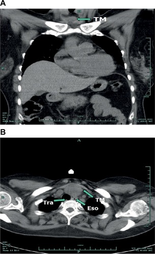
A 50-year-old female was admitted to our hospital after the diagnosis of infiltrative ductal carcinoma of the breast. She had received the diagnosis 2 months ago and had begun chemotherapy according to the EC schedule (epirubicin 140 mg/m2 and cyclophosphamide 800 mg/m2). The physical examination after admission revealed a remarkable abnormal finding, a palpable mass was examined at the anterior clavicle of the neck. Chest computed tomography (CT) findings demonstrated a mass in the anterosuperior mediastinum (). Blood tests including alpha-fetoprotein, acetylcholine receptor antibody (AchR-Ab) and titin antibody (Titin-Ab) as markers for myasthenia gravis, beta-human chorionic gonadotropin (β-HCG), CA19.9 and human immunodeficiency virus (HIV) were all within normal range. Abdominal CT, brain magnetic resonance imaging (MRI), colonoscopy, otolaryngological and gynecological examinations revealed no evidence of other abnormalities.
Figure 1 (A) Chest contrast-enhanced CT scan in the sagittal planes indicate the tumor. (B) CT in the coronal planes before surgery showing a mass lesion in the anterosuperior mediastinum.
Abbreviations: CT, computed tomography; Eso, espohagus; Tra, trachea; TM, tumor.
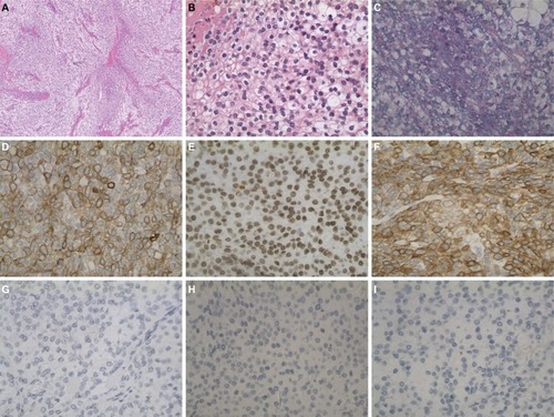
Partial thymectomy via median sternotomy and radical mastectomy were performed, and the surgically resected tumor was a 4.0 × 3.0 × 3.0 cm3 solid white mass obtained from the anterosuperior mediastinum. Histopathological examination with H&E staining showed the tumor with a lobulated architecture composed of undifferentiated clear cells, with the abundance and density of fibrous stroma (). The tumor appeared to have radially penetrated the peripheral adipose tissue. showed homogeneous neoplastic cells with abundant, clear cytoplasm, round or ovoid nuclei and a low nuclear/cytoplasmic ratio. Nuclear atypia was not marked, and nuclear chromatin was finely dispersed. Mucicarmine stain was negative in this case when tested, while the material was positive for Periodic acid Schiff (PAS) (). The neoplastic cells were positive for CKpan, p63 and 34βE12 (). In contrast, neoplastic cells were negative for TTF-1, GATA-3 and HMB45 (), and histological examination from left breast tumor demonstrated the typical breast infiltrating ductal carcinoma. Immunohistochemistry with estrogen receptor (ER) and progesterone receptor (PR) demonstrated negative cells in the breast tumor. The human epidermal growth factor (HER2/neu) IHC score was 3+, and the Ki-67 labeling index was 30% in the resected breast tumor.
Figure 2 (A) Microscopic appearance of the surgical specimen. Large lobules were composed of tumor cells closely spaced with fibrotic stroma of varying abundance and density (×40). Bar, 250 μm. (B) Uniform neoplastic cells with abundant, clear cytoplasm, nuclear atypia was not marked, and nuclear chromatin was finely dispersed, nucleoli were not prominent (×400). Bar, 25 μm. (C) Hyaline material is PAS-positive (×200). Bar, 50 μm. The neoplastic cells were positive for pan-cytokeratin (CKpan). (D) (×200), p63, (E) (×200), 34βE12, (F) (×200). Bar, 50 μm. In contrast, neoplastic cells were negative for TTF-1 (G) (×200), GATA-3 (H) (×200), HMB45 (I) (×200). Bar, 50 μm.
Abbreviations: PAS, Periodic acid Schiff; TTF-1, thyroid transcription factor 1; GATA-3, GATA binding protein 3; HMB45, human melanoma black 45.
We made a diagnosis of thymic clear cell carcinoma based on the histological and immunophenotypical findings. According to the Masaoka staging system, the tumor at the diagnosis presented in stage IIb. The patient was also evaluated as having stage IIIa (T3N1M0) breast cancer. Later, our patient was treated systemically with four cycles of chemotherapy which consisted of epirubicin, cyclophosphamide, docetaxel and trastuzumab (herceptin) after surgery. She underwent another operation for total thymectomy and the removal of surrounding adipose and received adjuvant radiotherapy administered to mediastinal lesion in the other hospital after our treatment. The patient performed well within 12 months after surgery, and the follow-up tests showed no symptoms of local recurrence or metastasis.
Systematic literature review
Our comprehensive review of reported literature of primary thymic clear cell carcinoma revealed only 18 cases. The detailed clinical and pathological information collected from all 19 cases (including our case) are summarized in . Eighteen patients had been diagnosed thymic clear cell carcinoma histologically either by biopsy or by surgical resection, and only one case was confirmed by cytology. Among the 19 patients, the reported age ranged between 33 and 84 years (average 52.6 years) with a slight male predominance (63.2%). The most common clinical manifestation was a mediastinum mass discovered incidentally. Eight patients were asymptomatic on admission, and five patients had chest pain. The reported tumor size ranged between 3 and 12 cm (average 7.8 cm). In 13 cases, including ours, complete resection was performed to obtain the diagnosis of thymic clear cell carcinoma. The treatment for thymic clear cell carcinoma was described in 18 cases, only six patients received chemoradiation therapy in which the tumor was resected. Of the 18 cases reported, eight had spread metastases, with survival ranging from 2 weeks to 13 years, while the other five patients remained alive without significant recurrence during their follow-up. In our case, the patient was in good health with no detectable recurrence at the time of writing (October 2017).
Table 1 The previously reported examples of thymic clear cell carcinoma
Discussion
Thymic epithelial tumors (TETs) are the second most common tumors of the mediastinum (21%) and the most predilection sites of the anterosuperior mediastinum.Citation15 However, thymic carcinoma is extremely rare and represents less than 1% of the thymic malignancies. Thymic carcinoma was defined as a malignant tumor that was completely different from thymoma in cell morphology and immunohistochemistry by Levine and Rosai in 1978.Citation16 Thymic carcinoma shows much more aggressive clinical behavior than thymoma, generally tends to spread radially into the adjacent thymus and adipose tissue and widespread metastases are common. Thymic carcinoma can be divided into further subtypes, i.e., squamous cell carcinoma, basaloid carcinoma, mucoepidermoid carcinoma, lymphoepithelioma-like carcinoma, clear cell carcinoma, sarcomatoid carcinoma, adenocarcinoma, NUT carcinoma, undifferentiated carcinoma and other rare thymic carcinoma according to the 4th edition of WHO classification of tumors of the lung, pleura, thymus and heart.Citation2 Several staging systems have been evolved over the years, the WHO staging system using the TNM classification and the Masaoka staging system are the most commonly used.Citation3,Citation15,Citation17
Thymic clear cell carcinoma, as histologic of cells with an optically clear cytoplasm, was first reported by Snover et al in 1982.Citation4 Microscopically, clear cell of the thymus is often not active in cytology, which is in contrast to its clinical invasion. Thymic clear cell carcinoma usually shows the lobulated structure, and the tumor cells are nested, lobed, and flaky, which are surrounded by hyperchromatic fibrous stroma. The blood sinus vascular structure of the metastatic lesion of renal cell carcinoma is lacking.Citation9 The tumors have a broad range of cytological features, ranging from uniform clear cells with minimal atypia to the pleomorphic neoplasm cells with obvious nucleoli. Cytoplasm is abundant, mostly transparent or granular, and can be mildly eosinophilic. In some cases, scattered lymphocytes, focal necrosis and transitions from areas of conventional squamous cell carcinoma can be observed. Although sometimes the tumor has well-defined periphery, it is characterized by the biological features of infiltrative, and surrounding fatty tissue or residual thymus around the mediastinal could be invaded.
The top differential diagnosis is metastatic renal cell carcinoma or a metastasis from other clear cell tumors.Citation18 Although renal cell carcinoma bears some similarities in cytologic and architectural, features such as glands with extensive luminal hemorrhage and sinusoidal vasculature are lacking in thymic clear cell carcinoma. Immunoreactivity for high-molecular weight keratins would support thymic clear cell carcinoma, and conversely, strong staining for PAX8 and vimentin supports metastatic renal cell carcinoma. In our case, the tumor cells were positive for 34βE12 and p63 and negative for PAX8 and vimentin. Additionally, a normal abdominal CT scan would exclude a renal primary. Primary lung and thyroid carcinoma both may show clear cell changes, frequently metastasize or extend into the mediastinum, but they exhibit histologic and immunohistochemical differences from thymic clear cell carcinoma.Citation9 Again, the radiographic exclusion of a lung mass at the time of diagnosis of thymic neoplasm is important. In our case, given the absence of a pulmonary mass on initial radiographic evaluation, the tumor cells were negative for TTF-1; a diagnosis of thymic clear cell carcinoma was rendered, excluding primary from metastatic lung.
Differential diagnosis should be discussed with other primary tumors, including mediastinal seminoma, parathyroid carcinoma, balloon cell melanoma and thymoma with clear cell component. Primary mediastinal seminomas, which occur almost exclusively in men, are morphologically similar to testicular seminomas, with small tumor lobules in a stroma containing a marked lymphocytic infiltrate.Citation19 Cytologically, seminoma cells are more pleomorphic, with vesicular nuclei and obvious nucleoli, in contrast to the bland nuclear feature of thymic clear cell carcinoma.Citation9 Seminomas in other extragonadal sites as well as primary testicular tumors are generally positive for placental alkaline phosphatase (PLAP).Citation20 In contrast, thymic clear cell carcinoma is focally positive for keratins and is generally unreactive for PLAP. Parathyroid carcinoma, although cytologically similar to thymic clear cell carcinoma, has broad trabecular, rather than lobular, architecture (or both) of endocrine differentiation and is more diffusely parathyroid hormone (PTH)-positive in the clear cells. Pure clear-cell variants of melanoma composed predominantly or entirely of cells with large amounts of clear, vacuolated cytoplasm, due to intracytoplasmic glycogen may be confused with thymic clear cell carcinoma. In such cases, immunohistochemical staining is a valuable diagnostic adjunct, as melanoma is uniformly positive for S-100, HMB-45, and Melan-A. The changes of clear cells were often found in WHO Type B3 thymoma and are almost focal lesions. In most tumors, these are gradual migrations of clear cell lesions, with the prominent traditional Type B3 thymoma region observed. Necrotic, hyperproliferative, fibroplasia or P53 overexpression were lacking and PAS were negative.
In recent years, a large number of studies have been published in the literature, attempting to identify specific immunohistochemical markers that can help diagnose and identify primary thymic carcinoma.Citation18 The use of immunomarkers is rather limited as there is not a single immunohistochemical stain that can unequivocally separate thymic clear cell carcinoma from other tumors. In general, CK (pan) is strongly positive in the tumor cells of thymic clear cell carcinoma. Variable staining has been described for CD5, c-kit and EMA, whereas Pax8, S-100, PLAP and TTF-1 are usually negative.Citation21 These markers are not specific, and especially, the clear cell variant of thymic carcinoma can show variable expression for CD5Citation22,Citation23 and is often negative for c-kit.Citation13,Citation24 In addition, tumor cells usually show strong cytoplasmic diastase-labile periodic acid-Schiff positivity, but the Mucicarmine stain is negative. All the immunophenotypic features described above are illustrated in our case, which supports the diagnosis of clear cell carcinoma of the thymus.
Clear cell carcinomas of the thymus are highly malignant, aggressive mediastinal neoplasms with frequent local recurrences and distant metastases. The specific criteria to confirm this tumor have not yet been established. However, based on its rarity, challenging histologic diagnosis, more cases should be collected to better define clinical behavior, histopathology, pathogenesis and other relevant information.
Disclosure
The authors report no conflicts of interest in this work.
References
- SusterSRosaiJThymic carcinoma. A clinicopathologic study of 60 casesCancer199167102510321991250
- HartmannCARothCMinckCNiedobitekGThymic carcinoma. Report of five cases and review of the literatureJ Cancer Res Clin Oncol199011669822179229
- KimDJYangWIChoiSSKimKDChungKYPrognostic and clinical relevance of the World Health Organization schema for the classification of thymic epithelial tumors: a clinicopathologic study of 108 patients and literature reviewChest200512775576115764754
- SnoverDCLevineGDRosaiJThymic carcinoma. Five distinctive histological variantsAm J Surg Pathol198264514707125053
- WolfeJTWickMRBanksPMScheithauerBWClear cell carcinoma of the thymusMayo Clin Proc1983583653706855274
- StephensMKhalilJGibbsARPrimary clear cell carcinoma of the thymus glandHistopathology1987117637653623438
- KuoTTChangJPLinFJWuWCChangCHThymic carcinomas: histopathological varieties and immunohistochemical studyAm J Surg Pathol19901424342294778
- TruongLDModyDRCaglePTJackson-YorkGLSchwartzMRWheelerTMThymic carcinoma. A clinicopathologic study of 13 casesAm J Surg Pathol1990141511661689123
- HasserjianRPKlimstraDSRosaiJCarcinoma of the thymus with clear-cell features. Report of eight cases and review of the literatureAm J Surg Pathol1995198358417793482
- OkudaMHuangCLHabaRYokomiseHClear cell carcinoma originating from ectopic thymusGen Thorac Cardiovasc Surg20095726927119440827
- NakanoTEndoSTsubochiHNokubiMWatanabeYKoyamaSThymic clear cell carcinomaGen Thorac Cardiovasc Surg2010589810020155348
- HsuY-HClear cell carcinoma of the thymusTzu Chi Med J201123151152 Available from: https://www.sciencedirect.com/science/article/pii/S1016319011000231Accessed March 03, 2018
- LaleSATiscornia-WassermanPGAzizMDiagnosis of thymic clear cell carcinoma by cytologyCase Rep Pathol2013201361781024175107
- BertocchiPMeriggiFZambelliCZorziFZaniboniAClear cell thymic carcinoma: a case reportTumori2015101e737425702670
- VenutaFAnileMDisoDThymoma and thymic carcinomaEur J Cardiothorac Surg201037132519615917
- LevineGDRosaiJThymic hyperplasia and neoplasia: a review of current conceptsHum Pathol19789495515361541
- TomaszekSWigleDAKeshavjeeSFischerSThymomas: review of current clinical practiceAnn Thorac Surg2009871973198019463649
- MoranCASusterSThymic carcinoma: current concepts and histologic featuresHematol Oncol Clin North Am20082239340718514123
- BerghNPGatzinskyPLarssonSLundinPRidellBTumors of the thymus and thymic region: III. Clinicopathological studies on teratomas and tumors of germ cell typeAnnals Thoracic Surg197825107111
- BentleyAJParkinsonMCHardingBNBainsRMLantosPLA comparative morphological and immunohistochemical study of testicular seminomas and intracranial germinomasHistopathology1990174434492076869
- WeissferdtAMoranCAImmunohistochemistry in the diagnosis of thymic epithelial neoplasmsAppl Immunohistochem Molec Morphol201522479487
- DorfmanDMShahsafaeiAChanJKThymic carcinomas, but not thymomas and carcinomas of other sites, show CD5 immunoreactivityAm J Surg Pathol1997219369409255257
- KuoTTChanJKThymic carcinoma arising in thymoma is associated with alterations in immunohistochemical profileAm J Surg Pathol199822147414819850173
- WeissferdtAMoranCAThymic carcinoma, part 1: a clinicopathologic and immunohistochemical study of 65 casesAm J Clin Pathol201213810311422706865