
Abstract
Purpose
The present study aimed to study the role of autophagy in the radiosensitivity of the radioresistant human nasopharyngeal carcinoma cell line CNE-2R.
Methods
Before being irradiated, CNE-2R cells were treated with the autophagy inhibitor chloroquine diphosphate (CDP) or the autophagy inducer rapamycin (RAPA). Microtubule-associated protein light chain 3 (LC3-II) and p62 were assessed using Western blotting analysis 48 hours after CNE-2R cells were irradiated. The percentage of apoptotic cells was assessed via flow cytometry. CNE-2R cell viability was evaluated using the Cell Counting Kit-8 (CCK8). The radiosensitivity of cells was assessed via clone formation analysis.
Results
The level of autophagy in CNE-2R cells improved as the radiation dose increased, reaching the maximum at a dose of 10 Gy. Autophagy was most significantly inhibited by 60 µmol/L CDP in CNE-2R cells, but was obviously enhanced by 100 nmol/L RAPA. Compared with the irradiation (IR) alone group, in the IR + CDP group, autophagy was significantly inhibited, viability was low, the rate of radiation-induced apoptosis was increased, and radiosensitivity was upregulated. In contrast, cells of the IR + RAPA group exhibited greater autophagy, higher viability, a lower rate of radiation-induced apoptosis, and downregulated radiosensitivity.
Conclusion
The autophagy level is negatively correlated with radiosensitivity for the radio-resistant human nasopharyngeal carcinoma cell line CNE-2R.
Introduction
Nasopharyngeal carcinoma (NPC) is endemic in southern China, southeast Asia, and northern Africa.Citation1,Citation2 According to a survey from the International Agency for Research on Cancer, there were an estimated 86,700 new cases and 50,800 NPC-related deaths in 2012.Citation1 Radiotherapy (RT) is the primary treatment option. Due to advances in disease management, diagnostic imaging, radiotherapy technology, and the broader application of systemic therapy, the prognosis of NPC has improved significantly.Citation3,Citation4 Nevertheless, local recurrence and distant metastasis after RT remain the main failure patterns in NPC and are closely associated with the radioresistance of NPC.Citation5,Citation6 Therefore, an urgent need exists to identify an approach that can eliminate or reverse radioresistance in NPC cells.
In cancer cells, autophagy is a conserved response to various anticancer therapies, including chemotherapy and radiotherapy.Citation7 Autophagy is characterized by the sequestration of bulk cytoplasm and organelles in double- or multimembrane autophagic vesicles as well as the delivery of these vesicles to, and subsequent degradation by, the lysosomal system of the cell. The process starts with the formation of the autophagosome or autophagic vacuole. Many studies have shown that autophagy is a basis for the radioresistance of various tumors, including lung cancer,Citation8 hepatocellular carcinoma,Citation9 and colorectal cancer.Citation10
In our previous study, we found that autophagic inhibition could enhance the radiosensitivity of the NPC cell line CNE-2.Citation11 In addition, we used fractionated radiation in vitro to construct a radioresistant NPC (CNE-2R) cell line.Citation12 To our knowledge, few studies have demonstrated the relationship between autophagy and the radioresistance of CNE-2R. We hypothesized that the level of autophagy had a negative correlation with the radiosensitivity of CNE-2R. In this study, we have examined the role of autophagy in the radiosensitivity of the radioresistant human NPC cell line CNE-2R.
Materials and methods
Cell culture
A human NPC cell line, CNE-2, which was constructed by the Cancer Hospital of Chinese Academy of Medical Sciences and Guangdong Medical University, was purchased from the Cancer Hospital of Shanghai Fudan University. CNE-2R, a radioresistant human NPC cell line, was previously constructed and maintained at the Cancer Laboratory of Guangxi Medical University.Citation12 CNE-2R cells were cultured in RPMI-1640 medium (HyClone, Logan, UT, USA) with 10% FBS (Thermo Fisher Scientifc, Waltham, MA, USA), penicillin (100 U/mL), and streptomycin (100 µg/mL) and incubated in a humidified 5% CO2 atmosphere at 37°C. The Ethics Committee of the Affiliated Tumor Hospital of Guangxi Medical University approved the study protocol.
Western blotting
Proteins were extracted 48 hours after the cells were irradiated. The quantitated cell lysates were separated on 8–12% SDS polyacrylamide gels and electroblotted onto polyvinylidene membranes. After blocking for 1.5 hours, the membranes were incubated sequentially with primary antibodies against LC3B (1:1,000 dilution; #L7543, Sigma-Aldrich Corp., St. Louis, MO, USA), P62 (1:1,000 dilution; #A0208, Abcam, Cambridge, UK), and GAPDH antibody (1:1000; #2118, Cell Signaling Technology, Boston, MA, USA) at 4°C overnight. The membranes were washed and incubated with a secondary antibody (1:15,000 dilution; #A0208, Beyotime Biotechnology, Shanghai, People’s Republic of China) for 1 hour at room temperature. Blotting images of the membranes were obtained using a BioRad Universal Hood III infrared fluorescence imaging system (BioRad Laboratories Inc., Hercules, CA, USA). GAPDH served as a control. All experiments were repeated three times.
Cell irradiation and CCK-8 assay
The Cell Counting Kit-8 (CCK-8) assay was used to assess cell viability after treatment with different doses of X-ray irradiation (IR). Cells were seeded in 96-well plates at 3 × 10³ cells/well and allowed to attach overnight. Then, they underwent IR with a 6 MV X-ray beam at 2, 4, 6, 8, or 10 Gy and were cultured for another 48 hours. After treatment, the cells were incubated with 10 µg/mL CCK-8 solution (Dojindo, Japan) for 1 hour in a humidified chamber containing 5% CO2 at 37°C. Absorbances were read on a microplate reader (BioRad) at 450 nm. Each group was assessed in five replicate wells, and three independent experiments were conducted. Cell survival was calculated using the following formula: survival rate (%) = OD/OD0h×100%.
Apoptosis assessment
Cells were irradiated with a 6 MV X-ray beam at a dose of 10 Gy and collected after 48 hours of culture. Then, the samples were stained with 5 µL Annexin V PE and 5 µL 7-aminoactinomycin D (BD Pharmingen, USA) according to the manufacturer’s instructions. Analysis was carried out immediately on an FC500 flow cytometry system. All samples were assessed in triplicate.
Colony-formation assay
Colony-formation assays were conducted to evaluate the radiosensitivity of cells after IR. Suspensions containing 200, 200, 400, 800, 1,000, and 2,000 cells were seeded into five of the six-well plates and individually exposed to doses of 0, 2, 4, 6, 8 and 10 Gy, respectively, with a 6 MV X-ray beam from an Elekta linear accelerator (Precise 1120; Elekta Instrument AB, Stockholm, Sweden) at a dose rate of 220 cGy/min. Then, the cells were incubated for another 14 days until colony appearance. Next, the colonies were fixed with carbinol for 15 minutes and stained with 0.1% Giemsa (AppliChem, Germany) for 30 minutes. Colonies with more than 50 cells were counted. All experiments were conducted three times. Dose–response curves were analyzed using the multitarget single-hit model in the GraphPad Prism 5.0 softwareCitation13,Citation14 with the formula: Survival fraction (SF)=1 − (1 − e −D/D0) N, where D0 (mean lethal dose) is the single dose of radiation that kills 63% of cells, N represents the number of intracellular radiosensitive areas; Dq (lnN*D0) is the required threshold for cell damage. When the Dq increases, the survival curve area is widened, and radioresistance is increased. SF2 is an important indicator of cell radiosensitivity; the greater the SF2, the stronger is the radioresistance.
Statistical analysis
Data were presented as mean ± SD. Statistical analyses were conducted using SPSS 17.0 (SPSS, Chicago, IL, USA) or the GraphPad Prism 5.0 software. Statistical differences were determined with a two-tailed t-test. ANOVA was used to assess the significance of the differences among the experimental groups. All cell-based experiments were conducted three or four times. A P-value <0.05 is indicated by *; A P-value <0.01 is indicated by **.
Results
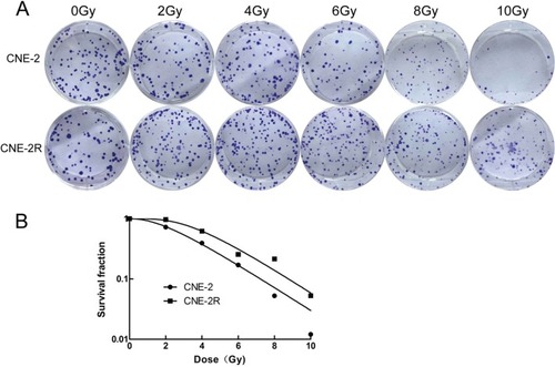
Radiosensitivity of the parental CNE-2 and radioresistant CNE-2R cell lines
Colony-formation assays were conducted to evaluate the radiosensitivity of the CNE-2R and CNE-2 cell lines (). There were more surviving CNE-2R colonies than CNE-2 colonies at doses of 2–10 Gy. Cell survival curves were fitted with the multitarget model in the GraphPad Prism 6.0 software. As shown in , CNE-2R cell survival was higher than CNE-2 survival at 2, 4, 6, 8, and 10 Gy. The main biological parameters associated with radiotherapy are shown in . Based on the Dq and survival fraction at 2 Gy (SF2) values, the repair capability of CNE-2R was stronger than that of CNE-2. Together, these data suggest that CNE-2R is radioresistant compared to CNE-2.
Table 1 Correlation parameters in the multitarget single-hit model
Figure 1 Colony-formation assays assessing the radiosensitivity of the parental CNE-2 and radioresistant CNE-2R cell lines.
Notes: (A) Colony-formation assays of the CNE-2 and CNE-2R cell lines were conducted to evaluate radiosensitivity. (B) The fit curves were generated with the GraphPad Prism 6.0 software. The results were reproduced in three independent experiments.
IR induces autophagy in CNE-2R cells
Western blotting was undertaken to examine the expression levels of autophagy-associated proteins in CNE-2R cells after 0, 2, 4, 6, 8, and 10 Gy IR. As the radiation dose increased, LC3-II protein expression in CNE-2R cells increased, and P62 expression gradually decreased; the levels of LC3-II and P62 became stable after 8–10 Gy IR (). These findings indicate that prompt radiation can activate autophagy in CNE-2R cells and that the level of autophagy reaches a plateau at 8–10 Gy. Therefore, in subsequent experiments, we used 10 Gy as the experimental IR dose.
Figure 2 Expression of LC3-I, LC3-II, and P62 in the CNE-2R cells of three groups.
Notes: (A) The CNE-2R cells exposed to different radiation doses. (B) The CNE-2R cells exposed to irradiation (IR; 10 Gy) with different concentrations of CDP. (C) The CNE-2R cells exposed to IR (10 Gy) with different concentrations of rapamycin. All experiments were repeated three times and produced identical findings.
Abbreviations: CDP, chloroquine diphosphate; IR, irradiation; LC3, light chain 3; RAPA, rapamycin.
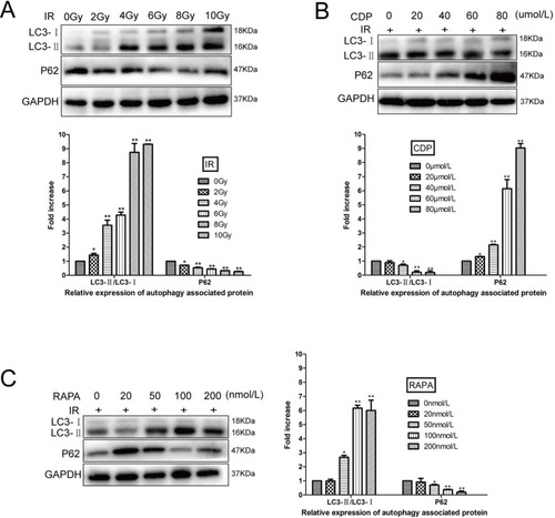
CDP inhibits the level of autophagy induced by IR (10 Gy) in CNE-2R cells, whereas RAPA increases the level of autophagy
Different concentrations of the autophagy inhibitor CDP (0, 20, 40, 60, and 80 µmol/L) and the autophagy activator RAPA (0, 20, 50, 100, and 200 nmol/L) were used for CNE-2R cells 12 hours before IR (10 Gy). After 48 hours, proteins were extracted, and Western blotting was undertaken to examine autophagy-related protein expression in CNE-2R cells. Our results showed that autophagy protein expression was inhibited by CDP. At 60–80 µmol/L CDP, the level of LC3-II decreased significantly, and P62 expression increased significantly, indicating that autophagy was clearly inhibited (). With regard to the responses to different concentrations of RAPA, the expression levels of LC3-II and P62 were most notably increased and decreased, respectively, at a concentration of 100 nmol/L, indicating that autophagy was significantly activated ().
Comparison of autophagy levels in response to factors related to different processes
In the aforementioned portion of the study, we found that 10 Gy radiation, 60 µmol/L CDP, and 100 nmol/L RAPA had the most significant effects on the autophagy of CNE-2R cells. Therefore, these conditions were used as the experimental conditions.
In our first analysis, we used untreated CNE-2R cells as the control group. Western blotting experiments were conducted to examine the expression levels of autophagy-associated proteins LC3-II and P62 in the control group, CDP alone group, and RAPA alone group. The levels of LC3-II in the three groups were low, and the levels of P62 protein in all groups were high, with no statistically significant differences (P>0.05; ). This indicated that the autophagy levels of these three groups were extremely low without IR.
Figure 3 Expression of LC3-I, LC3-II, and P62 in the CNE-2R cells of the different groups.
Notes: (A) The control group, CDP alone group, and rapamycin (RAPA) alone group. (B) The control group, irradiation (IR) alone group, IR+ CDP alone group, and IR+ RAPA group. The results were reproduced in three independent experiments.
Abbreviations: CDP, chloroquine diphosphate; IR, irradiation; LC3, light chain 3; RAPA, rapamycin.
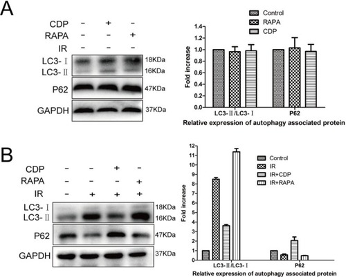
In our next analysis, we used untreated CNE-2R cells as the control group. Additionally, the 10-Gy radiotherapy group (the IR alone group), 10-Gy radiotherapy+60-µmol/L CDP group (the IR+ CDP group), and 10-Gy radiotherapy+100 nmol/L RAPA group (the IR+ RAPA group) were set up. Our results showed that LC3-II expression was significantly suppressed and P62 expression was obviously higher in the IR+ CDP group than in the IR alone group (P<0.05). These results demonstrated that IR-induced autophagy was significantly inhibited by CDP. However, the expression levels of LC3-II were clearly increased, and the expression levels of P62 were significantly decreased in the IR+ RAPA group as compared to the IR alone group, indicating that IR-induced autophagy was clearly enhanced by RAPA (all P<0.05; ).
The role of autophagy in the survival of CNE-2R cells
To assess the role of autophagy in CNE-2R cells, CCK-8 assays were conducted to assess CNE-2R cell survival after IR. As shown in , survival rates were significantly lower for the IR+ CDP group than for the control group at 2, 4, 6, 8, and 10 Gy (P<0.05 for all comparisons) but were significantly higher for the IR+ RAPA group than for the control group at 2, 4, 6, 8, and 10 Gy (P<0.05 for all comparisons). The results indicated that autophagy could increase the survival rate of CNE-2R cells.
Figure 4 The effects of autophagy on CNE-2R cell survival.
Notes: The CCK-8 assay was used to assess CNE-2R cell viability. The survival rates of the control, IR + CDP, and IR + RAPA groups were significantly different at the radiation doses of 2, 4, 6, 8, and 10 Gy (all P<0.05). The results were reproduced in three independent experiments.
Abbreviations: CDP, chloroquine diphosphate; RAPA, rapamycin.
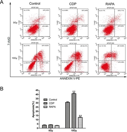
The effect of autophagy on the apoptosis of CNE-2R cells
Flow cytometry was used to determine the effects of autophagy on CNE-2R cell apoptosis (). The results showed that the incidence of apoptosis was low in the control, CDP alone, and RAPA alone groups without IR. The apoptosis rates for the control, CDP alone, and RAPA alone groups were 3.65±0.36, 4.04±0.19, and 3.35±0.41, respectively. There were no significant differences in the apoptosis rates of the three groups (all P>0.05). However, with 10 Gy IR for 48 hours, the apoptosis rates of the control, IR+ CDP, and IR+ RAPA groups were 30.65±0.68, 41.06±1.02, and 12.83±0.68, respectively (all P<0.01). These results indicated that the inhibition of autophagy in CNE-2R cells could increase the level of apoptosis, whereas the enhancement of autophagy could decrease the apoptosis rate.
Figure 5 The effects of autophagy on CNE-2R cell apoptosis.
Notes: Apoptotic cells were assessed with Annexin V–PE staining and then subjected to flow cytometry for analysis. (A) Representative images show the Annexin V–PE staining of the control group, CDP alone group, RAPA alone group, IR alone group, IR+ CDP alone group, and IR+ RAPA group. (B) Quantification of each group (**P<0.01). The results were reproduced in three independent experiments.
Abbreviations: CDP, chloroquine diphosphate; RAPA, rapamycin.
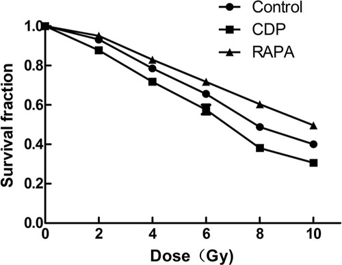
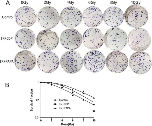
The role of autophagy in the radiosensitivity of CNE-2R cells
Colony-formation assays were conducted to evaluate the radiosensitivity of CNE-2R cells (). Cell survival curves were fitted with the multitarget model in the Graph-Pad Prism 6.0 software. The survival rate of the IR+ CDP group was lower than that of the control group, whereas the survival rate of the RAPA group was higher than those of the control and CDP groups at 2, 4, 6, 8, and 10 Gy. The main biological parameters associated with radiotherapy are shown in . Based on the Dq and SF2 values, we concluded that the higher the autophagy level was, the stronger the survival protection in CNE-2R cells against IR would be. Therefore, autophagy inhibition can increase the sensitivity of CNE-2R cells to IR, whereas autophagy enhancement can reduce radiosensitivity.
Table 2 Correlation parameters in the multitarget single-hit model
Figure 6 The role of autophagy in the radiosensitivity of CNE-2R cells.
Notes: (A) Effect of autophagy on the colony-formation ability of CNE-2R cells under different doses of IR. (B) Fit curves were generated with the GraphPad Prism 6.0 software. The results were reproduced in three independent experiments.
Abbreviations: CDP, chloroquine diphosphate; IR, irradiation; RAPA, rapamycin.
Discussion
The present study showed that the inhibition of autophagy with CDP increased the radiosensitivity of CNE-2R cells, whereas the activation of autophagy with rapamycin (RAPA) decreased the radiosensitivity of CNE-2R cells.
Autophagy is a cellular survival mechanism in which proteins and other cytoplasmic materials are recycled to support cell survival under metabolically stressful conditions, such as hunger, exposure to cytotoxic substances, and exposure to ionizing radiation. LC3 and p62 levels are often used to measure autophagy in cells.Citation15,Citation16 LC3 conversion (LC3-I to LC3-II) and the lysosomal degradation of LC3-II reflect the progression of autophagy. Another widely used autophagy marker, p62, binds directly to LC3 and GAB-ARAP (Atg8 orthologs) family proteins via a short LC3 interaction region (LIR). This may serve as a mechanism to deliver selective autophagic cargo for degradation by autophagy. When autophagy occurs, p62 decreases with protein degradation and intracytoplasmic soluble LC3-I interacts with phosphatidylethanolamine to form LC3-II, leading to a reduction in LC3-I. In the present study, when CNE-2R cells were exposed to IR, the levels of LC3-II/ LC3-I increased, and those of p62 decreased. Therefore, we concluded that autophagy was activated by IR. Moreover, our research showed that the levels of autophagy were boosted by increases in the radiation dose.
Autophagy can help tumor cells eliminate radiation damage, which is an important mechanism of radioresistance.Citation17,Citation18 Increasing evidence indicates that the inhibition of autophagy contributes to the sensitivity to anticancer treatments by promoting apoptosis and inhibiting cell proliferation.Citation9,Citation10 In 2017, Peng et al found that hepatocellular carcinoma cells with autophagic depletion proliferated much more slowly than control cells after IR exposure. Moreover, after autophagy inhibition, the levels of Bcl-2 remarkably decreased, and the levels of Bax and cleaved caspase-3 increased.Citation9 In 2018, Hu et al found that when compared to cells treated with IR alone, treatment with chloroquine, an autophagy inhibitor, sensitized colorectal cancer cells to radiation by increasing apoptosis and reducing the survival rate.Citation10 In the present study, we found that cell proliferation was reduced and that cell apoptosis was activated in CNE-2R cells that were exposed to IR when autophagy was inhibited with CDP. In contrast, the activation of autophagy leads to radioresistance during the treatment of cancer.Citation19,Citation20 In 2011, Zhuang et al found that RAPA activated autophagy in glioblastoma and that cell proliferation, viability, and clonogenic ability after radiation were reduced.Citation19 In 2016, Zhang et al found that the activation of autophagy could decrease the survival rate of non–small-cell lung cancer cells and thus lead to radioresistance.Citation20 Similarly, our study showed that the radiosensitivity of CNE-2R cells was decreased by the activation of autophagy, which could enhance proliferation and reduce apoptosis.
Recent studies have shown that chloroquine or RAPA can be applied to change the sensitivity of tumor cells to chemotherapy or radiotherapy via the modulation of autophagy.Citation21,Citation22 Due to its chemical properties as a weak base, chloroquine can inhibit H+-ATPase to increase the lysosomal pH, thus inhibiting the enzymatic activity of lysosomal hydrolases and preventing the fusion of the autophagosome and lysosome for lysosomotropic activities.Citation23,Citation24 Golden et al suggested that chloroquine blocked autophagy and triggered endoplasmic reticulum stress, thereby increasing the chemosensitivity of glioma cells to temozolomide.Citation25 In addition, Ye et al showed that chloroquine enhanced the radiosensitivity of glioma-initiating cells.Citation21 The mammalian target of rapamycin (mTOR) is an evolutionarily conserved serine/threonine kinase and the founding member of a signaling pathway that regulates many fundamental features of cell growth and division.Citation26 RAPA, a specific inhibitor of mTOR, can activate autophagy by inhibiting the mTOR pathway and plays a role in tumor inhibition.Citation27 Li et al found that RAPA-induced autophagy sensitizes lung cancer cells to radiation associated with DNA damage repair inhibition.Citation28 Moreover, the present study proved that both chloroquine and RAPA changed the radiosensitivity of CNE-2R cells by regulating autophagy. Moreover, we found that the autophagy of CNE-2R cells exposed to IR was significantly inhibited by 60 µmol/L CDP and was notably activated by 100 nmol/L RAPA. In a study by Wang et al, a low concentration of chloroquine (10 µM) was applied to investigate its influence on the radiosensitivity of bladder cancer cells.Citation29 However, the study by Ye et al found that a specific dose of chloroquine (20 nmol/L) clearly enhanced the radiosensitivity of glioma-initiating cells by inhibiting autophagy.Citation21 Dai et al confirmed that autophagy was significantly induced by RAPA (50 nmol/L) in pancreatic cancer cells.Citation30 Therefore, we speculate that, for the different kinds of cancer cells, CDP and RAPA might have different optimal drug concentrations to inhibit or activate autophagy.
Conclusion
We have demonstrated that the level of autophagy has a negative correlation with the radiosensitivity of the radio-resistant human NPC cell line CNE-2R. In addition, either chloroquine or RAPA can be utilized to change the sensitivity of CNE-2R to radiotherapy via the modulation of autophagy. Further animal experiments and clinical tests are warranted to verify our findings.
Acknowledgments
This work was sponsored by grants from the National Natural Science Foundation of China (grant nos. 81160285 and 81760544), the Basic Ability Enhancement Project of Young Teachers in Guangxi Zhuang Autonomous Region (grant no. 2017KY0114), the Youth Science Foundation of Guangxi Medical University (grant no. GXMUYSF201515), and the International Communication of Guangxi Medical University Graduate Education in 2018.
Author contributions
Xiao-Dong Zhu and Zhong-Guo Liang conceived and designed the experiments; Zhong-Guo Liang, Guo-Xiang Lin, and Bin-Bin Yu conducted the experiments; Fang Su, Ling Li, and Song Qu analyzed the data; and Zhong-Guo Liang and Guo-Xiang Lin wrote the paper. Zhong-Guo Liang, Guo-Xiang Lin, and Bin-Bin Yu contributed equally to this study and are co-first authors of this manuscript. All authors contributed to data analysis, drafting and revising the article, gave final approval of the version to be published, and agree to be accountable for all aspects of the work.
Disclosure
The authors report no conflicts of interest in this work.
References
- TorreLABrayFSiegelRLFerlayJLortet-TieulentJJemalAGlobal cancer statistics, 2012CA Cancer J Clin20156528710825651787
- TangLLChenWQXueWQGlobal trends in incidence and mortality of nasopharyngeal carcinomaCancer Lett20163741223026828135
- AuKHNganRKCNgAWYTreatment outcomes of nasopharyngeal carcinoma in modern era after intensity modulated radiotherapy (IMRT) in Hong Kong: A report of 3328 patients (HKNPCSG 1301 studyOral Oncol201877162129362121
- LeeAWMaBBNgWTChanATManagement of Nasopharyngeal Carcinoma: Current Practice and Future PerspectiveJ Clin Oncol201533293356336426351355
- KongFYingHduCPatterns of local-regional failure after primary intensity modulated radiotherapy for nasopharyngeal carcinomaRadiat Oncol201496024552293
- LiYQTianYMTanSHPrognostic Model for Stratification of Radioresistant Nasopharynx Carcinoma to Curative Salvage RadiotherapyJ Clin Oncol201836989189929412781
- HönscheidPDattaKMudersMHAutophagy: detection, regulation and its role in cancer and therapy responseInt J Radiat Biol201490862863524678799
- LiYQTianYMTanSHReducing autophagy and inducing G1 phase arrest by aloperine enhances radio-sensitivity in lung cancer cellsOncol Rep2017
- PengWXWanYYGongAHEgr-1 regulates irradiation-induced autophagy through Atg4B to promote radioresistance in hepatocellular carcinoma cellsOncogenesis201761e29228134935
- HuJLHeGYLanXLInhibition of ATG12-mediated autophagy by miR-214 enhances radiosensitivity in colorectal cancerOncogenesis2018721629459645
- ZhouZRZhuXDZhaoWPoly(ADP-ribose) polymerase-1 regulates the mechanism of irradiation-induced CNE-2 human nasopharyngeal carcinoma cell autophagy and inhibition of autophagy contributes to the radiation sensitization of CNE-2 cellsOncol Rep20132962498250623563481
- GuoYZhuXDQuSIdentification of genes involved in radioresistance of nasopharyngeal carcinoma by integrating gene ontology and protein-protein interaction networksInt J Oncol2012401859221874234
- GuoSI-YANZhuX-DGeL-YRNAi-mediated knockdown of the c-jun gene sensitizes radioresistant human nasopharyngeal carcinoma cell line CNE-2R to radiationOncol Rep20153331155116025571870
- LinHChenZTZhuXDSerum CD166: A novel biomarker for predicting nasopharyngeal carcinoma response to radiotherapyOncotarget2017838628586286728968954
- JiangPMizushimaNLC3- and p62-based biochemical methods for the analysis of autophagy progression in mammalian cellsMethods201575131825484342
- SchläfliAMBerezowskaSAdamsOLangerRTschanMPReliable LC3 and p62 autophagy marker detection in formalin fixed paraffin embedded human tissue by immunohistochemistryEur J Histochem2015592248126150155
- XinYJiangFYangCRole of autophagy in regulating the radiosensitivity of tumor cellsJ Cancer Res Clin Oncol2017143112147215728786037
- dasCKMandalMKögelDPro-survival autophagy and cancer cell resistance to therapyCancer Metastasis Rev2018
- ZhuangWLiBLongLChenLHuangQLiangZInduction of autophagy promotes differentiation of glioma-initiating cells and their radiosensitivityInt J Cancer2011129112720273121384342
- ZhangXJiJYangYZhangJShenLStathmin1 increases radioresistance by enhancing autophagy in non-small-cell lung cancer cellsOnco Targets Ther201692565257427199567
- YeHChenMCaoFHuangHZhanRZhengXChloroquine, an autophagy inhibitor, potentiates the radiosensitivity of glioma initiating cells by inhibiting autophagy and activating apoptosisBMC Neurol201616117827644442
- KaurASharmaSMammalian target of rapamycin (mTOR) as a potential therapeutic target in various diseasesInflammopharmacology201725329331228417246
- RubinszteinDCCodognoPLevineBAutophagy modulation as a potential therapeutic target for diverse diseasesNat Rev Drug Discov201211970973022935804
- Avniel-PolakSLeibowitzGRiahiYGlaserBGrossDJGrozinsky-GlasbergSAbrogation of Autophagy by Chloroquine Alone or in Combination with mTOR Inhibitors Induces Apoptosis in Neuroendocrine Tumor CellsNeuroendocrinology2016103672473726619207
- GoldenEBChoHYJahanianAChloroquine enhances temozolomide cytotoxicity in malignant gliomas by blocking autophagyNeurosurg Focus2014376E12
- LiuQThoreenCWangJSabatiniDGrayNSmTOR Mediated Anti-Cancer Drug DiscoveryDrug Discov Today Ther Strateg200962475520622997
- WeichhartTMammalian target of rapamycin: a signaling kinase for every aspect of cellular lifeMethods Mol Biol201282111422125056
- LiYLiuFWangYRapamycin-induced autophagy sensitizes A549 cells to radiation associated with DNA damage repair inhibitionThorac Cancer20167437938627385978
- WangFTangJLiPChloroquine Enhances the Radiosensitivity of Bladder Cancer Cells by Inhibiting Autophagy and Activating ApoptosisCell Physiol Biochem2018451546629316551
- DaiZ-JGaoJMaX-BAntitumor effects of rapamycin in pancreatic cancer cells by inducing apoptosis and autophagyInt J Mol Sci201214127328523344033