
Abstract
Background and purpose
Abundant studies have shown that lncRNA PANDAR plays an oncogenic role in human solid tumors. Although abnormal expression of PANDAR has been well investigated in solid tumors, it was rarely studied in hematologic diseases. Hence, the aim of this study was to determine the PANDAR expression level and its clinical significance in patients with acute myeloid leukemia (AML).
Materials and methods
For detecting the expression level of PANDAR in 119 AML patients and 26 controls, real-time quantitative PCR was used in this study. The prognostic values were evaluated by using Kaplan–Meier analysis, Cox regression analyses, and logistic regression analysis.
Results
PANDAR was significantly overexpressed in AML and might be a promising biomarker which could distinguish AML from normal samples (P<0.001). Patients with high expression of PANDAR (PANDARhigh) were older and showed higher bone marrow blasts than patients in PANDARlow group (P=0.029 and 0.032, respectively). Significant differences between these groups were also detected regarding risk group and karyotype finding (P=0.009 and 0.041, respectively). Importantly, PANDARhigh patients presented a significant lower complete remission rate compared to PANDARlow patients (P<0.001). Furthermore, Kaplan–Meier analysis showed that PANDARhigh patients had shorter overall survival compared to PANDARlow patients observing the whole AML cohort, and also in the non-M3 group of patients (P<0.001 and P=0.005, respectively). Multivariate analysis of Cox and logistic regression analysis confirmed that high PANDAR expression was an independent unfavorable risk factor for overall survival and complete remission in both observed patient groups.
Conclusion
These results revealed that PANDAR was overexpressed in AML, and that higher PANDAR expression was associated with poor clinical outcome. Our study therefore suggests that PANDAR expression is a promising biomarker for prognostic prediction for AML.
Introduction
Acute myeloid leukemia (AML) is a cytogenetically and molecularly heterogeneous disease which is marked by uncontrolled clonal expansion of blast cells.Citation1 Although the new treatment strategies based on molecular biology of AML have been adopted in recent years, the prognosis of the disease remains poor.Citation2–Citation4 It has become apparent that karyotype abnormalities have important value for AML diagnosis classification, prognostic evaluation, and guiding individual treatment.Citation5,Citation6 Cytogenetic aberrations together with several gene mutations including NPM1, CEBPA, TP53, TET2, DNMT3A, and FLT3-ITD have a strong impact on clinical outcome of AML patients.Citation7 In addition to genetic abnormalities, the aberrant expression of some genes, such as overexpression of ERG, BAALC, and EVI1, also has been proven to affect prognosis for AML patients.Citation7 These important findings open up a new field for discovering novel promising biomarkers for AML patients, especially for those who are at risk of poor outcome, so that these patients can be treated with optimized treatment strategies.
Long noncoding RNAs (lncRNAs) are regarded as a kind of noncoding RNA, which are longer than 200 nucleotides. Recently, many studies have reported that lncRNAs play vital roles in gene expression regulation through association with key transcription factors and microRNAs.Citation8,Citation9 lncRNAs could act as an important component in every step of cell biology, which includes the adjustments of transcription initiation and transcription and posttranscriptional level.Citation10 Recently, increasing number of research papers revealed that lncRNAs were relevant to many human diseases, especially to human cancers, and many studies began to explore the molecular mechanisms of lncRNA function in the pathogenesis of these disease or cancers.Citation11 With the deepening of the research, it is becoming increasingly apparent that most of the susceptibility to cancer is not caused by the variation of coding sequences of DNA but by the noncoding regulatory sequences, especially by lncRNAs.Citation10
lncRNA PANDAR, which is located at 6p21.2, plays a vital role in regulation of apoptosis by inhibiting the expression of proapoptotic genes through interaction with the transcription factor NF-YA.Citation12 To date, the abnormal expression of PANDAR has been reported in various solid cancers, such as hepatocellular carcinoma, gastric cancer, and breast cancer.Citation13 However, there are few reports about the expression of PANDAR in blood cancer. Therefore, we focused on exploring the PANDAR expression level and its connection with clinical implication in AML patients.
Materials and methods
Patients and treatment
A total of 119 de novo AML patients and 26 healthy donors were included in the present research, which was approved by the Ethics Committee and Institutional Review Board of the Affiliated People’s Hospital of Jiangsu University. Bone marrow (BM) was collected from all the participants after they signed the informed consents. BM mononuclear cells (BMMNCs) were extracted from BM specimen using Lymphocyte Separation Medium (TBD Sciences, Tianjin, People’s Republic of China). Treatment protocols for AML were described previously.Citation14
Cytogenetics and mutation analysis
By conventional R-banding method, karyotype was analyzed at the time of initial diagnosis. Risk classification based on the karyotype findings has been done as previously reported.Citation15 Mutations in C-KIT, NPM1, DNMT3A, N/K-RAS, and U2AF1 were detected by high-resolution melting analysis,Citation16–Citation20 whereas FLT3-ITD and CEBPA mutations were detected by direct DNA sequencing.Citation21,Citation22
RNA isolation and reverse transcription
Total RNA was extracted by using Trizol reagent (Invitrogen, Carlsbad, CA, USA). The specific procedure of reverse transcription was conducted as previously reported.Citation23
Real-time quantitative PCR
The primers for PANDAR are as follows: forward: 5′-CTCCATCATGCCAA GTTCTGC-3′ and reverse: 5′-GAAGGCAGGCAAGACTCGAA-3′. PANDAR expression was detected by real-time quantitative PCR using AceQ qPCR SYBR Green Master Mix (Vazyme Biotech Co., Pis-cataway, NJ, USA). The reaction condition of real-time quantitative PCR was conducted as reported earlier.Citation24,Citation25 Relative PANDAR expression levels were calculated by 2−ΔΔCT method.
Statistical analysis
SPSS software version 20.0 (IBM Corporation, Armonk, NY, USA) was used to carry out the statistical analysis. Meanwhile, receiver operating characteristic (ROC) curve and area under the ROC were applied to assess the value of PANDAR expression. Besides, Pearson’s chi-squared analysis was conducted to detect the difference of categorical variables between PANDARhigh group and PANDARlow group. Through Kaplan–Meier method and Cox regression analysis, the effect of PANDAR expression on prognosis was analyzed. Logistic regression analysis was used to identify the independent risk factors of complete remission (CR). In all tests, P<0.05 was defined as statistically significant.
Results
PANDAR expression in AML
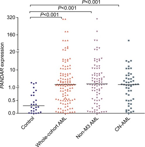
The expression level of PANDAR in controls ranged from 0.000 to 2.926 (median 0.294). PANDAR transcript level in AML patients ranged from 0.005 to 306.109 (median 1.862). Through nonparametric test, PANDAR was found to be significantly upregulated in AML (P<0.001, ). Besides this, significant upregulation of PANDAR was also found in non-M3-AML and cytogenetically normal AML subgroup of patients ().
Figure 1 Expression of PANDAR in controls, whole-cohort AML patients, non-M3 AML patients, and CN-AML patients.
Notes: The distributions of the PANDAR expression in controls, whole-cohort AML patients, non-M3 AML patients, and CN-AML patients are presented with scatter plots. The median level of PANDAR expression in each group is shown with horizontal line.
Abbreviations: AML, acute myeloid leukemia; CN-AML, cytogenetically normal AML.
Distinguishing capacity of PANDAR expression
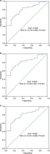
The ROC curve analysis was applied to evaluate whether PANDAR expression could be used as a biomarker for the diagnosis of AML. The results showed that area under the curve value was 0.800 (95% CI: 0.716–0.883), which suggested the PANDAR expression level might be a potential biomarker in discriminating AML from controls (P<0.001, ). In addition, when the cutoff value was 0.840, the sensitivity and specificity of diagnosis of AML were 65.5% and 80.8%. For non-M3-AML and CN-AML patients, significant differences also existed (, respectively).
Figure 2 Discriminative capacity of PANDAR expression by ROC curve analysis.
Notes: (A) For whole-cohort AML. (B) For non-M3 AML. (C) For CN-AML.
Abbreviation: AML, acute myeloid leukemia; AUC, area under the curve; CN-AML, cytogenetically normal AML; ROC, receiver operating characteristic.
The connection between PANDAR expression level and clinical characteristics in AML
By the set cutoff value based on the basis of ROC curve, the whole cohort of AML patients was divided into two groups. Clinical features and laboratory parameters representation between PANDARhigh and PANDARlow groups is separately shown in . No significant differences were observed in sex, white blood cells (WBCs), hemoglobin, and platelets between two groups (P>0.05). However, patients with PAN-DAR high expression were older than patients in the PANDAR low-expressed group (P=0.029). Patients in PANDARhigh group showed higher BM blasts than patients in PANDARlow group (P=0.032). Moreover, significant differences between these two groups were also detected regarding risk group and karyotype finding (P=0.009 and 0.041, respectively). Patients in PANDARhigh group had higher frequency of poor karyotypes (15%, 12/78) than patients in PANDARlow group (2%, 1/41). There was no correlation between PANDAR expression and the common gene mutations (, P>0.05).
Table 1 Comparison of clinical manifestations and laboratory features between AML patients with low and high PANDAR expression
Effect of PANDAR expression on chemotherapy response in AML
In order to explore the impact of PANDAR expression in clinical prognosis with AML patients, we analyzed 115 AML patients with available follow-up data. Compared with PANDARlow group, patients in PANDARhigh group had a lower CR rate (P<0.001, ). We then analyzed the expression level of PANDAR in AML patients who achieved CR and those without CR, and showed it in scatter plots (P<0.001, ). Additionally, clinical characteristics of patients with CR and non-CR were further compared. Significant differences were found in PANDAR expression, age, WBCs, BM blast, risk group, and karyotype finding (P<0.05, ). Logistic regression analysis including the most predictive factors was further performed which revealed that PANDAR expression was an independent risk factor that affected CR in whole-cohort AML and non-M3 AML patients (P=0.010 and 0.005, respectively, and ).
Table 2 Comparison of clinical manifestations and laboratory features between CR and non-CR in AML patients receiving induction therapy
Table 3 Univariate and multivariate analyses of variables for CR and OS in whole-cohort AML patients
Table 4 Univariate and multivariate analyses of variables for CR and OS in non-M3 AML patients
Figure 3 Expression of PANDAR in CR and non-CR AML patients receiving induction therapy.
Notes: The distributions of the PANDAR expression in CR and non-CR groups are illustrated with scatter plots. The median level of PANDAR expression in each group is shown with horizontal line.
Abbreviations: AML, acute myeloid leukemia; CR, complete remission.

The relationship between PANDAR expression and prognosis in AML patients
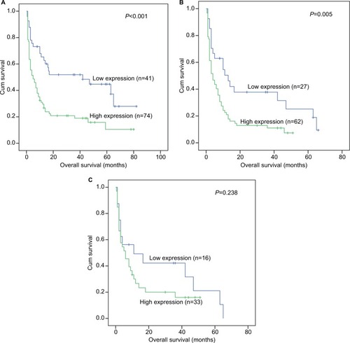
The survival analysis indicated that in the whole-cohort AML patients with high PANDAR expression had a shorter overall survival (OS) time than those who were in PANDAR low-expressed group (P<0.001, ). In non-M3 AML, patients with PANDAR high expression also had a shorter OS compared with those with PANDAR low expression (P=0.005, ). Regretfully, patients with PANDAR high expression did not presented a significant shorter OS than patients with PANDAR low expression among CN-AML (P=0.238, ). Multivariate analysis which included variables in univariate analysis with P<0.2 (WBC [≥30×109/L vs <30×109/L], age [≤60 vs >60 years], risk group [favorable vs intermediate vs poor], PANDAR expression [high vs low], gene mutations [mutant vs wild type]). Multivariate analysis further showed that PANDAR expression was a significant independent risk factor in affecting OS among whole-cohort AML patients and non-M3 AML patients (P=0.033 and 0.032, respectively, and ).
Figure 4 Prognostic value of PANDAR expression in AML.
Notes: (A) For whole-cohort AML patients. (B) For non-M3 patients. (C) For CN-AML patients. Overall survival was analyzed between PANDARhigh and PANDARlow groups and performed by Kaplan–Meier method.
Abbreviations: AML, acute myeloid leukemia; CN-AML, cytogenetically normal AML; cum, cumulative.
Discussion
Lately, more and more researchers are devoted to exploring noncoding RNA and AML.Citation26 Many studies have proved that lncRNAs indeed played an important regulatory role in human cancers, and it was closely related with the occurrence and the development of various tumors.Citation14,Citation27 Also, increasing number of research papers have shown that the abnormal expression of PANDAR was connected with the tumorigenesis of various solid tumors.Citation28–Citation32 In the first report published by Hung et al, it was indicated that PANDAR inhibited the expression of proapoptotic genes by interacting with the transcription factor NF-YA.Citation12 Thereafter, Li et alCitation28 found that PANDAR was upregulated in thyroid cancer. Further investigating the regulatory mechanism of PANDAR, Li et alCitation28 also found that knockdown of PANDAR could promote apoptosis of thyroid cells by reducing the expression of Bcl2 and activating Bax. In addition, Sang et alCitation30 also reported that PANDAR, which was obviously upregulated in breast cancer tissues and cell lines, could affect the cell cycle by regulating its downstream target p16INK4A. In summary, PANDAR played a significant role in various cancers, including in cancer initiation and progression, and it could serve as an oncogene in these cancers.
In the studies examining the expression level of PANDAR, many reports showed that PANDAR was associated with the prognosis of cancers. For instance, Li et al found that PANDAR was upregulated in thyroid cancer tissue and cell lines, and it could be a promising therapeutic target and important biomarker for thyroid cancer.Citation28 Similarly, an article reported that the expression level of PANDAR in hepatocellular carcinoma was crucially associated with the size of tumor nodule, vascular invasion, and TNM stage.Citation29 Moreover, overexpression of PANDAR was relevant to the poorer survival and shorter recurrence duration for the disease in hepatocellular carcinoma patients, and it could be recognized as a potential tumor biomarker and therapeutic target.Citation29 However, the effect of PANDAR expression on prognosis in blood malignancies remains poorly defined. Findings from our study demonstrated that high expression of PANDAR indicated a poor prognosis in AML patients. PANDAR expression level influenced CR rate, with the PANDARhigh group having lower CR rate in comparison to the PANDARlow group. Logistic regression analysis showed that PANDAR expression was an independent prognostic factor for CR. More importantly, Kaplan–Meier survival analyses clearly showed that patients with higher expression of PANDAR had a shorter OS than those patients with lower expression. Univariate and multivariate Cox regression analyses revealed the increased PANDAR expression was an independent unfavorable risk factor in AML patients.
Our study was the first to report that PANDAR was upregulated in AML and was also the first to demonstrate the prognostic value of PANDAR in AML.
Conclusion
Expression of PANDAR was frequently upregulated in AML, and high expression of PANDAR as an independent unfavorable risk factor for CR and OS in whole-cohort and non-M3 AML patients. Therefore, our findings indicated that PANDAR was a potential biomarker for AML and it might effectively predict the outcome of AML patients.
Acknowledgments
This work was supported by National Natural Science Foundation of China (81270630), Medical Innovation Team of Jiangsu Province (CXTDB2017002), 333 Project of Jiangsu Province (BRA2016131), Six Talent Peaks Project in Jiangsu Province (2015-WSN-115), Zhenjiang Clinical Research Center of Hematology (SS2018009), China Postdoctoral Science Foundation funded project (2016M601748), Youth Medical Talents Project of “Ke Jiao Qiang Wei” project of Jiangsu province (QNRC2016450, QNRC2016449), and Key Medical Talent Program of Zhenjiang City.
Disclosure
The authors report no conflicts of interest in this work.
References
- DöhnerHWeisdorfDJBloomfieldCDAcute Myeloid LeukemiaN Engl J Med2015373121136115226376137
- EsteyEDöhnerHAcute myeloid leukaemiaLancet200636895501894190717126723
- FerraraFUnanswered questions in acute myeloid leukaemiaLancet Oncol20045744345015231251
- AviviIRoweJMPrognostic factors in acute myeloid leukemiaCurr Opin Hematol2005121626715604893
- GrimwadeDThe clinical significance of cytogenetic abnormalities in acute myeloid leukaemiaBest Pract Res Clin Haematol200114349752911640867
- ByrdJCMrózekKDodgeRKPretreatment cytogenetic abnormalities are predictive of induction success, cumulative incidence of relapse, and overall survival in adult patients with de novo acute myeloid leukemia: results from Cancer and Leukemia Group B (CALGB 8461)Blood2002100134325433612393746
- MrózekKMarcucciGPaschkaPWhitmanSPBloomfieldCDClinical relevance of mutations and gene-expression changes in adult acute myeloid leukemia with normal cytogenetics: are we ready for a prognostically prioritized molecular classification?Blood2007109243144816960150
- HalleyPKadakkuzhaBMFaghihiMARegulation of the apolipoprotein gene cluster by a long noncoding RNACell Rep20146122223024388749
- GuttmanMDonagheyJCareyBWlincRNAs act in the circuitry controlling pluripotency and differentiationNature2011477736429530021874018
- CheethamSWGruhlFMattickJSDingerMELong noncoding RNAs and the genetics of cancerBr J Cancer2013108122419242523660942
- ZhangTJZhouJDZhangWH19 overexpression promotes leukemogenesis and predicts unfavorable prognosis in acute myeloid leukemiaClin Epigenetics2018104729643943
- HungTWangYLinMFExtensive and coordinated transcription of noncoding RNAs within cell-cycle promotersNat Genet201143762162921642992
- LiJLiZZhengWPANDAR: a pivotal cancer-related long non-coding RNA in human cancersMol Biosyst201713112195220128976505
- ZhouJDZhangTJLiXXEpigenetic dysregulation of ID4 predicts disease progression and treatment outcome in myeloid malignanciesJ Cell Mol Med20172181468148128452111
- GrimwadeDHillsRKMoormanAVRefinement of cytogenetic classification in acute myeloid leukemia: determination of prognostic significance of rare recurring chromosomal abnormalities among 5876 younger adult patients treated in the United Kingdom Medical Research Council trialsBlood2010116335436520385793
- LinJYaoDMQianJRecurrent DNMT3A R882 mutations in Chinese patients with acute myeloid leukemia and myelodysplastic syndromePLoS One2011610e2690622066015
- YangXQianJSunARAS mutation analysis in a large cohort of Chinese patients with acute myeloid leukemiaClin Biochem2013467–857958323313659
- QianJYaoDMLinJU2AF1 mutations in Chinese patients with acute myeloid leukemia and myelodysplastic syndromePLoS One201279e4576023029227
- LinJYaoDMQianJIDH1 and IDH2 mutation analysis in Chinese patients with acute myeloid leukemia and myelodysplastic syndromeAnn Hematol201291451952521997850
- LinJQianJYaoDMRapid and reliable detection of IDH1 R132 mutations in acute myeloid leukemia using high-resolution melting curve analysisClin Biochem20114410–1177978321539821
- WenXMLinJYangJDouble CEBPA mutations are prognostically favorable in non-M3 acute myeloid leukemia patients with wild-type NPM1 and FLT3-ITDInt J Clin Exp Pathol20147106832684025400766
- WenXMHuJBYangJCEBPA methylation and mutation in myelodysplastic syndromeMed Oncol201532719226025484
- ZhouJDYangLZhangYYOverexpression of BAALC: clinical significance in Chinese de novo acute myeloid leukemiaMed Oncol201532138625428390
- WangYXZhangTJYangDQReduced miR-215 expression predicts poor prognosis in patients with acute myeloid leukemiaJpn J Clin Oncol201646435035626802165
- ZhouJDLinJZhangTJHypomethylation-mediated H19 overexpression increases the risk of disease evolution through the association with BCR-ABL transcript in chronic myeloid leukemiaJ Cell Physiol201823332444245028776669
- ZhouJDZhangLCZhangTJDysregulation of miR-200s clusters as potential prognostic biomarkers in acute myeloid leukemiaJ Transl Med201816113529784043
- ShoreANHerschkowitzJIRosenJMNoncoding RNAs involved in mammary gland development and tumorigenesis: there’s a long way to goJ Mammary Gland Biol Neoplasia2012171435822402938
- LiZGaoBHaoSKnockdown of lncRNA-PANDAR suppresses the proliferation, cell cycle and promotes apoptosis in thyroid cancer cellsExcli J20171635436228507479
- PengWFanHLong non-coding RNA PANDAR correlates with poor prognosis and promotes tumorigenesis in hepatocellular carcinomaBiomed Pharmacother20157211311826054684
- SangYTangJLiSLncRNA PANDAR regulates the G1/S transition of breast cancer cells by suppressing p16INK4A expressionSci Rep201662236626927017
- MaPXuTHuangMShuYIncreased expression of LncRNA PANDAR predicts a poor prognosis in gastric cancerBiomed Pharmacother20167817217626898439
- LuMLiuZLiBWangGLiDZhuYThe high expression of long non-coding RNA PANDAR indicates a poor prognosis for colorectal cancer and promotes metastasis by EMT pathwayJ Cancer Res Clin Oncol20171431718127629879