
Abstract
Background
Helicobacter pylori (H. pylori) infection has been regarded as a main risk factor for gastric cancer. Nod-like receptor pyrin domain-containing protein 6 (NLRP6), a component of inflammasome, has been linked to colorectal tumorigenesis. Here, we aimed to evaluate NLRP6 expression profile and functions in gastric cancer.
Materials and methods
We examined NLRP6 expression in gastric cancer and adjacent normal gastric tissues. The biological functions and mechanism of NLRP6 overexpression in gastric cancer cells were investigated.
Results
Downregulated NLRP6 expression in human gastric cancer significantly correlated with H. pylori infection, tumor size, TNM stage, lymph node metastasis, and overall survival. NLRP6 overexpression in gastric cancer cells led to a significant decrease in cell proliferation, migration, and invasion, as well as a notable increase in cell apoptosis, whereas NLRP6 knockdown had opposing effects. In addition, NLRP6 overexpression significantly repressed STAT3 phosphorylation and the transcription of its target genes, Bcl-2 and MMP-2. Moreover, forkhead box O3 (FOXO3), a transcription factor regulated by H. pylori, was demonstrated as an upstream regulator of NLRP6 transcription.
Conclusion
Our study may provide insight into the understanding of NLRP6 as a tumor suppressor and implicate the potential application of NLRP6 for gastric cancer treatment.
Introduction
Gastric cancer is one of the most prevalent cancerCitation1 and the third leading cause of cancer death.Citation2 Currently, Helicobacter pylori (H. pylori) infection, living environment, diet, genetic and immune factors, and chronic gastritis are defined as main risk factors for gastric cancer.Citation3 In recent years, remarkable progresses have been made in surgery, chemotherapy, and radiotherapy for gastric cancer. However, the 5-year survival rate for gastric cancer remains less than 25%.Citation4 Therefore, more research is needed to identify more sensitive diagnosis and prognosis markers and to investigate molecular mechanisms of tumorigenesis.
Nod-like receptor pyrin domain-containing protein 6 (NLRP6), belonging to nod-like receptor (NLR) family of pattern recognition receptors, consists of a N-terminal pyrin domain (PYD), a central NOD domain, and C-terminal leucine-rich repeats (LRRs).Citation5 NLRP6 is known as a component of inflammasome and participates in inflammasome signaling.Citation6 NLRP6 plays critical roles in innate immunity and host defense. It negatively regulates MAPK and the canonical NF-κB pathway signaling and impedes clearance of intracellular bacterial pathogens Listeria monocytogenes, Salmonella typhimurium, and Escherichia coli.Citation7 Moreover, experimental evidence has linked NLR6 to tumorigenesis. NLRP6 exhibits enhanced levels in colorectal cancer tissues and has been proposed as a diagnostic marker for this malignancy.Citation8 Mice lacking NLRP6 are highly susceptible to chemically induced colitis and colitis-associated tumorigenesis, accompanied with reduced serum IL-18 level and increased expression of Wnt-target genes.Citation5,Citation9–Citation11 However, the expression pattern and biological functions of NLRP6 in gastric cancer remain poorly understood.
In the current study, we identified downregulated NLRP6 expression in gastric cancer. The protein expression of NLRP6 was associated with malignant tumor size, TNM stage, lymph node metastasis, and overall survival. We assessed NLRP6 function by overexpression and RNA interference, which suggested that NLRP6 played a critical role in the proliferation, apoptosis, migration, and invasion of gastric cancer cells. Further study identified forkhead box O3 (FOXO3) as a H. pylori-regulated transcription factor for NLRP6.
Materials and methods
Tissue samples
A total of 120 gastric cancer patients undergoing surgery at Department of Gastroenterology, Shanghai Sixth People’s Hospital (South), were enrolled in this study. All patients gave written informed consent. The study was approved by the independent ethics committee of Shanghai Sixth People’s Hospital (South, KY-201501). The tumor stage was determined according to the TNM classification. For RNA or protein extraction, 45 pairs of tumor tissue samples and adjacent nontumorous samples were obtained, frozen in liquid nitrogen immediately after surgical resection and stored at –80°C. For immunohistochemistry (IHC) analysis, formalin-fixed, paraffin-embedded gastric cancer specimens were collected from all patients. Tissue collection was conducted in accordance with the Declaration of Helsinki.
Quantitative reverse transcription (qRT)-PCR
Total RNA was extracted from normal/tumor paired tissues and cell lines using TRIzol Reagent (Thermo Fisher Scientific, Waltham, MA, USA) according to the manufacturer’s instructions. Reverse transcription (RT) reaction was performed with ProSTAR First-Strand RT-PCR kit (Stratagene, La Jolla, CA, USA). cDNA was used as a template for qRT-PCR with SYBR Green PCR kit (Thermo Fisher Scientific) on ABI 7300 (Thermo Fisher Scientific) thermal cycler. The specific primers were designed with Primer 5 software (Premier Biosoft, Palo Alto, CA, USA; ). The optimized PCR conditions were as follows: 95°C for 10 minutes, followed by 40 cycles of 95°C for 15 seconds and 60°C for 45 seconds. The relative gene expression was normalized to GAPDH expression. All data represent the average of three replicates.
Table 1 Primers for qRT-PCR
Western blot analysis
To determine protein expression levels, normal/tumor paired tissues and cell lines were lysed in precooled RIPA lysis buffer (Beyotime Biotechnology, Shanghai, China) with freshly added protease inhibitor cocktail (Hoffman-La Roche Ltd., Basel, Switzerland). Protein concentration was quantified by bicinchoninic acid assay kit (Thermo Fisher Scientific). Same amount of protein was separated by SDS-PAGE and subjected to Western blot analysis as previously described.Citation12 Antibodies against STAT3, phospho-STAT3, AKT, phosphor-AKT, phosphor-FOXO3, and GAPDH were purchased from Cell Signaling Technology (Danvers, MA, USA). Antibodies against NLRP6 and MMP-2 were from Abcam (Cambridge, MA, USA). Antibodies against Bcl-2 were from Santa Cruz Biotechnology Inc. (Dallas, TX, USA). Horseradish peroxidase (HRP)-conjugated secondary antibodies were from Beyotime Biotechnology. The experiments were repeated independently at least three times.
IHC analysis
IHC staining was performed as previously described.Citation9 Briefly, 5-μm-thick sections were deparaffinized and rehydrated. Antigen retrieval was performed by heating the slides in citrate buffer (pH 6.0) for 20 minutes. To block endogenous peroxidase activity, the slides were soaked in 3% hydrogen peroxide for 10 minutes. The slides were incubated with the polyclonal rabbit anti-NLRP6 (Novus Biologicals, Littleton, CO, USA; dilution: 1/50) for 1 hour and then with HRP-labeled secondary antibody for 1 hour. Sections were developed with 3,3′-diaminobenzidine substrate and counterstained with hematoxylin. The expression of NLRP6 was reviewed blindly by two pathologists independently. The specimens were graded into NLRP6 lower expression group and NLRP6 higher expression group using 20% of tumor cells positively stained as a cutoff.
Cell culture, transfection, lentiviral infection, and RNA interference
Seven cell lines derived from human gastric cancer, MKN-28, BGC-823, AGS, MGC-803, HGC-27, SGC-7901 and MKN-45, were obtained from the Institute of Biochemistry and Cell Biology, Chinese Academy of Sciences (Shanghai, China) and maintained in RPMI 1640 medium (HyClone, Logan, UT, USA) supplemented with 10% FBS and antibiotics at 37°C in a humidified incubator with 5% CO2. All the cell lines were authenticated by DNA fingerprinting analysis in 2015 and passaged in our laboratory for less than 6 months. Cell lines were tested routinely to ensure the absence of mycoplasma contamination.Citation13
Lentiviral constructs of GV348 empty vector (GeneChem, Shanghai, China) or GV348-NLRP6-Flag were cotransfected with the helper virus packaging systems into 293 T cells with Lipofectamine 2000 (Thermo Fisher Scientific) according to the manufacture’s instruction. At 48 hours posttransfection, viral supernatant was harvested and filtered through 0.45 μm filter. BGC-823 and HGC-27 cells were infected with NLRP6 lentivirus or control vector len-tivirus. Stable cells were obtained in the presence of 0.5 μg/mL puromycin (Sigma-Aldrich Co., St Louis, MO, USA).
NLRP6 siRNA (siNLRP6, GUGUCCGAGUACAAGAAGA) and negative control siRNA (siNC) were synthesized by GenePharma Co., Ltd (Shanghai, China). FOXO3 siRNA (siFOXO3; sc-37887) was obtained from Santa Cruz Biotechnology.
Cell proliferation assay
The Cell Counting Kit-8 (CCK-8) assay was performed by standard methods. Briefly, 3×10³ cells per well were seeded onto 96-well plates and treated as indicated. At indicated time point, CCK-8 solution was added to each well and incubated for 1 hour. Absorbance at wavelength 450 nm was measured using a microplate reader. All conditions were tested in three replicates.
Evaluation of cell apoptosis by flow cytometry
The percentage of cells in early and late apoptosis was determined by Annexin V-fluorescein isothiocyanate (FITC)/propidium iodide (PI) staining. Cells were harvested at 48 hours after treatment and double labeled with Annexin V-FITC and PI apoptosis detection kits (eBioscience, San Diego, CA, USA). Cell apoptosis was analyzed using a FACScan flow cytometry (BD Biosciences, San Jose, CA, USA). Early apoptosis and late apoptosis were defined by Annexin V+/PI– staining and Annexin V+/PI+ staining, respectively. The experiments were performed in triplicate.
In vivo tumorigenicity assay
Animal experiments were approved by Animal Care and Use Committee of Shanghai Sixth People’s Hospital (South) and performed in accordance with the principles and procedures outlined in the National Institute of Health Guide. Four-to five-week-old BALB/c nude mice were provided by SLAC Animal Co Ltd. (Shanghai, China), housed under specific pathogen-free conditions. BGC-823 stable cells (2×10⁶) were subcutaneously injected into the flank of each mouse. Tumor volume (mm3) was estimated using the following formula: 0.5×(the shortest diameter)Citation2×(the longest diameter) every 3 days. At 27 days after cell inoculation, the mice were sacrificed and the tumors were collected. Tumor xenografts were subjected to qRT-PCR analysis, TUNEL assay (Hoffman-La Roche Ltd), and IHC staining with anti-Ki-67 (Abcam).
Transwell assays
To evaluate the cell migration and invasive capacity of cells, Transwell assays were performed using Matrigel noncoated and Matrigel-coated Boyden chamber (BD Biosciences), respectively. Stable cells were serum-starved overnight, harvested, and resuspended in the serum-free medium. Cells (1×10⁵) were then plated to the upper chamber. The medium containing 10% FBS was added to the lower chamber. After incubation at 37°C for 24 hours, the migrant and invasion cells attached to the lower surface of the membrane were fixed with formalin, stained with 0.5% crystal violet, and counted using a microscope. The experiments were performed in triplicate.
Luciferase reporter assay for the activity of Bcl-2, MMP-2, and NLRP6 promoter
The full length of Bcl-2 promoter,Citation14 MMP-2 promoter,Citation15 or NLRP6 promoterCitation16 was inserted into pGL3 luciferase vector (Promega Corporation, Fitchburg, WI, USA) as previously described. Cells were transfected with pGL3-Bcl-2, pGLS-MMP-2, or pGLS-NLRP6. At 48 hours posttransfection, luciferase activity was determined using a luciferase assay kit (Promega Corporation), normalizing to protein concentration and then to a control sample transfected with pGL3.
H. pylori infection and LY294002 treatment
The CagA-positive/VacA-positive H. pylori strain (NCTC11637) was cultured and collected as previously described.Citation17 BGC-823 and HGC-27 cells were infected with H. pylori at a multiplicity of infection (MOI) of 0, 25, or 100. Cells were pretreated with PI3K inhibitor LY294002 (Calbiochem, San Diego, CA, USA) as indicated.
Statistical analyses
GraphPad Prism software Version 6.0 (San Diego, CA, USA) was used for statistical analysis. All data were presented as the mean±SD. Student’s t-test and ANOVA test followed by Tukey’s test were performed to assess the statistical significance between two groups and among more than two groups, respectively. Chi-square test was done to determine the relationship between NLRP6 protein expression and clinicopathological features. Overall survival time was analyzed by the Kaplan–Meier survival curves and log-rank test. P<0.05 was considered as statistically significant.
Results
Downregulated expression of NLRP6 in gastric cancer tissues
We analyzed the expression levels of NLR family proteins in gastric cancer tissues, as well as the association between their expression and the overall survival of gastric cancer patients using The Cancer Genome Atlas (TCGA; https://tcga-data.nci.nih.gov/tcga/) and Gene Expression Omnibus database. The results showed that NLRP6 expression was downregulated in gastric cancer tissues (), and its expression was related with the overall survival time of patients (). To elucidate whether NLRP6 is involved in gastric cancer progression, we performed gene set enrichment analysis (GSEA) on TCGA data set. Enrichment plots of GSEA showed that the genes of cell apoptosis and metastasis down pathways were more correlated with patients with NLRP6 lower expression than those with NLRP6 higher expression (), suggesting that NLRP6 may be a key regulator during gastric tumorigenesis.
Figure 1 NLRP6 is downregulated in gastric cancer tissues.
Notes: (A) NLRP6 expression was significantly decreased in gastric cancer tissues compared with that in normal gastric tissues based on the data from the TCGA data set (P<0.01). (B) Survival analysis of 254 patients from the GSE 66229 data set. The survival time in patients with higher NLRP6 expression patients was significantly longer than that in patients with lower NLRP6 expression. (C) qRT-PCR analysis of NLRP6 expression in 45 pairs of gastric cancer and its corresponding normal tissues (P<0.0001). (D) Western blot analysis of NLRP6 protein expression in 16 pairs of gastric cancer (T1–T16) and its corresponding normal tissues (N1–N16). (E) NLRP6 protein expression was assessed by IHC staining in gastric cancer tissues. Scale bar: 200 µm. (F) Survival analysis of 120 patients with gastric cancer.
Abbreviations: TCGA, The Cancer Genome Atlas; qRT, quantitative reverse transcription; IHC, immunohistochemistry.
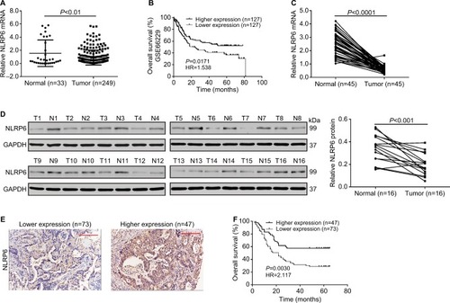
We then analyzed NLRP6 mRNA expression by qRT-PCR and showed that NLRP6 mRNA levels in tumor tissues decreased to ~76.9% (0.582±0.04) compared with adjacent normal gastric tissues (2.52±0.13, n=45; ). Our data were in line with the public-available data. We also examined the translational expression of NLRP6 in tissues from control and gastric cancer tissues by Western blot analysis and showed that NLRP6 protein levels were markedly reduced in tumor tissues ().
Decreased NLRP6 expression is associated with poor prognosis in gastric cancer patients
IHC staining was then performed and showed that 60.8% (73/120) gastric cancer tissues displayed negative or lower expression of NLRP6 (less than 20% of tumor cells were positively stained) (). Associations between NLRP6 expression and clinicopathologic characteristics were analyzed by chi-squared test. As shown in , there was a significant correlation between NLRP6 levels and H. pylori infection (P=0.0135), tumor size (P=0.0014), TNM stage (P=0.0179), and lymph node metastasis (P=0.0224). Furthermore, Kaplan–Meier analysis of our patient data () indicated that the overall survival time of patients with a lower expression of NLRP6 was significantly shorter than that of patients with a higher expression of NLRP6, suggesting that decreased NLRP6 expression may contribute to poor survival.
Table 2 Correlation of NLRP6 expression with patients’ features in gastric cancer
NLRP6 overexpression inhibits gastric cancer cell proliferation in vitro and in vivo
To explore the role of NLRP6 in gastric tumorigenesis, we stably overexpressed NLRP6 in BGC-823 and HGC-27 cells, which had relative lower protein and mRNA levels of NLRP6 (). For both cell lines, NLRP6 expression in cells transduced with NLRP6 overexpressing virus was significantly elevated compared to that in wild-type (WT) cells or cells with control vector virus (). The proliferation of BGC-823 and HGC-27 cells overexpressing NLRP6 was significantly suppressed at 24, 48, and 72 hours compared to that of control cells as measured by the CCK-8 assay (). Comparable expression levels of NLRP6 and cell proliferation rates were observed in the WT and vector cells. We then examined whether the proliferation inhibition is associated with cell apoptosis by flow cytometry analysis. Ectopic expression of NLRP6 markedly increased the early and late apoptotic ratios of cells compared to cells transducted with control vector virus (). We also knockdown NLRP6 expression in AGS (), a cell line with a higher NLRP6 expression (). Complementary to the results from NLRP6 overexpression, NLRP6 knockdown significantly promoted cell proliferation () and inhibited cell apoptosis ().
Figure 2 NLRP6 inhibits cell proliferation of gastric cancer cells.
Notes: (A) Overexpression of NLRP6 in BGC-823 and HGC-27 cells was confirmed by Western blot. Blots are representative of three separate experiments. (B) Cell proliferation was detected at 0, 24, 48, and 72 hours in BGC-823 and HGC-27 stable cells by CCK-8 assay (n=3). (C) Annexin V/PI assay showed that NLRP6 overexpression increased cell early and late apoptosis rates (n=3). (D) NLRP6 inhibits gastric cancer cell proliferation in vivo. BGC-823 cells stably expressed NLRP6 or control vector were subcutaneously inoculated into nude mice (six per group). Tumor volume was measured for 27 days. NLRP6-overexpressed BGC-823 cells grew much slower in nude mice than the control cells (left panels). At 27 days, mice were sacrificed and the tumors were isolated. IHC staining of Ki-67 and TUNEL assay were performed on the xenograft tumors (n=3, right panels). WT, wild-type cells; vector, cells stably expressed control vector; NLR6, cells stably expressed NLRP6. **P<0.01 and ***P<0.001.
Abbreviations: CCK-8, Cell Counting Kit-8; PI, propidium iodide; IHC, immunohistochemistry; FITC, fluorescein isothiocyanate.
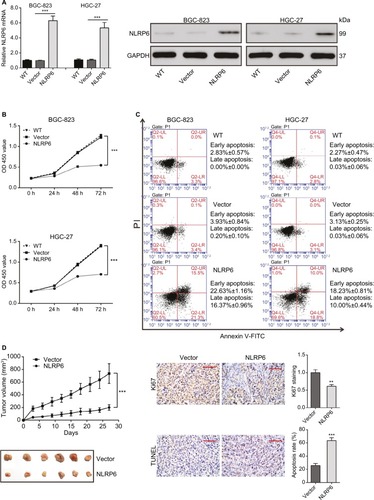
Next, we investigated the effect of NLRP6 on tumor growth in vivo. Tumor formation was examined after inoculation of BGC-823 cells stably expressed NLRP6 or control vector into the flank of nude mice. Tumor growth was much slower in mice with NLRP6 overexpressed cells compared to that in mice with cells expressing control vector (). Moreover, Ki-67-positive cells were significantly reduced, whereas apoptotic cells were notably increased in NLRP6 overexpression xenografts. These data indicated that NLRP6 may be a potential regulator of tumor growth in gastric cancer.
NLRP6 overexpression inhibits the migration and invasion of gastric cancer cells
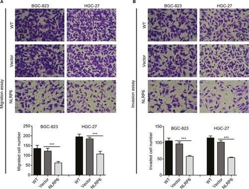
To determine whether NLRP6 affects migration and invasion of gastric cancer cells, transwell assay was performed. NLRP6 overexpression caused a significant reduction in cell migration and invasion compared to control cells (). Decreased migration and invasion of AGS cells were observed in AGS cells with NLRP6 knockdown ().
Figure 3 NLRP6 overexpression inhibits the migration and invasion of gastric cancer cells.
Notes: Transwell assays were performed to evaluate cell migration (A) and invasive abilities (B) (n=3). For invasion, the upper chamber was precoated with Matrigel. WT, wild-type cells; vector, cells stably expressed control vector; NLR6, cells stably expressed NLRP6. ***P<0.001.
Effects of NLRP6 on STAT3 signaling
Constitutively activated STAT3 is observed in various human cancers.Citation18 It is involved in cell survival, proliferation, and invasion of gastric cancer cell lines.Citation19,Citation20 Then, we tried to explore the effects of NLRP6 on STAT3 signaling. As shown in , NLRP6 overexpression in BGC-823 and HGC-27 cells significantly repressed the phosphoryla tion of STAT3. On the contrary, NLRP6 knockdown in AGS cells remarkably increased STAT3 phosphorylation (). The protein levels of Bcl-2Citation21 and MMP-2,Citation22,Citation23 two well-known target genes of STAT3, were also reduced by ectopic expression of NLRP6, which was consistent with the results of the biological function analyses.
Figure 4 Effects of NLRP6 on STAT3 signaling.
Notes: (A) Immunoblot of phosphorylated STAT3, STAT3, Bcl-2, and MMP-2. Blots are representative of three separate experiments. (B) mRNA levels of Bcl-2 and MMP-2 were assessed by qRT-PCR. (C) BGC-823 and HGC-27 cells were transfected with a Bcl-2 or an MMP-2 luciferase reporter plasmid. The cells were then cultured for 48 hours before determination of normalized luciferase activity. WT, wild-type cells; vector, cells stably expressed control vector; NLR6, cells stably expressed NLRP6. ***P<0.001.
Abbreviation: qRT, quantitative reverse transcription.
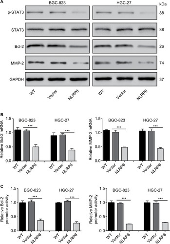
Furthermore, NLRP6 overexpression resulted in a significant downregulation of the amount of mRNA () and the promoter activity () of both Bcl-2 and MMP-2 genes. These data suggested that NLRP6 overexpression in gastric cancer cell lines reduced the STAT3 activity and repressed the transcription of its target gene.
H. pylori reduces NLRP6 expression
Considering that NLRP6 levels were strongly correlated with H. pylori infection (P=0.0135), we then determined whether NLRP6 expression was modulated by H. pylori infection. As illustrated in , H. pylori infection significantly decreased the protein and mRNA levels of NLRP6 in both BC-823 and HGC-27 cells at an MOI of 25 and 100.
Figure 5 Effect of H. pylori on NLRP6 expression.
Notes: (A and B) BGC-823 and HGC-27 cells were infected with H. pylori at an MOI of 0, 25, or 100 for 24 hours. The protein (A) and mRNA (B) levels of NLRP6 were detected. (C) The potential binding sites in the promoter of the NLRP6 genes for FOXO3 were shown based on the online program ALGGEN – PROMO. (D) The levels of p-AKT, AKT, p-FOXO3, and FOXO3 in gastric cancer cells with H. pylori infection were determined by Western blot. (E–G) BGC-823 and HGC-27 cells were transfected with siFOXO3 and siNC for 48 hours. Western blot (E), real-time PCR (F), and luciferase reporter assays (H) were performed. (H–J) Gastric cancer cells were treated with 20 µM of LY294002 of DMSO followed by infection with H. pylori (MOI of 100) for 24 hours. Western blot (H), real-time PCR (I), and luciferase reporter assays (J) were performed. *P<0.05, **P<0.01, and ***P<0.001.
Abbreviations: H. pylori, Helicobacter pylori; TSS, transcription start site; siFOXO3, FOXO3 siRNA; siNC, negative control siRNA; DMSO, dimethyl sulfoxide; MOI, multiplicity of infection; LY, LY294002.
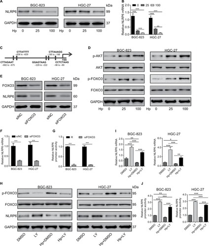
Role of AKT/FOXO3 on H. pylori-inhibited NLRP6 expression
We then tried to investigate which transcription factors were involved in H. pylori-mediated NLRP6 expression. A previ ous study has shown that H. pylori infection in gastric cells induces AKT activation, which phosphorylates and inactivates FOXO3,Citation24 a forkhead transcription factor and potential tumor suppressor.Citation25–Citation28 By utilizing the online program ALG-GEN – PROMO (based on the 8.3 version of TRANSFAC),Citation29 we found that FOXO3 was a potential transcription factor for NLRP6 gene (). As expected, H. pylori infection in both BC-823 and HGC-27 cells for 1 hour significantly induced the phosphorylation of AKT and FOXO3 but had no obvious effects on the total protein of AKT and FOXO3 ().
To further verify whether FOXO3 regulated the NLRP6 gene expression, gastric cancer cells were transfected with FOXO3 siRNA. FOXO3 knockdown led to a significant downregulation in the protein expression (), mRNA expression (), and promoter activity () of NLRP6. Furthermore, a PI3K inhibitor LY294002 was applied to gastric cancer cells exposed to H. pylori. As illustrated in , LY294002 treatment in gastric cancer cells remarkably suppressed the effects of H. pylori infection on the phosphorylation of FOXO3 and the protein levels of NLRP6. Furthermore, H. pylori led to a significant downregulation of the amount of mRNA () and the promoter activity () of NLRP6 gene, while LY294002 treatment displayed reversed effects. LY294002 treatment can rescue the effects of H. pylori infection. These data suggested that downregulation of NLRP6 expression in gastric cancers may be associated with H. pylori/AKT/FOXO3 signaling.
Discussion
NLRP6 has been proposed as a diagnostic marker for colorectal cancer.Citation8 NLRP6-deficient mice are highly susceptible to experimental colitis and colitis-induced tumorigenesis.Citation5,Citation9,Citation10 Here, we provided the first evidence of NLRP6 expression in gastric cancer, showing that NLRP6 was significantly downregulated in gastric cancer tissues compared to that in adjacent noncancerous tissues. In addition, a lower NLRP6 expression in gastric cancer tissues was associated with H. pylori infection, higher TNM grade, larger tumor size, higher incidence of lymph node metastasis, and poorer overall survival of patients ( and ), although multivariate analysis did not reveal NLRP6 expression to be an independent prognostic factor (). These data suggest that NLRP6 may be a novel prognostic marker for gastric cancer.
GSEA on TCGA data set showed that cell apoptosis and metastasis down pathways were negatively correlated with NLRP6 expression in gastric cancer patients, which was further validated by a series of functional experiments. The ectopic overexpression of NLRP6 in gastric cancer cells significantly suppressed cell growth, cell migration, and cell invasion but enhanced cell apoptosis, whereas siRNA-mediated knockdown displayed the reverse effects. The proliferation-suppressing and apoptosis-promoting effects of NLRP6 were also observed in xenograft tumors. These findings indicate that NLRP6 may work as a tumor suppressor in gastric cancer.
STAT3 is activated by growth factors and cytokines via the Janus kinase (JAK). Upon activation, it dimerizes and translocates to the nucleus to activate the transcription of target genes, such as Bcl-2 and MMP-2.Citation18,Citation21–Citation23 Multiple lines of evidence suggest a crucial role for STAT3 in the development and progression of various human cancers,Citation18 including gastric cancer.Citation19,Citation30 STAT3 expression in gastric cancer indicates a poor prognosis.Citation30 Constitutively, STAT3 signaling supports gastric cancer cell survival.Citation19 Suppression of Bcl-2 may contribute to cell apoptosis in gastric carcinomaCitation31 and gastric cancer cells.Citation32 MMP-2, which degrades the extracellular matrix, has been related to lymph node metastasis of gastric cancer.Citation33,Citation34 Here, NLRP6 overexpression in gastric cancer cells significantly repressed the phosphorylation of STAT3 as well as the transcription of Bcl-2 and MMP-2, suggesting that the apoptosis-promoting, anti-migration, and anti-invasion effects of NLRP6 were mediated by STAT3 signaling.
H. pylori-induced gastritis is a main risk factor for gastric cancer.Citation3 H. pylori activates the PI3K/AKT signaling pathway, which activates β-catenin signaling and upregulates the transcriptional activation of target genes that affect carcinogenesis.Citation35 Here, we found that NLRP6 levels were strongly correlated with H. pylori infection in patients with gastric cancer. H. pylori infection significantly decreased the levels of NLRP6 in gastric cancer cells. Furthermore, we tried to investigate the transcription factor for NLRP6. FOXO3 was phosphorylated by H. pylori infection in a PI3K/AKT-dependent manner in gastric cells as previously reported.Citation24 Knockdown of FOXO3 causes a significant decrease in the protein expression, mRNA expression, and promoter activity of NLRP6. H. pylori infection had the same effects as FOXO3 knockdown, and such inhibitory effects were also PI3K/AKT dependent. Overall, these results inferred that H. pylori infection downregulated the transcription of NLRP6 via AKT/FOXO3 signaling.
Collectively, our study suggests that NLRP6 acts as a tumor suppressor in gastric cancer, and the downregulation of NLRP6 expression is closely associated with patients’ poor prognosis. We provided key evidence that NLRP6 suppresses the proliferation, migration, and invasion of gastric cancer cells through regulating STAT3 signaling. H. pylori regulated the expression of NLRP6 via AKT/FOXO3 signaling.
Acknowledgments
This study was supported by the Natural Science Foundation of Shanghai (16ZR1429100).
Supplementary materials
Figure S1 NLRP6-associated pathways in gastric cancer.
Notes: GSEA was performed on TCGA data set by using GSEA version 2.0 from the Broad Institute at MIT as previously described.Citation1 Gene set permutations were performed 1,000 times, and the pathway set list was sorted by the NES. GSEA was performed using TCGA data set. The cell apoptosis (A) and metastasis (B) pathways were strongly associated with NLRP6 lower expression.
Abbreviations: GSEA, gene set enrichment analysis; TCGA, The Cancer Genome Atlas; NES, normalized enrichment score; ES, enrichment score.
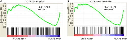
Figure S2 NLRP6 protein expression and mRNA expression in seven gastric cancer cell lines were determined by qRT-PCR (A) and Western blot (B) analysis, respectively.
Abbreviation: qRT, quantitative reverse transcription.
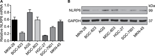
Figure S3 NLRP6 knockdown in AGS cells promoted cell proliferation, migration, and invasion but inhibited cell apoptosis. (A) qRT-PCR (upper panel) and Western blot (lower panel) analysis of knockdown efficiency in AGS cells. (B) CCK-8 assay showed that NLRP6 knockdown promoted cell proliferation. (C) Annexin V/PI assay showed that NLRP6 knockdown decreased cell early and late apoptosis. (D) Transwell assays showed that NLRP6 knockdown enhanced migration and invasive capacity. WT, wild-type cells. **P<0.01 and ***P<0.001.
Abbreviations: qRT, quantitative reverse transcription; CCK-8, Cell Counting Kit-8; PI, propidium iodide; siNC, negative control siRNA.
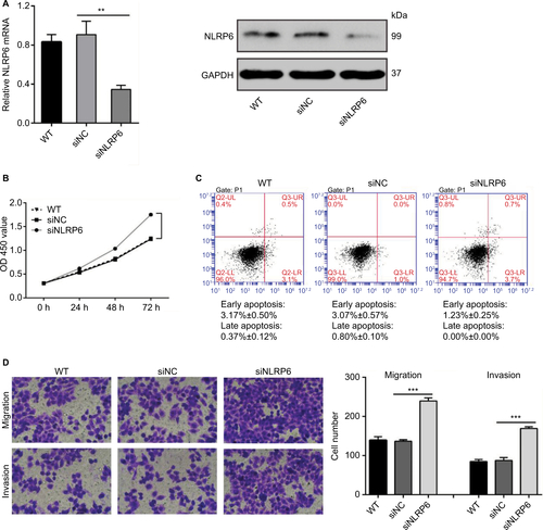
Figure S4 Effects of NLRP6 knockdown on STAT3 phosphorylation.
Notes: Blots are representative of three separate experiments. WT, wild-type cells; siNC, negative control siRNA.
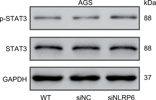
Table S1 Univariate Cox regression of prognostic parameters for survival in patients with gastric cancer
Table S2 Multivariate Cox regression of prognostic parameters for survival in patients with gastric cancer
Reference
- SubramanianAKuehnHGouldJTamayoPMesirovJPGSEA-P: a desktop application for Gene Set Enrichment AnalysisBioinformatics200723233251325317644558
Disclosure
The authors report no conflicts of interest in this work.
References
- Cancer IAfRoWorld Cancer ReportGenevaWHO2014
- LozanoRNaghaviMForemanKGlobal and regional mortality from 235 causes of death for 20 age groups in 1990 and 2010: a systematic analysis for the Global Burden of Disease Study 2010Lancet201238098592095212823245604
- CompareDRoccoANardoneGRisk factors in gastric cancerEur Rev Med Pharmacol Sci201014430230820496539
- OrdituraMGaliziaGSforzaVTreatment of gastric cancerWorld J Gastroenterol20142071635164924587643
- ChenGYLiuMWangFBertinJNúñezGA functional role for Nlrp6 in intestinal inflammation and tumorigenesisJ Immunol2011186127187719421543645
- GrenierJMWangLManjiGAFunctional screening of five PYPAF family members identifies PYPAF5 as a novel regulator of NF-kappaB and caspase-1FEBS Lett20025301–3737812387869
- AnandPKMalireddiRKLukensJRNLRP6 negatively regulates innate immunity and host defence against bacterial pathogensNature2012488741138939322763455
- AhmedFEVosPMolecular markers for human colon cancer in stool and blood identified by RT-PCRAnticancer Res20042464127413415736463
- NormandSDelanoye-CrespinABressenotANod-like receptor pyrin domain-containing protein 6 (NLRP6) controls epithelial self-renewal and colorectal carcinogenesis upon injuryProc Natl Acad Sci U S A2011108239601960621593405
- ElinavEStrowigTKauALNLRP6 inflammasome regulates colonic microbial ecology and risk for colitisCell2011145574575721565393
- HuBElinavEHuberSMicrobiota-induced activation of epithelial IL-6 signaling links inflammasome-driven inflammation with transmissible cancerProc Natl Acad Sci U S A2013110249862986723696660
- ZhangYHWangYYusufaliAHCytotoxic genes from traditional Chinese medicine inhibit tumor growth both in vitro and in vivoJ Integr Med201412648349425412666
- TojiLHLenchitzTCKwiatkowskiVASaramaJAMulivorRAValidation of routine mycoplasma testing by PCRIn Vitro Cell Dev Biol Anim19983453563589639095
- HuangHChevilleJCPanYRochePCSchmidtLJTindallDJPTEN induces chemosensitivity in PTEN-mutated prostate cancer cells by suppression of Bcl-2 expressionJ Biol Chem200127642388303883611495901
- BianJSunYTranscriptional activation by p53 of the human type IV collagenase (gelatinase A or matrix metalloproteinase 2) promoterMol Cell Biol19971711633063389343394
- KempsterSLBeltekiGForheadAJDevelopmental control of the Nlrp6 inflammasome and a substrate, IL-18, in mammalian intestineAm J Physiol Gastrointest Liver Physiol20113002G253G26321088234
- XiongHduWSunTTA positive feedback loop between STAT3 and cyclooxygenase-2 gene may contribute to Helicobacter pylori-associated human gastric tumorigenesisInt J Cancer201413492030204024127267
- YuHPardollDJoveRSTATs in cancer inflammation and immunity: a leading role for STAT3Nat Rev Cancer200991179880919851315
- KandaNSenoHKondaYSTAT3 is constitutively activated and supports cell survival in association with survivin expression in gastric cancer cellsOncogene200423284921492915077160
- WeiZJiangXQiaoHSTAT3 interacts with Skp2/p27/p21 pathway to regulate the motility and invasion of gastric cancer cellsCell Signal201325493193823333463
- BhattacharyaSRayRMJohnsonLRSTAT3-mediated transcription of Bcl-2, Mcl-1 and c-IAP2 prevents apoptosis in polyamine-depleted cellsBiochem J2005392Pt 233534416048438
- SeoJMParkSKimJHLeukotriene B4 receptor-2 promotes invasiveness and metastasis of ovarian cancer cells through signal transducer and activator of transcription 3 (STAT3)-dependent up-regulation of matrix metalloproteinase 2J Biol Chem201228717138401384922396544
- XieTXWeiDLiuMStat3 activation regulates the expression of matrix metalloproteinase-2 and tumor invasion and metastasisOncogene200423203550356015116091
- TabassamFHGrahamDYYamaokaYHelicobacter pylori-associated regulation of forkhead transcription factors FoxO1/3a in human gastric cellsHelicobacter201217319320222515357
- BullockMDBruceASreekumarRFOXO3 expression during colorectal cancer progression: biomarker potential reflects a tumour suppressor roleBr J Cancer2013109238739423828518
- HuMCLeeDFXiaWIkappaB kinase promotes tumorigenesis through inhibition of forkhead FOXO3aCell2004117222523715084260
- LiJLiPChenTExpression of microRNA-96 and its potential functions by targeting FOXO3 in non-small cell lung cancerTumour Biol201536268569225286764
- ParkSHLeeJHBerekJSHuMCMc-THAuranofin displays anticancer activity against ovarian cancer cells through FOXO3 activation independent of p53Int J Oncol20144541691169825096914
- MesseguerXEscuderoRFarréDNúñezOMartínezJAlbàMMPROMO: detection of known transcription regulatory elements using species-tailored searchesBioinformatics200218233333411847087
- KimDYChaSTAhnDHSTAT3 expression in gastric cancer indicates a poor prognosisJ Gastroenterol Hepatol200924464665119175826
- KoshidaYSaegusaMOkayasuIApoptosis, cell proliferation and expression of Bcl-2 and Bax in gastric carcinomas: immunohistochemical and clinicopathological studyBr J Cancer19977533673739020481
- ZhaoLGuoQLYouQDWuZQGuHYGambogic acid induces apoptosis and regulates expressions of Bax and Bcl-2 protein in human gastric carcinoma MGC-803 cellsBiol Pharm Bull2004277998100315256729
- MönigSPBaldusSEHenneckenJKExpression of MMP-2 is associated with progression and lymph node metastasis of gastric carcinomaHistopathology200139659760211903578
- MatsumuraSOueNNakayamaHA single nucleotide polymorphism in the MMP-9 promoter affects tumor progression and invasive phenotype of gastric cancerJ Cancer Res Clin Oncol20051311192515565457
- NakayamaMHisatsuneJYamasakiEHelicobacter pylori VacA-induced inhibition of GSK3 through the PI3K/Akt signaling pathwayJ Biol Chem200928431612161918996844