
Abstract
Background
Increased aberrant expression or activation of the epidermal growth factor receptor (EGFR) family members has been reported in a wide range of cancers, and the EGFR family of tyrosine kinases has emerged as an important therapeutic target in malignancies. However, the expression patterns and exact roles of each distinct EGFR family member, which contribute to tumorigenesis and progression of ovarian cancer (OC), are yet to be elucidated.
Materials and methods
In the current study, we report the distinct expression and prognostic value of EGFR family members in patients with OC by analyzing a series of databases including ONCOMINE, Gene Expression Profiling Interactive Analysis, Kaplan–Meier plotter, cBioPortal, and Database for Annotation, Visualization and Integrated Discovery .
Results
It was found that in patients with OC, mRNA expression levels of ERBB2/3/4 were significantly upregulated, whereas the transcription levels of EGFR were downregulated. Aberrant EGFR expression and ERBB2/3/4 mRNA levels were associated with OC prognosis.
Conclusion
These results suggest that EGFR and ERBB3/4 are distinct prognostic biomarkers and may be potential targets for OC. These results may be beneficial to better understand the molecular underpinning of OC and may be useful to develop tools for more accurate OC prognosis and for promoting the development of EGFR-targeted inhibitors for OC treatment.
Introduction
Ovarian cancer (OC) shows the highest cancer-related death rate among gynecological malignancies, with an estimated 204,000 cases and 125,000 deaths annually worldwide.Citation1,Citation2 Over 75% of patients are not diagnosed until the disease is advanced (stages III and IV). Current prognostic factors do not allow reliable prediction of response to chemotherapy and survival for individual OC patients. The poor rate of survival and the high rate of lethality are partly due to lack of effective biomarkers for prognosis. Therefore, there is a pressing need to find reliable predictive biomarkers for prognosis and to develop novel therapeutic strategies for OC patients.Citation2,Citation3
The epidermal growth factor receptor (EGFR) tyrosine kinase family consists of four members: EGFR, ERBB2, ERBB3, and ERBB4. These receptors are activated when a ligand binds to their extracellular ligand binding domain, which triggers receptor homodimerization or heterodimerization, resulting in the activation of several downstream cell signaling pathways and ultimately in tumor cell proliferation, reduced apoptosis, and tumor migration and invasion.Citation4–Citation6 In the past three decades, increased aberrant expression or activation of the EGFR family members has been reported in a wide range of cancers, and in some studies, has also been associated with poor prognosis and resistance to therapeutic options.Citation5,Citation7 Moreover, the EGFR family of tyrosine kinases has emerged as an important therapeutic target in malignancies, and to date, numerous antibodies, recombinant proteins, peptide mimetics, and small molecules, such as cetuximab, panitumumab, trastuzumab, gefitinib, erlotinib, and lapatinib, have been developed for targeting EGFR family receptors as therapeutic targets for many kinds of solid tumors.Citation4,Citation7 Recent reports have suggested that the functions of different EGFR members contribute to OC tumorigenesis. However, the clinicopathological and prognostic value and expression patterns of EGFR family members in OC remain controversial.Citation8–Citation10 In addition, the role of EGFR family members in OC and the underlying molecular mechanism responsible for its involvement in tumor development and progression are largely unknown.
The development of microarray and RNA-sequencing technology has revolutionized RNA and DNA research, which has become a crucial component of biology and biomedical research.Citation11,Citation12 In the current study, we extended the knowledge base related to OC based on a variety of large databases, with the purpose of determining the expression patterns, genetic alteration, potential functions, and distinct prognostic values of EGFR family members in OC.
Materials and methods
Ethics statement
This study was approved by the Academic Committee of the People’s Hospital of China Three Gorges University, and conducted according to the principles expressed in the Declaration of Helsinki. All the datasets were retrieved from the databases, so it was confirmed that written informed consent had been obtained from all patients.
ONCOMINE analysis
The gene expression array datasets of ONCOMINE (www.oncomine.org), which is a publicly accessible, online cancer microarray database helps facilitate research data from genome-wide expression analyses. ONCOMINE was used to analyze the mRNA levels of EGFR family members in OC.Citation13,Citation14 In this study, the Student’s t-test was used to generate P-values for comparison between cancer specimens and normal control datasets. The cutoff P-value and fold change were defined as 0.05 and 1, respectively.
Gene Expression Profiling Interactive Analysis (GEPIA) dataset analysis
GEPIA is an interactive web server for estimating mRNA expression data based on 9,736 tumors and 8,587 normal samples in The Cancer Genome Atlas (TCGA) and Genotype-Tissue Expression dataset projects. GEPIA provides key interactive and customizable functions including differential expression analysis, profiling plotting, correlation analysis, patient survival analysis, similar gene detection, and dimensionality reduction analysis.Citation15
The Kaplan–Meier plotter analysis
The prognostic value of the mRNA expression of EGFR family members was evaluated using an online database, Kaplan–Meier Plotter (www.kmplot.com), which contains gene expression data and survival information of 1,816 clinical OC patients. To analyze the overall survival (OS), progression-free survival (PFS), and post-progression survival (PPS) of patients with OC, patient samples were split into two groups by median expression (high vs low expression) and assessed by a Kaplan–Meier survival plot, with a HR with 95% CI and log-rank P-value.Citation16
TCGA and CBioPortal analysis
Gene alteration frequency of EGFR family member mRNA in OC was performed using CBioPortal for Cancer Genomics (http://www.cbioportal.org). The genomic profiles included mutations, putative copy-number alterations from GISTIC, mRNA expression z scores, and protein expression z scores.Citation17
Functional enrichment and bioinformatics analysis
GeneMANIA (http://www.genemania.org) is a flexible, user-friendly web interface for generating hypotheses about gene function, analyzing gene lists, and prioritizing genes for functional assays. GeneMANIA was used to conduct correlation analysis of EGFR family members at the gene level, which revealed relationships in pathways, shared protein domains, co-localization, and co-expression.Citation18 Finally, enrichment analysis was performed with The Database for Annotation, Visualization and Integrated Discovery (DAVID) (version 6.7) for EGFR family members and their neighboring genes. DAVID includes the gene ontology (GO) terms and Kyoto Encyclopedia of Genes and Genomes (KEGG) pathways.Citation19,Citation20
Results
Transcription levels of EGFR family members in patients with OC
Using ONCOMINE analysis, four EGFR family members have been identified in human cancers, including hematological malignancies and solid tumors (). ONCOMINE analysis revealed that the mRNA expression levels of ERBB3 were significantly upregulated in patients with OC in three datasets. In Hendrix’s dataset,Citation21 ERBB3 is overexpressed compared with that in the normal samples in all OC types – ovarian mucinous adenocarcinoma with a fold change of 2.355, ovarian clear cell adenocarcinoma with a fold change of 2.308, ovarian endometrioid adenocarcinoma with a fold change of 1.897, and ovarian serous adenocarcinoma with a fold change of 1.857. In Adib’s dataset,Citation22 ERBB3 is overexpressed in ovarian serous adenocarcinoma with a fold change of 1.807. In Lu’s dataset,Citation23 ERBB3 is overexpressed in ovarian endometrioid adenocarcinoma with a fold change of 1.635 and in ovarian serous adenocarcinoma with a fold change of 1.947 compared with that in the normal samples. The transcription levels of EGFR in ovarian serous adenocarcinoma were lower than that in normal ovarian tissues in two datasets (fold changes were −1.223 and −1.349, respectively)Citation22,Citation24 ().
Table 1 The transcription levels of EGFR family members between different types of OC and normal tissues (ONCOMINE)
Figure 1 The transcription levels of EGFR family members in different types of cancers (ONCOMINE).
Notes: The graphic demonstrated the numbers of datasets with statistically significant mRNA overexpression (red) or down-expression (blue) of the target gene. The threshold was designed with following parameters: P-value =0.001; fold-change =1.5 and data type, mRNA.
Abbreviations: EGFR, epidermal growth factor receptor; ERBB2, receptor tyrosine-protein kinase erbB-2; ERBB3, receptor tyrosine-protein kinase erbB-3; ErbB4, receptor tyrosine-protein kinase erbB-4.
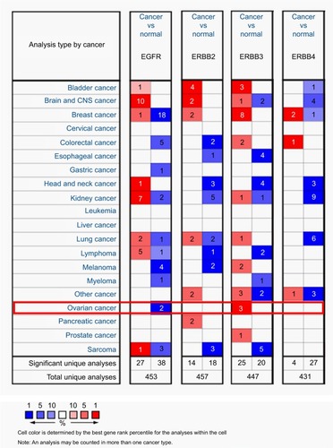
As shown in , the transcription levels of ERBB2 and ERBB4 in different pathological types of OC (eg, ovarian endometrioid adenocarcinoma, ovarian mucinous adenocarcinoma, ovarian serous adenocarcinoma, ovarian clear cell adenocarcinoma, ovarian serous surface papillary carcinoma, ovarian serous adenocarcinoma, and ovarian carcinoma) were also slightly higher than those in normal ovarian tissues, and their cutoff of P-value was >0.05.
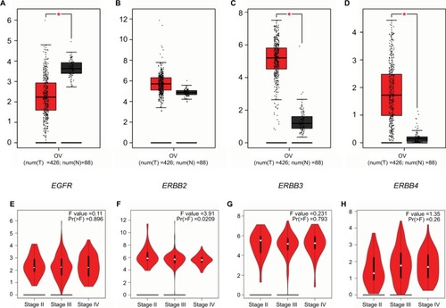
In addition, using the GEPIA dataset (http://gepia.cancer-pku.cn/), we compared the mRNA expression of EGFR family members between OC and normal tissues. The results demonstrated that the mRNA expression levels of ERBB3 and ERBB4 were significantly higher in OC tissues than in normal ovarian tissues, whereas the expression level of EGFR was significantly lower in the former than in the latter. We also analyzed the expression of EGFR family members in different tumor stages of OC. None of the EGFR family members varied in the different tumor stages ().
Figure 2 The expression of EGFR family members and tumor stage in OC patients (GEPIA).
Notes: Box plots derived from gene expression data in GEPIA comparing expression of a specific EGFR family member in OC tissue and normal tissues, the P-value was set up at 0.05. (A) The distribution of EGFR mRNA expression; (B) the distribution of ERBB2 mRNA expression; (C) the distribution of ERBB3 mRNA expression; (D) the distribution of ERBB3 mRNA expression between OC tissue and normal tissues; (E) correlation between EGFR expression and tumor stage; (F) correlation between ERBB2 expression and tumor stage; (G) correlation between ERBB3 expression and tumor stage; (H) correlation between ERBB4 expression and tumor stage in OC patients.
Abbreviations: EGFR, epidermal growth factor receptor; ERBB2, receptor tyrosine-protein kinase erbB-2; ERBB3, receptor tyrosine-protein kinase erbB-3; ERBB4, receptor tyrosine-protein kinase erbB-4; OC, ovarian cancer; T, tumor; N, normal.
Prognostic values of EGFR family members in all patients with OC
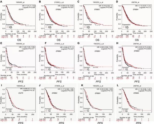
Using Kaplan–Meier plotter analysis, we initially assessed the prognostic significance of the EGFR family members in all OC patients. The Kaplan–Meier survival curves are demonstrated in . The increased EGFR mRNA level and the decreased ERBB2 and ERBB3 mRNA levels were strongly associated with the poor OS. However, high mRNA levels of EGFR or low mRNA levels of ERBB4 were predicted to have high PFS. In addition, the mRNA expression levels of EGFR, ERBB2, ERBB3, and ERBB4 were not correlated with PPS of all patients with OC.
Figure 3 The prognostic value of mRNA level of EGFR family members in OC patients (Kaplan–Meier plotter).
Notes: The OS, PFS, and PPS survival curve comparing the patient with high (red) and low (black) EGFR family members’ expression in OC were plotted from Kaplan–Meier plotter database as the threshold of P-value <0.05, respectively. OS curves of (A) EGFR (Affymetrix IDs: 1565483_at); (B) ERBB2 (Affymetrix IDs: 210930_s_at); (C) ERBB3 (Affymetrix IDs: 1563253_s_at); (D) ERBB4 (Affymetrix IDs: 206794_at). PFS curves of (E) EGFR (Affymetrix IDs: 1565483_at); (F) ERBB2 (Affymetrix IDs: 210930_s_at); (G) ERBB3 (Affymetrix IDs: 1563253_s_at); (H) ERBB4 (Affymetrix IDs: 206794_at). PPS curves of (I) EGFR (Affymetrix IDs: 1565483_at); (J) ERBB2 (Affymetrix IDs: 210930_s_at); (K) ERBB3 (Affymetrix IDs: 1563253_s_at); (L) ERBB4 (Affymetrix IDs: 206794_at).
Abbreviations: EGFR, epidermal growth factor receptor; ERBB2, receptor tyrosine-protein kinase erbB-2; ERBB3, receptor tyrosine-protein kinase erbB-3; ERBB4, receptor tyrosine-protein kinase erbB-4; OS, overall survival; PFS, progression-free survival; PPS, post-progression survival.
The prognostic value of EGFR family members was assessed in different pathological histology subtypes of OC, including serous and endometrioid. As shown in , high ERBB2 mRNA expression was correlated to longer OS in serous OC patients. The mRNA expression levels of EGFR and ERBB4 were associated with poor OS in serous OC patients. Increased EGFR, ERBB2, and ERBB3 mRNA expression levels were associated with poor PFS. In endometrioid OC, high ERBB4 mRNA expression level was associated with better OS. The rest of the EGFR family members were not related with prognosis in endometrioid OC.
Table 2 The prognostic values of EGFR family members in all and different pathological subtypes OC patients (Kaplan–Meier plotter)
Prognostic values of EGFR family members in OC patients with different clinicopathological features
To further assess the association of individual EGFR family members with other clinicopathological features, we assessed the correlation between them with pathological grades, clinical grades, and TP53 status of OC patients (). As shown in , high mRNA expression of ERBB4 was associated with better OS and PFS in pathological grade I OC patients. Elevated mRNA expression of ERBB3 was associated with better OS and PFS in grade II OC patients. In pathological grade III OC patients, high EGFR and ERBB4 mRNA expression was linked to poor OS or PFS, but high ERBB2 mRNA expression was found to be correlated to longer OS. None of the EGFR family members were related with prognosis in grade IV OC patients. In terms of clinical staging, as we can see from , increased mRNA expression of ERBB2 and ERBB4 was associated with longer PFS, but high mRNA expression of ERBB2 was linked to poor OS in clinical stage I and II patients. For clinical stage III and IV OC patients, high mRNA expression of EGFR and ERBB2 was associated with poor PFS in this subgroup. Additionally, also shows that high mRNA expression levels of ERBB2 and ERBB3 were associated with poor OS and PFS, and elevated mRNA expression of EGFR was associated with poor PFS in mutated-TP53-type OC. However, high mRNA expression level of ERBB4 was associated with better PFS in this subgroup.
Table 3 The prognostic values of EGFR family members in OC patients with different clinicopathological features (Kaplan–Meier plotter)
Genetic alteration and neighbor genes of EGFR family members in OC
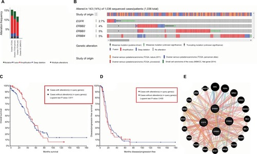
We analyzed the genetic alterations of EGFR family members by using the cBioPortal online tool for OC. A total of 839 patients from three datasets of ovarian serous cystadenocarcinoma and 12 patients from one dataset of small cell carcinoma were analyzed. Among 4°C datasets analyzed, alterations ranging from 10.3% (58/563) to 13.7% (83/606) were found for the gene sets submitted for analysis (). The percentages of genetic alterations in EGFR family members for OC varied from 2.7% to 5.0% for individual genes (EGFR, 2.7%; ERBB2, 4%; ERBB3, 5%; and ERBB4, 5%) (). After cBioPortal, Kaplan–Meier plotter and log-rank test, the results indicated that there are no significant difference in OS and disease-free survival (DFS) in cases with or without alterations in one of the EFGR family genes (P-values, 0.454 and 0.321, respectively) (). We then constructed a network for EGFR family members with the structure or function of neighboring genes using Gen-eMANIA. The results showed that 20 genes – ABL1, ABL2, ANKS1A, ANKS1B, BTC, CRK, EREG, GRAP2, GRB2, GRB7, NRG1, NRG2, PIK3R2, PIK3R3, PLCG2, PTK6, SHC1, SHC4, TGFA, and TNS3 – were closely associated with EGFR family members (). GeneMANIA also was used to conduct correlation analysis of EGFR family members at the gene level. There were relationships between EGFR and ERBB2 in co-expression, pathway, physical interactions, and shred protein domains. There were also relationships between EGFR and ERBB3 in pathway, physical interactions, and shred protein domains. There were physical interactions, prediction, and shared protein domains between EGFR and ERBB4. In addition, there were relationships in co-expression, co-localization, pathway, physical interactions, shared protein domains, and prediction between ERBB2 and ERBB3. There were relationships between EEBB2 and ERBB4 in pathway, physical interactions, prediction, and shred protein domains. ERBB3 and ERBB4 shared physical interactions, prediction, and shred protein domains. Detailed results are presented in .
Figure 4 Alteration frequency of EGFR familymembers and neighbor genes network in OC (cBioPortal and GeneMANIA).
Notes: (A) Summary of alteration in EGFR family members. (B) OncoPrint visual summary of alteration on a query of EGFR family members. (C) Kaplan–Meier plots comparing OS in cases with/without EGFR family members gene alterations. (D) Kaplan–Meier plots comparing disease-free survival (DFS) in cases with/without EGFR family member alterations. (E) Gene–gene interaction network among EGFR family members.
Abbreviations: EGFR, epidermal growth factor receptor; ERBB2, receptor tyrosine-protein kinase erbB-2; ERBB3, receptor tyrosine-protein kinase erbB-3; ERBB4, receptor tyrosine-protein kinase erbB-4; OS, overall survival.
Significant functions and pathway enrichment analysis of EGFR family members in OC
The functions of EGFR family members and their neigh-boring genes were predicted by analyzing GO and KEGG in DAVID. Based on DAVID, a total of 58 GO functions were enriched. The enrichment items were classified into three functional groups: biological process (BP) group (10 items), molecular function (MF) group (41 items), and cellular component (CC) group (7 items). As shown in , the EGFR family members and their neighboring genes were mainly enriched in the following BP: transmembrane receptor protein tyrosine kinase signaling pathway, EGFR signaling pathway, insulin receptor signaling pathway, positive regulation of cell proliferation, and cell differentiation. The MF that these genes were mainly associated with are receptor binding, non-membrane spanning protein tyrosine kinase activity, manganese ion binding, ATP binding, and receptor tyrosine kinase binding; the CC that these genes were associated with are the extrinsic component of the cytoplasmic side of the plasma membrane, extracellular space, and phosphatidylinositol 3-kinase complex, and the focal adhesion and receptor complex.
Table 4 The GO function enrichment analysis of EGFR family members and neighbor genes in OC (DAVID)
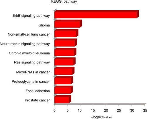
Next, 51 pathways related to the functions of EGFR family members were found through KEGG analysis. The top ten KEGG pathways for EGFR family members are shown in . Among these pathways, the ErbB signaling pathway, neurotrophin signaling pathway, Ras signaling pathway, microRNAs in cancer, proteoglycans in cancer, and focal adhesion were found to be involved in OC tumorigenesis and pathogenesis.
Figure 5 The KEGG pathway enrichment analysis of EGFR family members and neighbor genes in OC (DAVID).
Notes: The graphic demonstrated the functions of EGFR family and genes significantly associated with EGFR family alterations were predicted by analysis of the KEGG. The length of the X-axis represents −log (P-value).
Abbreviations: OC, ovarian cancer; EGFR, epidermal growth factor receptor; KEGG, Kyoto Encyclopedia of Genes and Genomes.
Discussion
Accumulative studies have determined that aberrant expression or activation of the EGFR family members is a common feature in human cancers, and the functions of different EGFR family members are associated with tumorigenesis and progression of solid tumors.Citation4,Citation5,Citation7,Citation25–Citation27 However, the patterns of expression and the exact roles the distinct EGFR family members play in contributing to OC are yet to be elucidated.Citation8–Citation10,Citation28 In the current study, we comprehensively explored the expression patterns, prognostic values (OS, PFS, and PPS), genetic alteration, and potential functions of different EGFR family members based on a variety of large databases.
Among the EGFR family members, EGFR is the most studied in OC since it was first identified in the 1970s.Citation29 Till date, various cancer cells are characterized by EGFR hyper-activation, overexpression, or mutants with dysregulated signaling. EGFR and its signaling activity have been targets for developing novel therapeutic drugs to treat a variety of cancers.Citation30–Citation33 Recent studies confirmed that amplification and overexpression of EGFR have been reported in several solid cancers, and a growing body of research interests has focused on the prognostic value and therapeutic potential of EGFR for OC.Citation8–Citation10,Citation28 In our study, ONCOMINE and GEPIA datasets revealed that the mRNA expression of EGFR was lower in OC than in normal tissues. This inconsistent expression pattern might be because ONCOMINE and GEPIA only represent mRNA data, which only correlate to ~40% of the total protein levels.Citation34 Consistent with the results of most previous studies, our results demonstrated that EGFR expression was not correlated with the clinical stage of the patients with OC, and an increased EGFR expression was significantly associated with poor OS and PPS in the patients with OC, especially in serous and advanced OC. However, several different studies suggest that EGFR is not a reliable marker of survival in OC.Citation29,Citation35,Citation36 The utility of EGFR expression as an independent prognostic indicator in OC patients is yet to be confirmed.Citation37,Citation38
ERBB2 is a tyrosine kinase receptor in the EGFR family and plays a pivotal role in cell proliferation and tumor cell metastasis.Citation39,Citation40 Previous studies have demonstrated that ERBB2 overexpression or mutations in human malignant cancers correlate with poor prognosis and chemo-resistance.Citation40–Citation42 Until now, the association between ERBB2 expression and OC has been widely studied, however, its relationship with disease stage, grade, and response to treatment remains controversial.Citation43–Citation45 A recent meta-analysis study showed that HER2 expression can be used as a prognostic biomarker in OC patients.Citation46 Our results demonstrated that the transcription levels of ERBB2 in different pathological types of OC were not remarkably higher than those in normal tissues, and increased ERBB2 mRNA levels were significantly associated with the better OS, especially in clinical stage I and II OC patients. Interestingly, high expression of ERBB2 was associated with poor PFS in clinical stage III and IV OC patients and with poor OS and PFS in mutated-TP53-type OC patients.
The third member of the EGFR family, ERBB3, unlike the other EGFR family members that are activated through autophosphorylation upon binding with the ligand, lacks an intracellular tyrosine kinase domain.Citation47 Therefore, ERBB3 must act as an allosteric activator. It forms heterodimers with other EGFR family members, thus stimulating downstream growth and signaling pathways.Citation48 ERBB3 has been shown to be overexpressed in several human carcinomas, and somatic mutations have been found scattered throughout the ERBB3 gene in subsets of breast cancers, gastric cancers, and OC.Citation49–Citation51 In addition, ERBB3 has been recently characterized as having a significant role in mediating resistance to EGFR- and ERBB2-directed therapies in solid malignancies, suggesting that ERBB3 also has a role in mediating resistance to PI3K/AKT pathway inhibitors.Citation48,Citation52,Citation53 Our study showed that the mRNA expression levels of ERBB3 were considerably upregulated in patients with OC in three datasets, and increased ERBB3 mRNA levels were associated with the better OS, especially in pathological grade II OC patients. High expression of ERBB2 was associated with poor PFS in serous OC patients and poor OS and PFS in mutated-TP53-type OC patients.
ERBB4 is one of the four members in the EGFR subfamily of receptor tyrosine kinases. Unlike ERBB2, which cannot directly bind a ligand, and ERBB3, which does not have a functional kinase domain, ERBB4 is a fully functional receptor tyrosine kinase capable of signaling, both as a homodimer and as a heterodimer.Citation54 Among different EGFR family members, the role of ERBB4 in cancer is probably the least understood.Citation55 ERBB4 is necessary for the development of the heart, mammary gland, and the central nervous system, and mutations in ERBB4 have been identified in various cancer types including melanoma, lung adenocarcinoma, and medulloblastoma. These results suggest that ERBB4 can be a potential biomarker for malignant tumors.Citation54,Citation56 Our results showed that increased expression of ERBB4 might indicate better PFS in all OC patients and longer OS in endometrioid OC patients; however, increased ERBB4 expression may correlate with worse OS in serous OC patients.
Mutations, gene amplification, and protein overexpression of EGFR family members are all linked to carcinogenesis.Citation47 Mutant EGFR family members cause a gain-of-function phenotype and are involved in tumorigenesis, invasion, and metastasis.Citation57 In our current analysis, we found that the percentages of alterations in EGFR family members among OC varied from 2.7% to 5.0% for individual genes, but there is no significant difference in OS and DFS in cases with or without alterations in one of the EFGR family genes (P-values, 0.454 and 0.321, respectively). To further clarify the carcinogenic mechanism of the EGFR family members, we constructed a network for EGFR family members and 20 neighboring genes. The results of GO and KEGG analysis indicated that these genes are mainly enriched in tumor-related pathways, including the ErbB signaling pathway, neurotrophin signaling pathway, and Rams signaling pathway, and in microRNAs and proteoglycans in cancer, and during focal adhesion. Our study adds to the growing evidence regarding the complexity of the EGFR family members and their associated signaling pathways, which offer clues into the rational development of dual targeting with anti-EGFR or HER2 and downstream pathway inhibitors.
To the best of our knowledge, this is the first bioinformatics analysis exploring the distinct expression and prognostic value of EGFR family members in OC. There were some limitations to this study that need to be addressed. First, this is an in silico and bioinformatics analysis based on functional genomics using data from several large databases, which may introduce background heterogeneity. To address these issues, we are planning functional verification studies in well designed in vitro and in vivo models in the near future. In addition, the sample size of the study cohort was limited, and a small fraction of the clinical data was missing. As such, larger studies are needed to clarify these findings. Finally, no multivariable analyses were included; therefore, it is impossible to identify any potential association with other important prognostic factors, such as the FIGO stage, patient age, residual tumor after initial surgery, lymph node metastasis, vascular invasion, cancer antigen 125, and Human epididymis protein 4, BRCA, Risk of Malignancy Index II, and Risk of Malignancy Algorithm. Therefore, future research is still needed to address these issues.
Conclusion
In summary, the mRNA expression levels of ERBB2/3/4 were significantly upregulated, whereas the transcription levels of EGFR were low in patients with OC. Aberrant EGFR expression and ERBB2/3/4 mRNA levels were all found to be associated with the prognosis of OC. These results suggest that EGFR and ERBB 3/4 may be prognostic biomarkers and potential targets for OC. These results may help us better understand the molecular foundations of OC. They may also be useful for the development of tools that can be used for OC prognosis and may help promote the development of EGFR-targeted inhibitors for the treatment of OC.
Disclosure
This study is funded by the Yichang Medical and Health Research Project (No. A17-301-12) to Quan Zhou. The authors report no other conflicts of interest in this work.
References
- TorreLABrayFSiegelRLFerlayJLortet-TieulentJJemalAGlobal cancer statistics, 2012CA Cancer J Clin20156528710825651787
- TorreLATrabertBDesantisCEOvarian cancer statistics, 2018CA Cancer J Clin201868428429629809280
- HolschneiderCHBerekJSOvarian cancer: epidemiology, biology, and prognostic factorsSemin Surg Oncol200019131010883018
- RoskoskiRThe ErbB/HER family of protein-tyrosine kinases and cancerPharmacol Res201479347424269963
- Appert-CollinAHubertPCrémelGBennasrouneARole of ErbB Receptors in cancer cell migration and invasionFront Pharmacol2015628326635612
- HymanDMPiha-PaulSAWonHHER kinase inhibition in patients with HER2- and HER3-mutant cancersNature2018554769118919429420467
- HynesNEMacDonaldGErbB receptors and signaling pathways in cancerCurr Opin Cell Biol200921217718419208461
- ShengQLiuJThe therapeutic potential of targeting the EGFR family in epithelial ovarian cancerBr J Cancer201110481241124521364581
- WilkenJABadriTCrossSEGFR/HER-targeted therapeutics in ovarian cancerFuture Med Chem20124444746922416774
- GuiTShenKThe epidermal growth factor receptor as a therapeutic target in epithelial ovarian cancerCancer Epidemiol201236549049622818908
- SealfonSCChuTTRNA and DNA microarraysMethods Mol Biol201167133420967621
- RaghavachariNBarbJYangYA systematic comparison and evaluation of high density exon arrays and RNA-seq technology used to unravel the peripheral blood transcriptome of sickle cell diseaseBMC Med Genomics201252822747986
- RhodesDRYuJShankerKONCOMINE: a cancer micro-array database and integrated data-mining platformNeoplasia2004611615068665
- RhodesDRKalyana-SundaramSMahavisnoVOncomine 3.0: genes, pathways, and networks in a collection of 18,000 cancer gene expression profilesNeoplasia20079216618017356713
- TangZLiCKangBGaoGLiCZhangZGEPIA: a web server for cancer and normal gene expression profiling and interactive analysesNucleic Acids Res201745W1W98W10228407145
- GyorffyBLánczkyASzállásiZImplementing an online tool for genome-wide validation of survival-associated biomarkers in ovarian-cancer using microarray data from 1287 patientsEndocr Relat Cancer201219219720822277193
- CeramiEGaoJDogrusozUThe cBio cancer genomics portal: an open platform for exploring multidimensional cancer genomics dataCancer Discov20122540140422588877
- MontojoJZuberiKRodriguezHBaderGDMorrisQGeneMANIA: fast gene network construction and function prediction for CytoscapeF1000Res2014315325254104
- Huang daWShermanBTLempickiRASystematic and integrative analysis of large gene lists using DAVID bioinformatics resourcesNat Protoc200941445719131956
- Huang daWShermanBTLempickiRABioinformatics enrichment tools: paths toward the comprehensive functional analysis of large gene listsNucleic Acids Res200937111319033363
- HendrixNDWuRKuickRSchwartzDRFearonERChoKRFibroblast growth factor 9 has oncogenic activity and is a downstream target of Wnt signaling in ovarian endometrioid adenocarcinomasCancer Res20066631354136216452189
- AdibTRHendersonSPerrettCPredicting biomarkers for ovarian cancer using gene-expression microarraysBr J Cancer200490368669214760385
- LuKHPattersonAPWangLSelection of potential markers for epithelial ovarian cancer with gene expression arrays and recursive descent partition analysisClin Cancer Res200410103291330015161682
- YoshiharaKTajimaAKomataDGene expression profiling of advanced-stage serous ovarian cancers distinguishes novel subclasses and implicates ZEB2 in tumor progression and prognosisCancer Sci200910081421142819486012
- KhelwattySAEssapenSSeddonAMModjtahediHPrognostic significance and targeting of HER family in colorectal cancerFront Biosci (Landmark Ed)20131839442123276932
- MontemurroFScaltritiMBiomarkers of drugs targeting HER-family signalling in cancerJ Pathol2014232221922924105684
- MoorcraftSYChauIInvestigational therapies targeting the ErbB family in oesophagogastric cancerExpert Opin Investig Drugs2014231013491363
- ReyesHDThielKWCarlsonMJComprehensive profiling of EGFR/HER receptors for personalized treatment of gynecologic cancersMol Diagn Ther201418213715124403167
- MehnerCObergALGoergenKMEGFR as a prognostic biomarker and therapeutic target in ovarian cancer: evaluation of patient cohort and literature reviewGenes Cancer201785–658959928740577
- SasakiTHirokiKYamashitaYThe role of epidermal growth factor receptor in cancer metastasis and microenvironmentBiomed Res Int20132013546318823986907
- LiuXFengCLiuJThe importance of EGFR as a biomarker in molecular apocrine breast cancerHum Pathol20187711029409930
- CorcoranRBAndréTAtreyaCECombined BRAF, EGFR, and MEK inhibition in patients with BRAFV600E-mutant colorectal cancerCancer Discov20188442844329431699
- WangYNLeeHHChouCKAngiogenin/ribonuclease 5 Is an EGFR ligand and a serum biomarker for erlotinib sensitivity in pancreatic cancerCancer Cell201833475276929606349
- SchwanhäusserBBusseDLiNGlobal quantification of mammalian gene expression controlNature2011473734733734221593866
- SkirnisdóttirISorbeBSeidalTThe growth factor receptors HER-2/neu and EGFR, their relationship, and their effects on the prognosis in early stage (FIGO I-II) epithelial ovarian carcinomaInt J Gynecol Cancer200111211912911328410
- WangKLiDSunLHigh levels of EGFR expression in tumor stroma are associated with aggressive clinical features in epithelial ovarian cancerOnco Targets Ther2016937738626855586
- de GraeffPCrijnsAPTen HoorKAThe ErbB signalling pathway: protein expression and prognostic value in epithelial ovarian cancerBr J Cancer200899234134918628764
- de GraeffPCrijnsAPde JongSModest effect of p53, EGFR and HER-2/neu on prognosis in epithelial ovarian cancer: a meta-analysisBr J Cancer2009101114915919513073
- PerezEACortésJGonzalez-AnguloAMBartlettJMHER2 testing: current status and future directionsCancer Treat Rev201440227628424080154
- WolffACHammondMEHicksDGRecommendations for human epidermal growth factor receptor 2 testing in breast cancer: American Society of Clinical Oncology/College of American Pathologists clinical practice guideline updateJ Clin Oncol201331313997401324101045
- BallingerTJSandersMEAbramsonVGCurrent HER2 testing recommendations and clinical relevance as a predictor of response to targeted therapyClin Breast Cancer201515317118025516402
- RakhaEAPigeraMShaabanANational guidelines and level of evidence: comments on some of the new recommendations in the American Society of Clinical Oncology and the College of American Pathologists human epidermal growth factor receptor 2 guidelines for breast cancerJ Clin Oncol2015331113011302
- TannerBKreutzEWeikelWPrognostic significance of c-erB-2 mRNA in ovarian carcinomaGynecol Oncol19966222682778751560
- MedenHKuhnWOverexpression of the oncogene c-erbB-2 (HER2/neu) in ovarian cancer: a new prognostic factorEur J Obstet Gynecol Reprod Biol19977121731799138962
- TomićSIlić ForkoJBabićDSundovDKuretSAndelinovićSc-erbB-2, p53, and nm23 proteins as prognostic factors in patients with epithelial ovarian carcinomaCroat Med J200344442943412950146
- LuoHXuXYeMShengBZhuXThe prognostic value of HER2 in ovarian cancer: a meta-analysis of observational studiesPLoS One2018131e019197229381731
- WangZErbB receptors and cancerMethods Mol Biol2017165233528791631
- ZhangNChangYRiosAAnZHER3/ErbB3, an emerging cancer therapeutic targetActa Biochim Biophys Sin (Shanghai)2016481394826496898
- NielsenTOFriis-HansenLPoulsenSSFederspielBSorensenBSExpression of the EGF family in gastric cancer: down regulation of HER4 and its activating ligand NRG4PLoS One201494e9460624728052
- HamburgerAWThe role of ErbB3 and its binding partners in breast cancer progression and resistance to hormone and tyrosine kinase directed therapiesJ Mammary Gland Biol Neoplasia200813222523318425425
- TannerBHasencleverDSternKErbB-3 predicts survival in ovarian cancerJ Clin Oncol200624264317432316896008
- MaJLyuHHuangJLiuBTargeting of erbB3 receptor to overcome resistance in cancer treatmentMol Cancer20141310524886126
- MujooKChoiBKHuangZZhangNAnZRegulation of ERBB3/HER3 signaling in cancerOncotarget2014521102221023625400118
- Muraoka-CookRSFengSMStrunkKEEarpHSErbB4/HER4: role in mammary gland development, differentiation and growth inhibitionJ Mammary Gland Biol Neoplasia200813223524618437540
- HollménMEleniusKPotential of ErbB4 antibodies for cancer therapyFuture Oncol201061375320021208
- WaliVBGilmore-HebertMMamillapalliROverexpression of ERBB4 JM-a CYT-1 and CYT-2 isoforms in transgenic mice reveals isoform-specific roles in mammary gland development and carcinogenesisBreast Cancer Res201416650125516216
- MishraRHankerABGarrettJTGenomic alterations of ERBB receptors in cancer: clinical implicationsOncotarget201786911437111439229371993