
Abstract
Nicotinamide adenine dinucleotide phosphate (NADPH) biogenesis is an essential mechanism by which both normal and cancer cells maintain redox balance. While antitumor approaches to treat cancers through elevated reactive oxygen species (ROS) are not new ideas, depleting specific NADPH-biogenesis pathways that control recovery and repair pathways are novel, viable approaches to enhance cancer therapy. However, to elicit efficacious therapies exploiting NADPH-biogenic pathways, it is crucial to understand and specifically define the roles of NADPH-biogenesis pathways used by cancer cells for survival or recovery from cell stress. It is equally important to select NADPH-biogenic pathways that are expendable or not utilized in normal tissue to avoid unwanted toxicity. Here, we address recent literature that demonstrates specific tumor-selective NADPH-biogenesis pathways that can be exploited using agents that target specific cancer cell pathways normally not utilized in normal cells. Defining NADPH-biogenesis profiles of specific cancer-types should enable novel strategies to exploit these therapeutic windows for increased efficacy against recalcitrant neoplastic disease, such as pancreatic cancers. Accomplishing the goal of using ROS as a weapon against cancer cells will also require agents, such as NQO1 bioactivatable drugs, that selectively induce elevated ROS levels in cancer cells, while normal cells are protected.
Introduction
Reduced nicotinamide adenine dinucleotide phosphate (NADPH) is a necessary cofactor for anabolic reactions, such as lipid and nucleic acid biosynthesis. Additionally, NADPH provides reducing power to oxidation–reduction reactions necessary for protecting cancer cells against the accumulation of reactive oxygen species (ROS) produced during rapid cellular proliferation.Citation1
While increased ROS in cancer cells may be an important initiating event in carcinogenesis, excessive levels of ROS can be toxic and lead to cell death by causing irreversible damage to DNA, lipids, and proteins.Citation1–Citation3 Many chemotherapeutic agents act by inducing excessive ROS damage in cancer cells, but lack the ability to differentiate between normal and tumor tissues, leading to a narrow therapeutic window.Citation4,Citation5 In addition, some cancers in advanced stages may become resistant to intrinsic oxidative stress and can up-regulate canonical antioxidant defenses to protect against ROS-inducing agents. Reduced glutathione (GSH) and thioredoxin (TRX) are essential ROS scavenging molecules in cancer and in normal cells.Citation6 GSH and TRX are necessary for peroxidases, thioreductases, and peroxiredoxins to detoxify ROS. GSH and TRX rely on continuous reduction from NADPH to sustain their function as ROS scavengers.Citation6 Therefore, the strategies to inhibit NADPH-biogenesis may dramatically alter the ROS scavenging abilities of cancer cells and sensitize them to oxidative damage. However, to achieve therapeutic selectivity, NADPH must be modulated through tumor-specific NADPH-biogenesis pathways that are necessary for cancer cells, but expendable in normal cells. To this end, this review describes cancer-selective alterations in NADPH biogenesis, defines potential therapies that exploit these pathways to sensitize cancer to ROS damage, and provides a method to predict cancer-specific NADPH-biogenesis profiles. We will not focus on pharmacological modulation of de novo GSH and/or TRX pathways, as these topics have been comprehensively reviewed elsewhere.Citation7–Citation9
NADPH-biogenesis pathways in normal vs cancer cells
Oxidative pentose phosphate pathway (PPP)
A key mechanism of NADPH generation in normal cells is through the oxidative arm of the PPP. The PPP consists of two phases: the oxidative phase and the non-oxidative phase. The non-oxidative phase produces ribose from glucose, while the oxidative phase generates two NADPH molecules for every glucose entering the pathway ().Citation10 NADPH produced from the oxidative PPP is essential for protection against ROS damage arising from mitochondrial respiration, ionizing radiation, and various xenobiotic agents.Citation11 In this pathway, glucose 6-phosphate dehydrogenase (G6PD) and 6-phosphogluconate dehydrogenase (6PGD) reduce NADP+ to NADPH while oxidizing glucose-6-phosphate (G6P) and carboxylating 6-phosphogluconate (6PG), respectively ().Citation12,Citation13
Figure 1 NADPH production from the oxidative PPP and one-carbon serine catabolism pathway.
Abbreviations: PPP, pentose phosphate pathway; NADPH, nicotinamide adenine dinucleotide phosphate; G6PD, glucose-6-phosphate dehydrogenase; 6PGD, 6-phosphogluconate dehydrogenase; 6-AN, 6-aminonicotinamide; NMN, nicotinamide mononucleotide; NAMPT, nicotinamide phosphoribosyltransferase; NADK, NAD+-kinase; ROS, reactive oxygen species; TIGAR, TP53-induced glycolysis and apoptosis regulator; PKM2, pyruvate kinase 2; G6P, glucose-6-phosphate; 6PG, 6-phosphogluconate; R5P, ribulose-5-phosphate; F16BP, fructose-1,6-bisphosphate; PEP, phosphoenolpyruvate; FDA, food and drug administration; NAD, nicotine adenine dinucleotide.
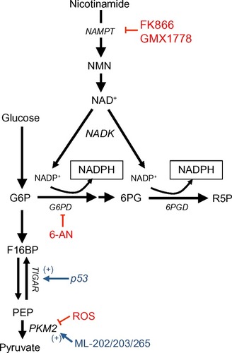
Pyruvate kinase (PK) is an essential glycolytic enzyme for conversion of phosphoenolpyruvate (PEP) to pyruvate (). The M2 isoform of PK (PKM2) is found in many cancer cells and self-renewing cells, but is expressed in an inactive state in normal adult tissues.Citation14 In many human cancers, PKM2 can be inactivated by ROS, which diverts glycolytic flux back into the oxidative PPP to generate NADPH and detoxify ROS ().Citation15 After ROS stress, PKM2 is an essential in cancer, but not normal, cells to maintain cell viability via redox scavenging. It could provide a potential efficacious antitumor therapeutic window for ROS-inducing agents.Citation15 PKM2 overexpression ensures that rapidly proliferating cancer cells create enough NADPH to match oxidative metabolism-generating ATP production, protecting the cell from attack by oxidative damage.Citation16,Citation17,Citation11
The tumor suppressor, p53, can also regulate flux into the oxidative PPP. During genotoxic stress, p53 induces TP53-induced glycolysis and apoptosis regulator (TIGAR), which encodes a protein that degrades fructose-2,6-bisphosphate ().Citation18 Low fructose-2,6-bisphosphate levels inhibit the activity of phosphofructokinase 1 (PFK1), a rate-limiting enzyme in glycolysis that leads to shuttling of earlier glycolytic metabolites into the oxidative PPP to generate NADPH. Overexpression of TIGAR was observed in colon, breast, and glioblastoma cancers.Citation19–Citation21 Consisten with the enzyme’s role in redox balance, TIGAR knockdown dramatically sensitized glioma cells to ionizing radiation.Citation22
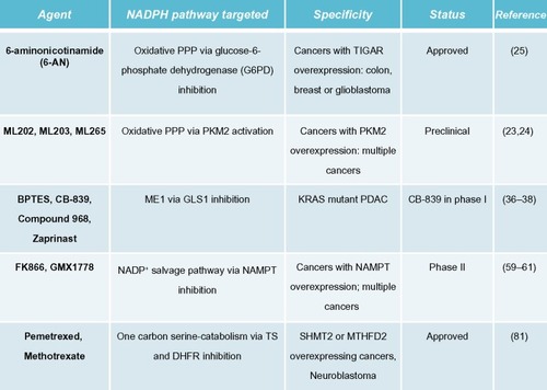
In cancers that overexpress PKM2, activating PKM2 with ML202, ML203, or other PKM2 activators blocks inhibition of PKM2 from ROS-inducing agents and decreases the flux of glucose through the oxidative PPP. This attenuates production of NADPH during oxidative damage, thereby sensitizing cancer cells to ROS-inducing agents. In contrast, normal cells that have inactive PKM2 are not sensitized to ROS-inducing agents.Citation15,Citation23,Citation24 Alternatively, the FDA-approved G6PD inhibitor, 6-aminonicotinamide (6-AN), may be utilized in cancers with PKM2 or TIGAR overexpression, thus directly inhibiting NADPH production via the oxidative PPP pathway. This, in turn, also sensitizes cancer cells to ROS-inducing agents ( and ).Citation25 The utility of this latter strategy needs to be empirically determined as G6PD is a major NADPH source in normal cells as well and toxicity concern will be a major factor in its efficacy.
Figure 2 Agents targeting specific NADPH-biogenesis pathways.
Serine catabolism
Serine-driven, one-carbon metabolism was recently shown to be a major source of NADPH in dividing cells.Citation26 Serine is metabolized in the cytoplasm or mitochondria to methylene-tetrahydrofolate (methylene-THF) by serine hydroxymethyltransferase (SHMT) 1 or 2 (cytoplasmic and mitochondrial, respectively), which then forms 10- formyl-THF via methylenetetrahydrofolate dehydrogenase (MTHFD) 1 or 2 (cytoplasmic and mitochondrial, respectively). The flux through MTHFD generates NADPH in the cytoplasm or mitochondria. 10-formyl-THF is an essential for purine biogenesis, and MTHFD’s most important function was thought to facilitate purine biosynthesis. However, the NADPH generated from this reaction is also an integral source of cellular reducing power in dividing cells (), including normal tissues that turn over quickly, like the colon.Citation26
Figure 3 One-carbon serine catabolism pathway.
Abbreviations: NADPH, nicotinamide adenine dinucleotide phosphate; MTHFD2, methylenetetrahydrofolate dehydrogenase; THF, tetrahydrofolate; DHF, dihydrofolate; DHFR, dihydrofolate reductase; PEM, pemetrexed; MTX, methotrexate; dUMP, deoxyuridine monophosphate; TS, thymidylate synthase; SHMT2, serine hydroxymethyltransferase 2.
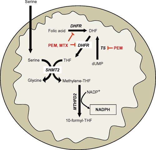
Recently, it was reported that this serine catabolism pathway can regulate mitochondrial redox control during hypoxia in Myc-driven cancers.Citation27 Specifically, SHMT2 was essential in maintaining mitochondrial NADPH and reduced GSH levels during hypoxia. SHMT2 expression was transcriptionally regulated by the coordinated activities of Myc and HIF-1α. Indeed, silencing SHMT2 in neuroblastoma cell lines significantly decreased growth in vitro under hypoxic conditions, and in a xenograft model of neuroblastoma ().Citation27 Additionally, the authors demonstrated that high SHMT2 levels correlated with a poorer prognostic outcome in neuroblastoma patients, providing a clinical context for targeting this pathway. While SHMT2 is a source of NADPH in normal dividing cells, inhibiting SHMT2 in normal cells should not significantly alter NADPH-biogenesis, since normal cells have robust compensatory mechanisms for redox balance, unlike cancer cells.Citation26
In Myc-driven neuroblastoma, inhibiting SHMT2 or MTHFD2 would decrease NADPH biogenesis derived from one-carbon serine catabolism. While there are currently no known specific inhibitors of SHMT2 or MTHFD2, targeting production of serine’s obligate reaction partner, THF, may offer a strategy to decrease NADPH production from serine catabolism in a tumor-selective manner. For example, inhibiting dihydrofolate reductase (DHFR) with the anti-folate methotrexate (MTX) will decrease THF production, thereby decreasing the flux through SHMT2 and MTHFD2. This would attenuate NADPH levels specifically in cancer cells with elevated SHMT2 expression ( and ). Indeed, MTX exposure leads to cytostasis in overactive inflammatory cells seen in autoimmune diseases by decreasing GSH production, presumably due to decreased NADPH-biogenesis.Citation28,Citation29 Alternatively, the new-generation anti-folate, pemetrexed (PEM), can also attenuate NADPH production from THF/serine catabolism by inhibiting both thymidylate synthase (TS) and DHFR, enzymes essential in THF synthesis ( and ).Citation30 Utilizing anti-folate drugs in SHMT2 overexpressing cancers in combination with ROS-inducing agents may provide a robust antitumor therapeutic window to exploit using these agents.
Malic enzymes
Another source of cellular NADPH is the NADP-dependent family of malic enzymes. This family of enzymes catalyzes the oxidative decarboxylation of malate to generate CO2 and pyruvate, while reducing NAD+ or NADP+ to NADH or NADPH in the process ().Citation31 Three isoforms were identified in mammalian systems: cytosolic NADP+- dependent (ME1), mitochondrial NAD(P)+-dependent (ME2), and mitochondrial NADP+-dependent malic enzyme (ME3).Citation32
Figure 4 KRAS-reprogrammed glutamine metabolism in pancreatic cancer.
Abbreviations: GOT2, mitochondrial aspartate transaminase; GLUD1, glutamate dehydrogenase 1; Asp, aspartate; NADPH, nicotinamide adenine dinucleotide phosphate; ME1, malic enzyme 1; ME2, malic enzyme 2; BPTES, bis-2-(5-phenylacetamido-1,2,4-thiadiazol-2-yl)ethyl sulfide 3; GLS1, glutaminase 1; MDH1, malate dehydrogenase 1; GOT1, cytosolic aspartate transaminase; OAA, oxaloacetate; αKG, α-ketoglutarate; TCA, tricarboxylic acid cycle.
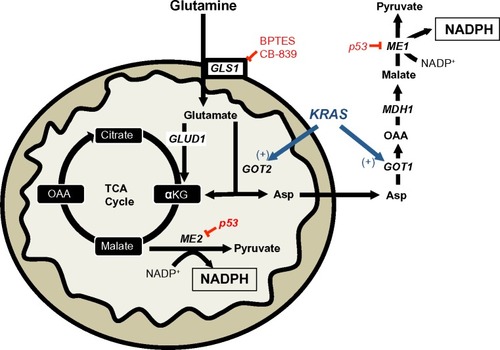
A recent report demonstrated the requirement of the cytosolic malic enzyme (ME1) in utilizing glutamine as an upstream metabolite to generate NADPH and to maintain redox balance in KRAS-mutated pancreatic ductal adenocarcinomas (PDAC), but not in normal pancreatic tissue ().Citation33 The canonical metabolism of glutamine generates α-ketoglutarate (αKG) via the upstream activity of glutamate dehydrogenase 1 (GLUD1) to drive anaplerosis to replenish the tricarboxylic acid cycle.Citation34 However, in KRAS-mutated PDAC, glutamine flux is primarily driven through mitochondrial aspartate transaminase (GOT2) to generate mitochondrial αKG and aspartate from glutamate and oxaloacetate (OAA). Aspartate is then shuttled to the cytoplasm and then acted on by cytosolic aspartate transaminase (GOT1), which is converted back to OAA (). OAA is then converted to malate by malate-dehydrogenase 1 (MDH1) and then to pyruvate and NADPH by ME1 (). The depletion of ME1 in these PDAC cancer cells suppressed cell line growth and tumor growth in vivo by ROS accumulation from loss of NADPH.Citation33 Furthermore, the inhibition of these enzymes in normal pancreatic cells did not significantly alter NADPH concentrations. Intriguingly, KRAS-mutated PDACs have dramatically decreased glucose flux into the oxidative PPP, suggesting that this non-canonical glutamine pathway is compensated for decreased NADPH production from the oxidative PPP.Citation35 In PDAC with activating KRAS mutations (which is ~90% of all PDACs), inhibiting ME1 decreases the utilization of glutamine for NADPH production and sensitizes cells to oxidative damage. While there are currently no known ME1 inhibitors, inhibiting the upstream utilization of glutamine via glutaminase 1 (GLS1) with bis-2-(5-phenylacetamido-1,2,4-thiadiazol-2-yl)ethyl sulfide 3 (BPTES), Compound 968, CB-839, or other GLS1 inhibitors would sensitize KRAS-mutated PDAC to ROS-inducing agents in a tumor-specific manner ( and ).Citation36–Citation38
A recent report demonstrated that a subset of lung tumors overexpress ME2 relative to normal lung tissue. A similar overexpression of ME2 was observed in melanoma vs normal skin, suggesting an important role for ME2 in these cancer-types.Citation39 Indeed, when ME2 was knocked down in the A549 lung cancer cell line, the cellular NADPH/NADP+ ratio decreased three-fold compared to non-targeting control cells, indicative of a pro-oxidant state in the absence of ME2.
It was recently shown that ME1 and ME2 are negatively regulated by wild-type p53, and that the absence of a functional p53 led to a dramatic up-regulation of ME1/2 expression.Citation40 Consistent with this finding, the authors demonstrated that ME1/2 enzymes were essential for NADPH maintenance in the absence of functional p53.Citation40 In the context of cancer, this is an important observation as p53 is a commonly mutated tumor suppressor and the loss of its function may lead to a cancer cell-specific mechanism of NADPH biogenesis via ME1/2 de-repression.
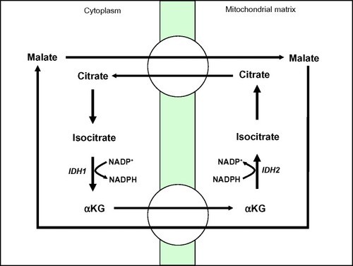
Isocitrate dehydrogenases
NADPH production can also be driven by the conversion of isocitrate to αKG by NADP+-dependent cytosolic isocitrate dehydrogenase 1 (IDH1) and mitochondrial isocitrate dehydrogenase 2 (IDH2) ().Citation41,Citation42 While NADPH generation has well known roles in the reduction of ROS, αKG can also detoxify ROS by scavenging hydrogen peroxide (H2O2) through non-enzymatic decarboxylation to form water and succinate.Citation43,Citation44
Figure 5 NADPH biogenesis via IDH1.
Abbreviations: IDH1, isocitrate dehydrogenase 1; NADPH, nicotinamide adenine dinucleotide phosphate; IDH2, isocitrate dehydrogenase 2; αKG, α-ketoglutarate.
IDH1 and IDH2 are mutated in ~80% of cases of adult glioma and secondary glioblastoma, and in 30% of cases of acute myeloid leukemia (AML).Citation45,Citation46 It was originally believed that these mutants led to enzymatic loss of function through dominant-negative inhibition of wild-type IDH1 and IDH2.Citation46–Citation48 However, it is now believed that IDH1 and IDH2 mutants confer to these enzymes the ability to convert αKG to the novel oncometabolite, 2-hydroxyglutarate (2-HG).Citation49 This change causes mutated IDH1 and IDH2 enzymes to consume rather than produce NADPH, altering the cellular redox balance and leading to a pro-oxidant state in the cancer cell.Citation50,Citation51 Additionally, overexpression of the IDH1 mutant protein in glioma cell lines sensitizes these cells to the ROS-inducing effects of ionizing radiation.Citation52
Glioma patients with IDH1R132 mutations have prolonged survival compared to patients with wild-type IDH1.Citation47,Citation50,Citation53 A hypothesis for this observation could be that IDH1 mutants are defective in generating protective concentrations of NADPH to maintain reduced GSH and thus are more sensitive to oxidative damage. Thus, glioma or AML patients with a R132 mutation in IDH1 might benefit from ROS-inducing agents early during the course of treatment.
Nicotinamide phosphoribosyltransferase (NAMPT)
NADPH generation can be driven through the NAD+ salvage pathway via NAMPT, which catalyzes the transfer of the phosphoribosyl group from 5-phosphoribosyl-1-pyrophosphate to nicotinamide, forming nicotinamide mononucleotide (NMN), and pyrophosphate ().Citation54 NAD+ generation can then be coupled with NAD+ kinase (NADK) activity to generate NADP+ that can then be reduced to NADPH through the enzymes discussed above ().Citation55
Increased NAMPT expression was reported in colorectal, non-small cell lung (NSCL), prostate, and pancreatic cancers.Citation56–Citation60 In these contexts, NAMPT has been shown to be an important source of reducing equivalents for redox balance within the cancer cell.Citation56 In fact, knockdown of NAMPT sensitized prostate and head and neck cancer cell lines to ROS induction from ionizing radiation.Citation56,Citation61–Citation63
NAMPT inhibitors are undergoing clinical trials as single-agent therapies, but recent results have, unfortunately, not been promising ().Citation64 The NAMPT inhibitors, FK866, and GMX1778, may have the greatest efficacy when combined with ROS-inducing agents that take advantage of the pro-oxidant state of NAMPT-inhibited tumors. Indeed, pre-clinical studies have validated this strategy utilizing GMX1778 to sensitize breast cancer cells against the ROS production from ionizing radiation therapy, FK866 to sensitize prostate cancer cell against H2O2, and FK866 to sensitize neuroblastoma cells against cisplatin.Citation56,Citation61,Citation65 To enhance the selectivities of NAMPT inhibitors, NADPH:quinone oxidoreductase 1 (NQO1) bioactivatable drugs can be used in combination treatments. This results in cancer-specific lethality of cells that overexpress NQO1,Citation66 such as in pancreatic, NSCL, breast, prostate, and head and neck cancers.
Predicting tumor-specific NADPH-biogenesis profiles from publicly available datasets
Known NADPH-biogenesis pathways can be useful when combined with publically available cancer gene expression and patient outcomes data to generate hypotheses for tumor-specific NADPH-biogenesis profiles.
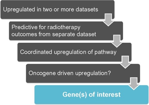
Here, we present a conservative set of criteria for determining candidate genes () using the following approach:
Determine if the genes of interest are significantly up-regulated in patient tumor tissue vs associated normal tissue in two or more independent datasets for the cancer-type in question. Only genes with P-values of <1×10⁻⁴ in each dataset will be considered for further analyses.
Of the up-regulated genes found in #1, determine which genes significantly predict poor outcomes in patients after radiation treatment.
Of the candidates from #2, determine if genes in the same NADPH biogenesis pathway as the candidate gene are coordinately up-regulated. Only positive correlation values of 0.5 and above will be considered for further analysis.
Determine if common oncogenic drivers of the tumor type in question drive genes from #3.
Figure 6 Work flow to identify NSCLC-specific NADPH-biogenesis genes in a cancer-specific NADPH-biogenesis screen.
Utilizing these criteria, we attempted to assess the NADPH-biogenesis profile of non-small cell lung cancer (NSCLC). Using the Oncomine webtool (http://www.oncomine.com), we first determined which NADPH-biogenesis related genes were overexpressed in NSCLC tissue vs associated normal tissue from patient samples. To ensure the robustness of our results, a gene was considered a potential hit only if it was found to be up-regulated in two or more datasets with a P-value <1×10⁻⁴ in each dataset. This narrowed our initial list of ten genes down to four potential genes ( and ). Next, to determine if our potential targets were clinically relevant in the context of ROS-inducing therapy, we used the KMPLOT software and stratified NSCLC patients into high- and low-expressers of the genes of interest, and compared overall survival outcomes between these groups after radiation therapy ( and ).Citation67
Figure 7 A cancer-specific NADPH-biogenesis screen.
Abbreviations: NADPH, nicotinamide adenine dinucleotide phosphate; NSCLC, non-small cell lung cancer; G6PD, glucose-6-phosphate dehydrogenase; PKM2, pyruvate kinase 2; TIGAR, TP53-induced glycolysis and apoptosis regulator; SHMT2, serine hydroxymethyltransferase 2; MTHFD2, methylenetetrahydrofolate dehydrogenase; ME1, malic enzyme 1; ME2, malic enzyme 2; IDH1, isocitrate dehydrogenase 1; IDH2, isocitrate dehydrogenase 2; NAMPT, nicotinamide phosphoribosyltransferase.
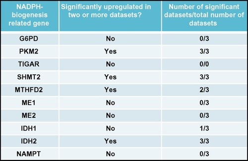
Figure 8 Cancer-specific NADPH-biogenesis screen continued.
Abbreviations: NADPH, nicotinamide adenine dinucleotide phosphate; NSCLC, non-small cell lung cancer; MTHFD2, methylenetetrahydrofolate dehydrogenase; PKM2, pyruvate kinase 2; SHMT2, serine hydroxymethyltransferase 2; IDH2, isocitrate dehydrogenase 2; G6PD, glucose-6-phosphate dehydrogenase; TS, thymidylate synthase.
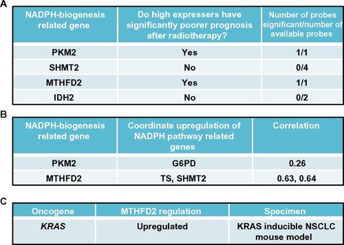
Figure 9 Screen results for MTHFD2 in NSCLC patients.
Abbreviations: MTHFD2, methylenetetrahydrofolate dehydrogenase; NSCLC, non-small cell lung cancer; NADPH, nicotinamide adenine dinucleotide phosphate; SHMT2, serine hydroxymethyltransferase 2; TS, thymidylate synthase; HR, hazard ratio.
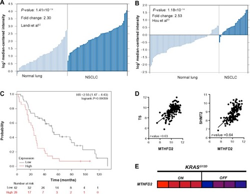
From this analysis, we selected the genes whose high expression in NSCLC patients led to significantly decreased survival after radiation therapy, suggesting that these genes may confer tumor protection from radiation-induced ROS, presumably through enhanced NADPH biogenesis.Citation68 Using this cutoff, we were able to narrow down the gene list to PKM2, which regulates NADPH biogenesis via the oxidative PPP, and MTHFD2, which generates NADPH from serine catabolism (). We then determined if these genes were coordinately up-regulated with other enzymes in their respective NADPH-biogenesis pathways. Utilizing the r2 Genomics Analysis platform’s (r2.amc.nl) co-expression analysis feature in NSCLC patient samples, we found that G6PD was coordinately up-regulated with PKM2 in patient samples with a correlation value of 0.26, and that TS and SHMT2 were co-expressed with MTHFD2 with correlation values of 0.63 and 0.64, respectively ( and ). TS and SHMT2 also appear to be co-expressed with a correlation value of 0.65. Given that we defined our correlation cutoff at 0.5 and above, we considered MTHFD2 as the top candidate for NSCLC ( and ).
Next, we investigated whether mutant-KRAS, the most common oncogenic driver in NSCLC,Citation69 might regulate MTHFD2. Such an association would provide insight into regulatory mechanisms of MTHFD2 and additional therapeutic targets. For this, we utilized publically available gene expression datasets from the National Center for Biotechnology Information Gene Expression Omnibus (GEO). To determine if mutant-KRAS regulates MTHFD2, we analyzed the mRNA expression-profiling data from a transgenic mouse model of NSCLC expressing doxycycline-inducible KRASG12D in the respiratory epithelium (GSE40606).Citation70 When administered doxycycline, the mice develop lung tumors that are dependent on constitutive KRASG12D expression. Within 48 hours of doxycycline withdrawal, KRASG12D expression was extinguished and whole-genome gene expression analyses of lung tumors were performed. The expression levels of MTHFD2 were significantly up-regulated when KRASG12D was induced vs 48 hours after KRAS extinction with doxycycline withdrawal, indicating a positive regulatory role for mutant-KRAS in MTHDF2 expression ( and ).
If this hypothesis is validated through RNAi and redox balance studies, it may suggest that the utilization of serine catabolism inhibitors, such as MTX or PEM, may provide an effective therapeutic strategy to target NADPH biogenesis, specifically in KRAS-mutated NSCLC. This analysis also reveals that more than one NADPH-biogenesis pathway may also be regulating NADPH as observed from the PKM2 data. However, validation experiments will need to be conducted to determine which pathway predominates in NSCLC.
Modulating NADPH biogenesis as a mechanism to potentiate NQO1-bioactivatable drugs
To exploit the metabolic vulnerabilities within a cancer as mentioned above, ROS-inducing agents with the capacity to specifically target tumors would be ideal. We believe that NQO1-bioactivatable drugs represent an ideal class of agents to combine with metabolic inhibitors. NQO1 is an inducible phase II detoxifying enzyme overexpressed in breast, lung, pancreatic, and colon cancers. The two-electron oxidoreductase is capable of reducing quinones by forming stable hydroquinones of the parent quinone.Citation71,Citation72 β-Lapachone (β-lap, in clinical trials as ARQ761), IB-DNQ, and other NQO1-bioactivatable drugs are unique quinones that are metabolized by NQO1 into an unstable hydroquinone that spontaneously oxidizes back to the parental compound, generating a futile redox cycle in which 1 mole of β-lap generates ~120 moles of superoxide within 2 minutes, consuming >60 moles of NADH and/or NADPH.Citation73,Citation74 The superoxide (O2−) radicals formed are quickly metabolized by superoxide dismutase (SOD) into H2O2.Citation73,Citation75 The massive levels of H2O2 formed causes extensive oxidative DNA damage that hyperactivates poly(ADP-ribosyl) polymerase 1 (PARP1), resulting in a dramatic loss of the intracellular NAD+/ATP pools and an inability to repair DNA damage.Citation76–Citation78 Cell death is stimulated by caspase-independent NAD+-Keresis, a form of programmed necrosis.Citation66,Citation73 Cancer cells with >100 units of NQO1 enzyme activity are sensitive to β-lap lethality, while normal tissues that lack, or express low levels of, NQO1 are spared.Citation79 While this class of drugs represents an attractive antitumor strategy, dose-limiting methemoglobinemia caused by non-specific ROS generation at high doses may limit its efficacy in monotherapy.Citation80 Strategies for increasing cancer cell- cytotoxicity while maintaining NQO1 specificity could greatly enhance the efficacy of β-lap for use in solid cancers that overexpress NQO1. Thus, combining β-lap with metabolic inhibitors that target cancer-specific NADPH-biogenesis pathways, such as FK866, CB-839, 6-AN, PEM or MTX, may synergistically expand the antitumor therapeutic window for NQO1 bioactivatable drugs, while increasing the tumor selectivity of metabolic inhibitors.
Conclusion
Cancer cells need to tightly regulate NADPH biogenesis to protect themselves against oxidative damage.Citation81 To sustain protective levels of NADPH, cancer cells rely on various NADPH-biogenesis pathways, including oxidative PPP, serine catabolism, glutamine metabolism, and NAD+ salvage pathways (, , and ). Strategies to inhibit NADPH biogenesis may dramatically alter the ROS scavenging abilities of cancer cells and sensitize them to oxidative damage. However, to achieve therapeutic selectivity, NADPH must be modulated through tumor-specific NADPH-biogenesis pathways that are necessary for cancer cells but are expendable in normal cells. Thus, by rigorously studying these unique pathways in the context of a specific cancer, we will be able to create novel patient-specific antitumor therapeutic strategies that exploit the ROS balances of tumor tissue while sparing normal tissue in the process. Here, we summarized our current understanding of known cancer-specific NADPH-biogenesis pathways, drugs to specifically target these pathways, and an example of using publically available databases to predict cancer-type specific NADPH-biogenesis genes. It is our belief that studying these pathways and comprehensively profiling tumors based on this understanding will be an essential step forward in designing cancer-specific ROS combination therapies, such as the use of NQO1-bioactivatable drugs.
Acknowledgments
The authors thank Dr Ralph J DeBerardinis for his assistance in reviewing this work. Support for use of Shared Resources within the Simmons Comprehensive Cancer Center was funded by NIH/NCI grant 5P30CA142543. Support for these studies was provided by grant NIH/NCI R01 CA102972, and a grant from the Pancreatic Cancer Action Network (PanCan) to DAB. This work is dedicated to the memories of Mrs Sarah Hildebrand and Rosemary Bouley for their strong support of our research.
Disclosure
The authors declare no conflicts of interest in this work.
References
- SchumackerPTReactive oxygen species in cancer cells: live by the sword, die by the swordCancer Cell20061017517616959608
- BehrendLHendersonGZwackaRMReactive oxygen species in oncogenic transformationBiochem Soc Trans2003311441144414641084
- WuWSThe signaling mechanism of ROS in tumor progressionCancer Metastasis Rev20062569570517160708
- TrachoothamDAlexandreJHuangPTargeting cancer cells by ROS-mediated mechanisms: a radical therapeutic approach?Nat Rev Drug Discov2009857959119478820
- GorriniCHarrisISMakTWModulation of oxidative stress as an anticancer strategyNat Rev Drug Discov20131293194724287781
- FruehaufJPMeyskensFLJrReactive oxygen species: a breath of life or death?Clin Cancer Res20071378979417289868
- BalendiranGKDaburRFraserDThe role of glutathione in cancerCell Biochem Funct20042234335215386533
- TraversoNRicciarelliRNittiMRole of glutathione in cancer progression and chemoresistanceOxid Med Cell Longev2013201397291323766865
- MahmoodDFAbderrazakAEl HadriKSimmetTRouisMThe thioredoxin system as a therapeutic target in human health and diseaseAntioxid Redox Signal2013191266130323244617
- KrugerNJvon SchaewenAThe oxidative pentose phosphate pathway: structure and organisationCurr Opin Plant Biol2003623624612753973
- NathanCDingASnapShot: reactive oxygen intermediates (ROI)Cell2010140951–951e952
- TianWNBraunsteinLDPangJImportance of glucose-6-phosphate dehydrogenase activity for cell growthJ Biol Chem199827310609106179553122
- PatraKCHayNThe pentose phosphate pathway and cancerTrends Biochem Sci201439834735425037503
- ZwerschkeWMazurekSMassimiPBanksLEigenbrodtEJansen-DurrPModulation of type M2 pyruvate kinase activity by the human papillomavirus type 16 E7 oncoproteinProc Natl Acad Sci U S A199996129112969990017
- AnastasiouDPoulogiannisGAsaraJMInhibition of pyruvate kinase M2 by reactive oxygen species contributes to cellular antioxidant responsesScience20113341278128322052977
- SchneiderJNeuKGrimmHVelcovskyHGWeisseGEigenbrodtETumor M2-pyruvate kinase in lung cancer patients: immunohistochemical detection and disease monitoringAnticancer Res20022231131812017309
- CerwenkaHAignerRBacherHTUM2-PK (pyruvate kinase type tumor M2), CA19-9 and CEA in patients with benign, malignant and metastasizing pancreatic lesionsAnticancer Res19991984985110216504
- BensaadKTsurutaASelakMATIGAR, a p53-inducible regulator of glycolysis and apoptosisCell200612610712016839880
- CheungECAthineosDLeePTIGAR is required for efficient intestinal regeneration and tumorigenesisDev Cell20132546347723726973
- SinhaSGhildiyalRMehtaVSSenEATM-NFkappaB axis-driven TIGAR regulates sensitivity of glioma cells to radiomimetics in the presence of TNFalphaCell Death Dis20134e61523640457
- WonKYLimSJKimGYRegulatory role of p53 in cancer metabolism via SCO2 and TIGAR in human breast cancerHum Pathol20124322122821820150
- Pena-RicoMACalvo-VidalMNVillalonga-PlanellsRTP53 induced glycolysis and apoptosis regulator (TIGAR) knockdown results in radiosensitization of glioma cellsRadiother Oncol201110113213921864926
- WalshMJBrimacombeKRAnastasiouDML265: a potent PKM2 activator induces tetramerization and reduces tumor formation and size in a mouse xenograft modelProbe Reports from the NIH Molecular Libraries ProgramBethesda (MD)National Center for Biotechnology Information2010
- AnastasiouDYuYIsraelsenWJPyruvate kinase M2 activators promote tetramer formation and suppress tumorigenesisNat Chem Biol2012883984722922757
- BelfiCAChatterjeeSGoskyDMBergerSJBergerNAIncreased sensitivity of human colon cancer cells to DNA cross-linking agents after GRP78 up-regulationBiochem Biophys Res Commun199925736136810198218
- FanJYeJKamphorstJJShlomiTThompsonCBRabinowitzJDQuantitative flux analysis reveals folate-dependent NADPH productionNature201451029830224805240
- YeJFanJVennetiSSerine catabolism regulates mitochondrial redox control during hypoxiaCancer Discov20144121406141725186948
- PhillipsDCWoollardKJGriffithsHRThe anti-inflammatory actions of methotrexate are critically dependent upon the production of reactive oxygen speciesBr J Pharmacol200313850151112569075
- SungJYHongJHKangHSMethotrexate suppresses the interleukin-6 induced generation of reactive oxygen species in the synoviocytes of rheumatoid arthritisImmunopharmacology200047354410708808
- AdjeiAAPemetrexed (ALIMTA), a novel multitargeted antineoplastic agentClin Cancer Res2004104276s4280s15217974
- BaggettoLGDeviant energetic metabolism of glycolytic cancer cellsBiochimie1992749599741477140
- PongratzRLKibbeyRGShulmanGIClineGWCytosolic and mitochondrial malic enzyme isoforms differentially control insulin secretionJ Biol Chem200728220020717102138
- SonJLyssiotisCAYingHGlutamine supports pancreatic cancer growth through a KRAS-regulated metabolic pathwayNature201349610110523535601
- DeBerardinisRJLumJJHatzivassiliouGThompsonCBThe biology of cancer: metabolic reprogramming fuels cell growth and proliferationCell Metab20087112018177721
- YingHKimmelmanACLyssiotisCAOncogenic Kras maintains pancreatic tumors through regulation of anabolic glucose metabolismCell201214965667022541435
- ElhammaliAIppolitoJECollinsLCrowleyJMarasaJPiwnica- WormsDA high-throughput fluorimetric assay for 2-hydroxyglutarate identifies zaprinast as a glutaminase inhibitorCancer Discov2014482883924740997
- ShuklaKFerrarisDVThomasAGDesign, synthesis, and pharmacological evaluation of bis-2-(5-phenylacetamido-1,2,4-thiadiazol-2-yl)ethyl sulfide 3 (BPTES) analogs as glutaminase inhibitorsJ Med Chem201255105511056323151085
- GrossMIDemoSDDennisonJBAntitumor activity of the glutaminase inhibitor CB-839 in triple-negative breast cancerMol Cancer Ther20141389090124523301
- RenJGSethPClishCBKnockdown of malic enzyme 2 suppresses lung tumor growth, induces differentiation and impacts PI3K/AKT signalingSci Rep20144541424957098
- JiangPDuWMancusoAWellenKEYangXReciprocal regulation of p53 and malic enzymes modulates metabolism and senescenceNature201349368969323334421
- NekrutenkoAHillisDMPattonJCBradleyRDBakerRJCytosolic isocitrate dehydrogenase in humans, mice, and voles and phylogenetic analysis of the enzyme familyMol Biol Evol199815167416849866202
- LeonardiRSubramanianCJackowskiSRockCOCancer-associated isocitrate dehydrogenase mutations inactivate NADPH-dependent reductive carboxylationJ Biol Chem2012287146151462022442146
- MaillouxRJSinghRBrewerGAugerCLemireJAppannaVDAlpha-ketoglutarate dehydrogenase and glutamate dehydrogenase work in tandem to modulate the antioxidant alpha-ketoglutarate during oxidative stress in Pseudomonas fluorescensJ Bacteriol20091913804381019376872
- FedotchevaNISokolovAPKondrashovaMNNonezymatic formation of succinate in mitochondria under oxidative stressFree Radic Biol Med200641566416781453
- MardisERDingLDoolingDJRecurring mutations found by sequencing an acute myeloid leukemia genomeN Engl J Med20093611058106619657110
- IchimuraKPearsonDMKocialkowskiSIDH1 mutations are present in the majority of common adult gliomas but rare in primary glioblastomasNeuro Oncol20091134134719435942
- BalssJMeyerJMuellerWKorshunovAHartmannCvon DeimlingAAnalysis of the IDH1 codon 132 mutation in brain tumorsActa Neuropathol200811659760218985363
- ParsonsDWJonesSZhangXAn integrated genomic analysis of human glioblastoma multiformeScience20083211807181218772396
- WardPSPatelJWiseDRThe common feature of leukemia-associated IDH1 and IDH2 mutations is a neomorphic enzyme activity converting alpha-ketoglutarate to 2-hydroxyglutarateCancer Cell20101722523420171147
- BleekerFEAtaiNALambaSThe prognostic IDH1(R132) mutation is associated with reduced NADP+-dependent IDH activity in glioblastomaActa Neuropathol201011948749420127344
- AtaiNARenkema-MillsNABosmanJDifferential activity of NADPH-producing dehydrogenases renders rodents unsuitable models to study IDH1R132 mutation effects in human glioblastomaJ Histochem Cytochem20115948950321527585
- LiSChouAPChenWOverexpression of isocitrate dehydrogenase mutant proteins renders glioma cells more sensitive to radiationNeuro Oncol201315576823115158
- YanHParsonsDWJinGIDH1 and IDH2 mutations in gliomasN Engl J Med200936076577319228619
- GartenAPetzoldSKornerAImaiSKiessWNampt: linking NAD biology, metabolism and cancerTrends Endocrinol Metab20092013013819109034
- YingWNAD+/NADH and NADP+/NADPH in cellular functions and cell death: regulation and biological consequencesAntioxid Redox Signal20081017920618020963
- WangBHasanMKAlvaradoEYuanHWuHChenWYNAMPT overexpression in prostate cancer and its contribution to tumor cell survival and stress responseOncogene20113090792120956937
- SrivastavaMKhuranaPSugadevRLung cancer signature biomarkers: tissue specific semantic similarity based clustering of digital differential display (DDD) dataBMC Res Notes2012561723122428
- HuftonSEMoerkerkPTBrandwijkRde BruineAPArendsJWHoogenboomHRA profile of differentially expressed genes in primary colorectal cancer using suppression subtractive hybridizationFEBS Lett1999463778210601642
- BiTQCheXMLiaoXHOverexpression of Nampt in gastric cancer and chemopotentiating effects of the Nampt inhibitor FK866 in combination with fluorouracilOncol Rep2011261251125721743967
- ChiniCCGuerricoAMNinVTargeting of NAD metabolism in pancreatic cancer cells: potential novel therapy for pancreatic tumorsClin Cancer Res20142012013024025713
- CernaDLiHFlahertySTakebeNColemanCNYooSSInhibition of nicotinamide phosphoribosyltransferase (NAMPT) activity by small molecule GMX1778 regulates reactive oxygen species (ROS)-mediated cytotoxicity in a p53- and nicotinic acid phosphoribo-syltransferase1 (NAPRT1)-dependent mannerJ Biol Chem2012287224082241722570471
- KatoHItoEShiWEfficacy of combining GMX1777 with radiation therapy for human head and neck carcinomaClin Cancer Res20101689891120103674
- OkumuraSSasakiTMinamiYOhsakiYNicotinamide phosphoribo-syltransferase: a potent therapeutic target in non-small cell lung cancer with epidermal growth factor receptor-gene mutationJ Thorac Oncol20127495622089115
- von HeidemanABerglundALarssonRNygrenPSafety and efficacy of NAD depleting cancer drugs: results of a phase I clinical trial of CHS 828 and overview of published dataCancer Chemother Pharmacol2010651165117219789873
- TravelliCDragoVMaldiEReciprocal potentiation of the antitumoral activities of FK866, an inhibitor of nicotinamide phospho-ribosyltransferase, and etoposide or cisplatin in neuroblastoma cellsJ Pharmacol Exp Ther201133882984021685314
- MooreZChakrabartiGLuoXNAMPT inhibition sensitizes pancreatic adenocarcinoma cells to tumor-selective, PAR-independent metabolic catastrophe and cell death induced by beta-lapachoneCell Death Dis20156e159925590809
- GyorffyBSurowiakPBudcziesJLanczkyAOnline survival analysis software to assess the prognostic value of biomarkers using transcript-tomic data in non-small-cell lung cancerPLoS One20138e8224124367507
- DiehnMChoRWLoboNAAssociation of reactive oxygen species levels and radioresistance in cancer stem cellsNature200945878078319194462
- RobertsPJStinchcombeTEKRAS mutation: should we test for it, and does it matter?J Clin Oncol2013311112112123401440
- FisherGHWellenSLKlimstraDInduction and apoptotic regression of lung adenocarcinomas by regulation of a K-Ras transgene in the presence and absence of tumor suppressor genesGenes Dev2001153249326211751631
- CaoLLiLSSpruellCTumor-selective, futile redox cycle-induced bystander effects elicited by NQO1 bioactivatable radiosensitizing drugs in triple-negative breast cancersAntioxid Redox Signal20142123725024512128
- AwadallahNSDehnDShahRJNQO1 expression in pancreatic cancer and its potential use as a biomarkerAppl Immunohistochem Mol Morphol200816243118091324
- BeyEAReinickeKESrougiMCCatalase abrogates beta-lapachone-induced PARP1 hyperactivation-directed programmed necrosis in NQO1-positive breast cancersMol Cancer Ther2013122110212023883585
- PinkJJWuerzberger-DavisSTagliarinoCActivation of a cysteine protease in MCF-7 and T47D breast cancer cells during beta-lapachone-mediated apoptosisExp Cell Res200025514415510694431
- TagliarinoCPinkJJDubyakGRNieminenALBoothmanDACalcium is a key signaling molecule in beta-lapachone-mediated cell deathJ Biol Chem2001276191501915911279125
- BentleMSReinickeKEDongYBeyEABoothmanDANonhomologous end joining is essential for cellular resistance to the novel antitumor agent, beta-lapachoneCancer Res2007676936694517638905
- BoothmanDAGreerSPardeeABPotentiation of halogenated pyrimidine radiosensitizers in human carcinoma cells by beta-lapachone (3,4-dihydro-2,2-dimethyl-2H-naphtho[1,2-b]pyran- 5,6-dione), a novel DNA repair inhibitorCancer Res198747536153663652040
- BoothmanDATraskDKPardeeABInhibition of potentially lethal DNA damage repair in human tumor cells by beta-lapachone, an activator of topoisomerase ICancer Res1989496056122535961
- LiLSBeyEADongYModulating endogenous NQO1 levels identifies key regulatory mechanisms of action of beta-lapachone for pancreatic cancer therapyClin Cancer Res20111727528521224367
- HartnerLPLRMHMendelsonDPhase 2 dose multi-center, open-label study of ARQ 501, a checkpoint activator, in adult patients with persistent, recurrent or metastatic leiomyosarcoma (LMS)J Clin Oncol20072520521
- Vander HeidenMGCantleyLCThompsonCBUnderstanding the Warburg effect: the metabolic requirements of cell proliferationScience20093241029103319460998
- LandiMTDrachevaTRotunnoMGene expression signature of cigarette smoking and its role in lung adenocarcinoma development and survivalPLoS One20083e165118297132
- HouJAertsJden HamerBGene expression-based classification of non-small cell lung carcinomas and survival predictionPLoS One20105e1031220421987
- TalbotSGEstiloCMaghamiEGene expression profiling allows distinction between primary and metastatic squamous cell carcinomas in the lungCancer Res2005653063307115833835