
Abstract
Background
Hyperlipidemia is a worth-mentioning risk factor in quickly expanding cardiovascular diseases, including myocardial infarction and, furthermore, in stroke.
Methods
The present work describes the synthesis of phenolic derivatives 4a–e and 6a–c with the aim of developing antihyperlipidemic agents. The structures of the synthesized compounds were confirmed by spectroscopic data. The in silico docking studies were performed against human 3-hydroxy-3-methylglutaryl-coenzyme A (HMG CoA) reductase enzyme (PDB ID: 1HWK), and it was observed that compounds 4a and 6a exhibited maximum binding affinity with target protein having binding energies −8.3 and −7.9 kcal, respectively.
Results
Compound 4a interacts with amino acids Val805 with distance 1.89 Å and Met656, Thr558, and Glu559 with bonding distances 2.96, 2.70, and 2.20 Å, respectively. The in vivo antihyperlipidemic activity results revealed that compound 4a indicated minimum weight increment, ie, 20% compared with 35% weight increment with standard drug atorvastatin during 6 weeks of treatment. Moreover, increment in high-density lipoprotein cholesterol and decrease in total cholesterol, low-density lipoprotein cholesterol, and triglyceride levels were more prominent in case of 4a compared to atorvastatin with P<0.05. The synthesized compounds were nontoxic and well tolerated because none of the mice were found to suffer from any kind of morbidity and death during 6 weeks of dosing.
Conclusion
Based on our pharmacological evaluation, we may propose that compound 4a may act as a lead structure for the design and development of more potent antihyperlipidemic drugs.
Introduction
Human beings are persistently confronting life-threatening morbidities such as cardiovascular disorders and cancer. Cardiovascular diseases account for ~17 million deaths annually worldwide. Above all, atherosclerosis is the major factor for the pathogenesis of myocardial and cerebral infarctions. Increased plasma low-density lipoproteins and triglycerides along with low levels of high-density lipoproteins are often the major root cause of atherosclerosis and ultimately myocardial and cerebral infarctions. Correspondingly, obesity has a direct linkage with atherosclerosis and myocardial infarction. Exact etiology of obesity is relatively unclear, but still the major contributors are genetic, environmental, and dietary factors.Citation1
To counter these morbidities, an efficacious and safer pharmacological solution is of most extreme significance. The process of drug discovery is fastidious and expensive. The quest for new effective molecules is challenging yet the accessibility of various docking tools and computer-aided methodologies has limited the gap to a specific level.Citation2,Citation3 Antioxidants have phenolic flavonoids that scavenge oxygen radicals and are involved in the inhibition of enzyme activity. Phenolic compounds are auxiliary metabolites present in fruits and vegetables.Citation4,Citation5 They possess diverse pharmacological activities including cardiovascular,Citation6 antioxidant,Citation5–Citation9 antiatherogenic,Citation10,Citation11 anti-inflammatory,Citation12–Citation16 and antithrombotic.Citation17–Citation19 Various studies on the experimental animals reported the effect of phenolic compounds such as quercetin and rutinosides on the lipid metabolism causing a decrease in the serum lipid levels.Citation20,Citation21
Derivatives of natural poly-phenols have shown remarkable bioactivity in various disease areas both in prevention and treatment. Among numerous classes of polyphenols, hydroxycinnamic acid derivative is the renowned group of phenols found in cereals, roasted coffee, and green vegetables and is reported as a potential antioxidant.Citation22–Citation24 Sinapic acid and ferulic acid are the most studied hydroxycinnamic acid derivatives and were found to be highly effective as antioxidant,Citation25,Citation26 and in recent data, they are found to be preventive and therapeutic agents in several other diseases including atherosclerosis, inflammatory injury, and cancer.Citation27,Citation28 Caffeic acid derivatives are another class of biologically active phenolic derivatives as antioxidantCitation29,Citation30 and anticancer.Citation31–Citation33
Recently, few studies have demonstrated polyphenolic moieties as potential antihyperlipidemic agents by inhibiting 3-hydroxy-3-methylglutaryl-coenzyme A (HMG CoA) reductase in in silico models.Citation34 Considering such enormous pharmacological potential, derivatives of natural phenols are exciting prospect for future drug development especially in antihyperlipidemic and cardiovascular therapeutic domains. The use of computational analysis to establish the binding affinity of the drug-like compounds with different enzymes, receptors, and proteins is categorized in in silico pharmacology.Citation35 The in silico methods have been frequently used in the discovery and optimization of novel molecules with affinity to a target and in the optimization of pharmacokinetic parameters.Citation36,Citation37 Various studies have been reported in which the in silico assessment of the drug-like compounds showed the binding affinity with enzymes, proteins, transporter proteins, and receptors and is very well correlated with their in vitro and in vivo pharmacological effects.Citation38 Consequently, the in silico pharmacology paradigm is ongoing and presents a vast platform for the discovery of new drug targets as well as screening of new drug-like compounds that can be considered as drug.
In this perspective, current study is aimed to develop potent antihyperlipidimic agents. The phenolic derivatives 4a–e and 6a–c have been synthesized and characterized; the in vivo antihyperlipidemic evaluation was carried out. The in silico docking studies against HMG CoA reductase enzyme (PDB ID: 1HWK) were also performed to compare the dry laboratory results with wet laboratory findings.
Materials and methods
Drugs and chemicals
Atorvastatin was obtained from Global Pharmaceuticals (Islamabad, Pakistan), and mice were received as gift from National Institute of Health (Islamabad, Pakistan).
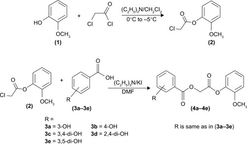
Synthesis of 2-methoxyphenyl chloroacetate (2)
The 2-methoxyphenol (1) (0.01 mol) was dissolved in anhydrous dichloromethane (25 mL), and triethylamine (0.01 mol) was added slowly. The resulting solution was then cooled in an ice salt mixture to 0 to −5°C. The chloroacetyl chloride (0.01 mol) in dry dichloromethane was added drop wise to the reaction mixture with constant stirring over a period of 1 h maintaining the temperature constant. The reaction mixture was then stirred at room temperature for further 5 h and washed with 5% HCl and 5% sodium hydroxide solution. The organic layer was washed with saturated aqueous NaCl, dried over anhydrous magnesium sulfate and filtered, and the solvent was removed under reduced pressure. The crude product was purified by silica gel column to afford the corresponding 2-methoxyphenyl chloroacetate (2): melting point, 56°C–58°C; reaction time, 4 h; yield, 78%; Rf, 0.62 (n-hexane:ethyl acetate 3:1); FTIR νmax (1/cm): 2,903 (sp2 C−H), 2,863 (sp3 C−H), 1,719 (C=O ester), 1,589 (C=C aromatic), 1,164 (C−O ester).
General procedure for the synthesis of title compounds 4a–4e and 6a–6c
The substituted benzoic acids (3a–3e) (0.01 mol), triethyl amine (0.01 mol), and potassium iodide (0.01 mol) were mixed in dimethylformamide (25 mL) and stirred at room temperature. The 2-methoxyphenyl chloroacetate intermediate (2) was then added to the reaction mixture slowly and at the same temperature overnight (). The final products (4a–4e) were then extracted with ethyl acetate (3×25 mL). The combined ethyl acetate layer was then washed with 5% HCl, 5% sodium carbonate, and finally brine solution. The organic layer was dried over anhydrous magnesium sulfate and filtered, and the solvent was removed under reduced pressure to afford the crude products (4a–4e). The title compounds (4a–4e) were purified by silica gel column chromatography (n-hexane:ethyl acetate 3:1). The same procedure was used for the preparation of final products (6a–6c) ().
2-(2-Methoxyphenoxy)-2-oxoethyl 3-hydroxybenzoate (4a): solid; melting point, 81°C–83°C; yield, 76%; Rf, 0.56 (n-hexane:ethyl acetate 3:1); FTIR νmax (1/cm): 3,248 (O−H), 3,965 (sp2 C−H), 2,864 (sp3 C−H), 1,723 (C=O ester), 1,595 (C=C aromatic), 1,164 (C−O ester); 1H NMR (CDCl3, δ ppm): 7.69 (m, 1H, H-6), 7.58 (dd, J=1.6, 1.6 Hz, 1H, H-2), 7.33 (dd, J=4.8, 4.4 Hz, 1H, H-5), 7.24 (m, 1H, H-4), 7.12 (dd, J=6.4, 1.6 Hz, 1H, H-6′), 7.07 (dd, J=3.6, 1.8 Hz, 1H, H-3′), 6.95–6.99 (m, 2H, H-4′, H-5′), 5.65 (s, 1H, −OH), 5.10 (s, 2H, −CH2), 3.85 (s, 3H, −OCH3); 13C NMR (CDCl3, δ ppm); 166.1 (C=O ester), 165.7 (C=O ester), 155.8 (C-3), 150.9 (C-2′), 139.0 (C-1′), 130.4 (C-5), 129.7 (C-6), 127.3 (C-2), 122.6 (C-1), 122.3 (C-4), 121.4 (C-4′), 120.8 (C-6′), 116.6 (C-3′), 114.5 (C-5′), 61.0 (−CH2), 55.8 (−OCH3); anal calcd for C16H14O6: C, 63.57; H, 4.63; found C, 63.64; H, 4.71.
2-(2-Methoxyphenoxy)-2-oxoethyl 4-hydroxybenzoate (4b): solid; melting point, 126°C–128°C; yield, 78%; Rf, 0.53 (n-hexane:ethyl acetate 3:1); FTIR νmax (1/cm): 3,149 (O−H), 2,932 (sp2 C−H), 2,842 (sp3 C−H), 1,718 (C=O ester), 1,593 (C=C aromatic), 1,148 (C−O, ester); 1H NMR (DMSO-d6, δ ppm): 7.90 (d, J=8.8 Hz, 2H, H-2, H-6), 7.26 (ddd, J=6.8, 1.6, 1.4 Hz, 1H, H-4′), 7.11–7.16 (m, 2H, H-3′, H-6′), 6.97 (ddd, J=6.4, 1.6, 1.21H, H-5′), 6.90 (d, J=8.8 Hz, 2H, H-3, H-5), 5.15 (s, 2H, −CH2), 3.78 (s, 3H, −OCH3), 3.33 (s, 1H, −OH); 13C NMR (DMSO-d6, δ ppm): 166.8 (C=O ester), 165.4 (C=O ester), 162.9 (C-4), 151.1 (C-2′), 139.0 (C-1′), 132.2 (C-2, C−), 127.7 (C-5′), 123.0 (C-1), 121.3 (C-3′), 119.6 (C-4′), 116.0 (C-3, C-5), 113.5 (C-6′), 60.9 (−CH2), 56.3 (−OCH3); anal calcd for C16H14O6: C, 63.57; H, 4.63; found C, 63.66; H, 4.73.
2-(2-Methoxyphenoxy)-2-oxoethyl 3,4-dihydroxy-benzoate (4c): solid; melting point, 154°C–156°C; yield, 72%; Rf, 0.48 (n-hexane:ethyl acetate 3:1); FTIR νmax (1/cm): 3,166 (O−H), 2,935 (sp2 C−H), 2,837 (sp3 C−H), 1,722 (C=O ester), 1,600 (C=C aromatic), 1,148 (C−O ester); 1H NMR (DMSO-d6, δ ppm): 7.43 (d, J=2.0 Hz, 1H, H-2), 7.39 (dd, J=6.0, 2.0 Hz, 1H, H-6), 7.25 (ddd, J=3.6, 2.0, 1.6 Hz, 1H, H-4′), 7.12–7.16 (m, 2H, H-3′, H-6′), 6.97 (ddd, J=4.0, 2.4, 1.6 Hz, 1H, H-5′), 6.84 (d, J=8.0 Hz, 1H, H-5), 5.13 (s, 2H, −CH2), 3.80 (s, 3H, −OCH3), 3.42 (s, 2H, −OH); 13C NMR (DMSO-d6, δ ppm): 171.4 (C=O ester), 166.8 (C=O ester), 165.5 (C-4), 151.4 (C-3), 151.1 (C-2′), 148.1 (C-1′), 139.0 (C-5′), 127.9 (C-6), 123.2 (C-4′), 122.7 (C-3′), 121.1 (C-6′), 119.8 (C-2), 116.9 (C-5), 113.5 (C-1), 60.9 (−CH2), 56.1 (−OCH3); anal calcd for C16H14O7: C, 60.37; H, 4.40; found C, 60.44; H, 4.46.
2-(2-Methoxyphenoxy)-2-oxoethyl 2,4-dihydroxybenzoate (4d): solid; melting point, 109°C–111°C; yield, 76%; Rf, 0.46 (n-hexane:ethyl acetate 3:1); FTIR νmax (1/cm): 3,263 (O−H), 2,942 (sp2 C−H), 2,864 (sp3 C−H), 1,726 (C=O ester), 1,606 (C=C aromatic), 1,162 (C−O ester); 1H NMR (DMSO-d6, δ ppm): 7.73 (d, J=8.8 Hz, 1H, H-6), 7.24–7.29 (m, 2H, H-3′, H-6′), 7.15 (ddd, J=6.4, 5.6, 1.6, Hz, 1H, H-4′), 6.98 (ddd, J=5.2, 3.6, 2.0, Hz, 1H, H-5′), 6.43 (dd, J=6.4, 2.4 Hz, 1H, H-3), 6.34 (d, J=2.0 Hz, 1H, H-5), 5.20 (s, 2H, −CH2), 3.78 (s, 3H, −OCH3), 3.44 (s, 2H, −OH); 13CNMR (DMSO-d6, δ ppm): 168.3 (C=O ester), 166.5 (C=O ester), 165.1 (C-2), 163.2 (C-4), 151.1 (C-2′), 139.0 (C-1′), 132.5 (C-5′), 127.8 (C-6), 123.2 (C-4′), 121.3 (C-3′), 119.6 (C-6′), 116.0 (C-3), 109.0 (C-5), 103.1 (C-1), 61.1 (−CH2), 56.1 (−OCH3); anal calcd for C16H14O7: C, 60.37; H, 4.40; found C, 60.41; H, 4.48.
2-(2-Methoxyphenoxy)-2-oxoethyl 3,5-dihydroxybenzoate (4e): solid; melting point, 173°C–175°C; yield, 84%; Rf, 0.45 (n-hexane:ethyl acetate 3:1); FTIR νmax (1/cm): 3,134 (O−H), 2,919 (sp2 C−H), 2,838 (sp3 C−H), 1,729 (C=O ester), 1,608 (C=C aliphatic), 1,598 (C=C aromatic), 1,145 (C−O, ester); 1H NMR (DMSO-d6, δ ppm): 7.27 (ddd, J=5.6, 3.6, 1.6 Hz, 1H, H-4′), 7.12–7.15 (m, 2H, H-3′, H-6′), 6.96 (ddd, J=6.4, 6.4, 2.4 Hz, 1H, H-5′), 6.89 (d, J=2.4 Hz, 2H, H-2, H-6), 6.48 (dd, J=2.4, 1.6 Hz, 1H, H-4), 5.17 (s, 2H, −CH2), 3.78 (s, 3H, −OCH3), 3.33 (s, 2H, −OH); 13C NMR (DMSO-d6, δ ppm); 166.6 (C=O ester), 165.6 (C=O ester), 159.1 (C-3, C-5), 151.1 (C-2′), 139.0 (C-1′), 130.7 (C-5′), 127.8 (C-2, C-6), 123.0 (C-4′), 121.1 (C-3′), 113.5 (C-6′), 108.2 (C-4), 107.8 (C-1), 61.3 (−CH2), 56.3 (C-1″); anal calcd for C16H14O7: C, 60.37; H, 4.40; found C, 60.43; H, 4.47.
2-(2-Methoxyphenoxy)-2-oxoethyl (2E)-3-phenylprop-2-enoate (6a): solid; melting point, 76°C–78°C; yield, 85%; Rf, 0.56 (n-hexane:ethyl acetate 3:1); FTIR νmax (1/cm): 2,938 (sp2 C−H), 2,835 (sp3 C−H), 1,719 (C=O ester), 1,599 (C=C aromatic), 1,164 (C−O, ester); 1H NMR (CDCl3, δ ppm): 7.85 (d, J=16.0 Hz, 1H, H-2), 7.58 (dd, J=4.4, 1.6 Hz, 2H, H-2′, 6′), 7.41–7.43 (m, 3H, H-3′, H-4′, H-5′), 7.24 (dd, J=6.0, 2.4 Hz, 1H, H-3″), 7.13 (dd, J=6.4, 2.4 Hz, 1H, H-4″), 6.96–6.99 (m, 2H, H-4″, H-5″), 6.56 (d, J=16.0 Hz, 1H, H-1), 5.04 (s, 2H, −CH2), 3.86 (s, 3H, −OCH3); 13C NMR (CDCl3, δ ppm); 167.7 (C=O ester), 166.1 (C=O ester), 150.9 (C-2″), 146.3 (C-2), 139.2 (C-1″), 134.1 (C-5″), 130.0 (C-2′, C-6′), 128.9 (C-3′, C-5′), 128.2 (C-4′), 127.2 (C-1′), 122.6 (C-3″), 120.7 (C-4″), 116.7 (C-6″), 112.5 (C-1), 60.5 (−CH2), 55.9 (−OCH3); anal calcd for C18H16O5: C, 69.23; H, 5.12; found C, 69.29; H, 5.17.
2-(2-Methoxyphenoxy)-2-oxoethyl (2E)-3-(4- hydroxyphenyl)prop-2-enoate (6b): solid; melting point, 101°C–103°C; yield, 78%; Rf, 0.47 (n-hexane:ethyl acetate 3:1); FTIR νmax (1/cm): 3,132 (−OH), 2,931 (sp2 C−H), 2,849 (sp3 C−H), 1,728 (C=O), 1,609 (C=C aromatic), 1,148 (C−O ester); 1H NMR (DMSO-d6, δ ppm): 7.70 (d, J=16.0 Hz, 1H, H-2), 7.62 (d, J=8.8 Hz, 2H, H-2′, 6′), 7.26 (ddd, J=7.6, 6.0, 1.6 Hz, 1H, H-4″), 7.11–7.16 (m, 2H, H-3″, H-6″), 6.98 (ddd, J=7.6, 6.0, 1.6 Hz, 1H, H-5″), 6.80 (d, J=8.8 Hz, 2H, H-3′, H-5′), 6.54 (d, J=16.0 Hz, 1H, H-1), 5.06 (s, 2H, −CH2), 3.78 (s, 3H, −OCH3), 3.33 (s, 1H, −OH); 13C NMR (DMSO-d6, δ ppm): 166.1 (C=O ester), 165.8 (C=O ester), 150.9 (C-2″), 144.8 (C-2), 139.1 (C-1″), 132.6 (C-4′), 129.5 (C-2′, C-6′), 129.3 (C-3′, C-5′), 127.2 (C-5″), 122.7 (C-1′), 117.4 (C-3″), 112.6 (C-4″), 112.5 (C-6″), 60.6 (−CH2), 55.9 (−OCH3); anal calcd for C18H16O6: C, 65.85; H, 4.87; found C, 65.93; H, 4.93.
2-(2-Methoxyphenoxy)-2-oxoethyl (2E)-3-(4-chlorophenyl)prop-2-enoate (6c): solid; melting point, 73°C–75°C; yield, 83%; Rf, 0.55 (n-hexane:ethyl acetate 3:1); FTIR νmax (1/cm): 2,919 (sp2 C−H), 2,862 (sp3 C−H), 1,725 (C=O ester), 1,611 (C=C aromatic), 1,156 (C−O ester); 1H NMR (CDCl3, δ ppm): 7.79 (d, J=16.0 Hz, 1H, H-2), 7.50 (d, J=7.6 Hz, 2H, H-2′, 6′), 7.43 (d, J=7.6 Hz, 2H, H-3′, H-5′), 7.24 (dd, J=6.0, 1.6 Hz, 1H, H-3″), 7.11 (dd, J=6.4, 1.6 Hz, 1H, H-6″), 6.95–6.99 (m, 2H, H-4″, H-5″), 6.57 (d, J=16.0 Hz, 1H, H-1), 5.06 (s, 2H, −CH2), 3.86 (s, 3H, −OCH3); 13C NMR (CDCl3, δ ppm); 166.1 (C=O ester), 165.8 (C=O ester), 151.0 (C-2″), 144.9 (C-2), 139.1 (C-1″), 136.5 (C-4′), 132.6 (C-2′, C-6′), 129.5 (C-3′, C-5′), 127.3 (C-5″), 122.7 (C-1′), 121.6 (C-3″), 120.7 (C-4″), 117.3 (C-6″), 60.6 (−CH2), 56.0 (−OCH3); anal calcd for C18H15O5Cl: C, 62.33; H, 4.32; found C, 62.41; H, 4.39.
Antihyperlipidemic studies
An aggregate of 66 mice (Balb C, male, 25–30 g, aged 4–6 weeks) was obtained, kept in National Institute of Health, and left to be acclimatized for 1 week before starting the investigation. Animal experiments were performed in accordance with the Institute of Laboratory Animal Resources, Commission on Life Sciences, National Research Council (1996), approved by Ethical Committee of Riphah Institute of Pharmaceutical Sciences (ref no REC/RIPS/2017/001). Mice were divided into 11 groups, each having six mice. Mice were kept in normal laboratory conditions of temperature 23°C±1°C and ambient humidity 55%±5%. Mice were housed in stainless steel cages individually and kept in an isolated room. Lights were maintained on an artificial 12 h light–dark cycle. All mice were weighed weekly during 6 weeks of experimental period. Animal care was in accordance with the guidelines established and approved by the National Institute of Health. Mice were fed with high-fat diet (HFD) mentioned in containing cholesterol, cholic acid, peanut oil, and normal laboratory diet for 6 weeks. Standard drug (atorvastatin in this study) and test compounds from 4a–e and 6a–c were mixed with HFD, and the composition of the test compounds was 10 mg/kg. Experimental compounds from 4a–e and 6a–c were mixed with HFD, and the dose of the test compounds was 10 mg/kg. Group I received normal laboratory diet, Group II received HFD, Group III received standard drug (atorvastatin) along with HFD, and Groups IV–XI received test compounds from 4a–e and 6a–c, respectively, along with HFD. Mice had free access to feed and water throughout the study. The specified quantity of test compounds and standard was dissolved in 1 mL of DMSO and mixed in pellet diet. All mice were weighed weekly during 6 weeks of experimental period. The activity was continued for 6 weeks under controlled laboratory conditions (temperature 23°C±1°C and ambient humidity 55%±5%).Citation39
Table 1 Fat diet composition
Blood collection and analysis
At the end of the experiment, the animals were anesthetized. Blood samples were collected from the retro orbital vein. The serum was separated by centrifugation at 2,500× g for 15 min at 4°C. The amount of serum triglyceride, total cholesterol, high-density lipoprotein (HDL) cholesterol, and low-density lipo-protein (LDL) cholesterol were assayed automatically using an ADVIA 1650 lipid analyzer (Bayer, Wuppertal, Germany).
Histopathological evaluation
Animals were euthanized under ether anesthesia, and the liver was dissected out immediately and weighed. For histopathological analysis, the liver and adipose tissues were fixed in 10% formalin at room temperature. Adipose tissues used were collected from subcutaneous fat of the abdomen of mice. The tissue was embedded in paraffin, sectioned into 3–4 μm thickness and mounted on the glass microscope slides using standard histological techniques. The sections were stained with hematoxylin–eosin and examined using light microscopy at 200× magnitudes. These light microscopic fields were assessed by an image analyzer on each section.Citation40
Docking studies
Acquisition and preparation of target protein
Crystal structure of HMG CoA reductase was acquired from Protein Data Bank as PDB format (PDB ID: 1HWK-Homosapien) (www.rcsb.org/pdb).Citation41 Crystal structure of target protein was validated using the MolProbity server.Citation42 Ligands and water molecules were removed from crystal structure using the Accelrys Discovery Studio, and the energy of protein was minimized using the UCSF Chimera.Citation43 Active binding site studies were viewed using the PyMOL (Python molecular visualization) software.Citation44
Preparation of ligand
Structures of phenolic experimental compounds were drawn in MarvinSketch tools and were cleaned into 3D using Marvin Space and were downloaded in the PDB file extension.Citation45
Docking run
Docking is a virtual screening tool used to predict in silico interaction of ligand with enzyme and its possible inhibition. Both ligand and target protein were converted from PDB to PDBQT files after the addition of hydrogen and removal of water molecules. Energies of ligand and enzyme were minimized using the UCSF Chimera. These prepared molecules were uploaded as ligand, macromolecules in PyRx, a Python-based AutoDock Vina-based scoring function screening tool, autogrid dimension were calculated for HMG CoA reductase (X=147.451, Y=87.546, and Z=3.341), and inhibitor and target were geometrically optimized and docked.Citation46,Citation47
Statistical analysis
For the determination of change in body weight, liver weight, plasma lipids, and HDL and LDL-cholesterol levels, individual mouse was considered as an experimental unit. All data were analyzed by IBM SPSS 20 package for one-way analysis of variance (ANOVA) testing; post hoc Tukey test was used with P-value <0.05 considering as statistically significant.Citation48
Results and discussion
Body weights
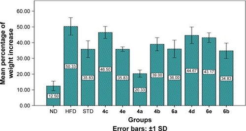
Among dietary factors, an increase in carbohydrate and fats in diet has been associated with obesity.Citation49 Similarly, few researchers have reported that the differences among strains with the ability of HFD to promote obesity have been demonstrated in both rats and mice.Citation32 Some strains of mice such as A/J and C57BL do develop obesity when fed on HFD. Meanwhile, SWR mice were resistant to develop obesity when fed on similar HFD. In the present study, mice fed with a HFD demonstrated predictable weight, cholesterol, triglycerides, and LDL increment with a decrease in HDL levels after 6 weeks in the hyperlipidemic control group (HFD group) (). After 6 weeks, mice fed with 4a experimental compound indicated least weight increase, ie, 20% compared with 35% weight increase with atorvastatin. Even 4e, 6a, and 6b showed comparable weight increment vs atorvastatin, ie, 36, and 35%, respectively (). These results propose that weight increment in case of 4a was significantly (P<0.05) less than reference standard and increase in mice weight in case of 4e, 6a, and 6b was insignificant but comparable with reference standard ().
Figure 1 Impact of phenolic derivatives on body weight changes on mice supplemented with high-fat diet.
Abbreviations: ND, normal diet; HFD, high-fat diet; Std, reference standard (atorvastatin).
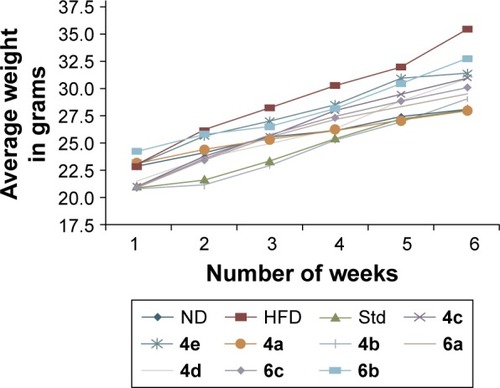
Figure 2 Effect of phenolic derivatives on body weight of mice fed on high-fat diet compared with standard (Atorvastatin) and high-fat diet alone.
Abbreviations: ND, normal diet; HFD, high-fat diet; STD, reference standard (atorvastatin).
Plasma lipid profile
As already discussed, an increase in serum cholesterol and LDL levels may lead to atherosclerosis and ultimately to myocardial infarction. Raised serum triglycerides (TGs) have also been reported to have a role in the aggravation of atherosclerosis.Citation50 Meanwhile various studies have shown that drugs decreasing serum TG, LDL, and cholesterol levels reduce the risk of coronary artery diseases.Citation51 Various phenols including gallic acid and linoleic acid have been found to be antiatherogenic especially in in vivo green tea extracts reducing serum TGs and cholesterol as well as stimulating energy expenditure, fat oxidation, and fecal lipid oxidation.Citation52
In this study, it was observed that all experimental molecules were able to decrease cholesterol, LDL, and triglycerides and increase HDL in hyperlipidemic mice, but in particular, 4a showed highly significant reduction in serum cholesterol compared to standard with P<0.01, highly significant increase in HDL compared to standard with P<0.01, and significant reduction in LDL compared to standard with P<0.05 (). In order to unveil dose-dependent effect of 4a, it was evaluated in same hyperlipidemic mice model with three different doses, ie, high dose (10 mg/kg), medium dose (5 mg/kg) and low dose (2.5 mg/kg). Effect of 4a reduced with decrease in dose when compared with high dose 10 mg/kg, and this decrease was statistically significant (). In vivo outcomes clearly showing the superiority of antihyperlipidemic potential of 4a compared with other phenolic derivatives and even compared to standard atorvastatin. Furthermore, after 42 days of treatment none of the mice showed any kind of morbidity or mortality, which further advocates that all experimental phenolic compounds were well tolerated and were fairly safe during the whole experiment. These astounding outcomes establish the commendable and plausible antihyperlipidemic potential of our experimental natural phenolic derivatives.
Table 2 Effect of phenolic derivatives on lipid lowering efficiency in mice (n=6, mean ± SD) compared with normal control, hyperlipidemic control, and standard drug (atorvastatin)
Table 3 Dose-dependent antihyperlipidemic effect of 4a (n=6, mean ± SD)
Histopathological examination
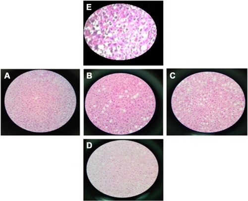
Fatty liver is gaining importance in investigations and now is known to be most prevalent cause of abnormal liver. Fatty liver initially starts with simple steatosis, which later may develop into advanced fibrosis and cryptogenic cirrhosis via steatohepatitis and ultimately to hepatocellular carcinoma.Citation53 Obesity is the single main causative factor in the development of fatty liver both in children and adults.Citation54 In our current investigation, 4a fed group of mice showed significant histopathological changes in liver sections under microscopic evaluation (). A small loss of nucleus and membrane disruption was observed when compared with the normal diet (ND) group, but no fat droplet was observed in case of 4a group when compared with the HFD group. Histology of adipocytes showed that difference in size was evidently less in case of 4a group compared with the HFD group ().
Figure 3 Histopathological evaluation of liver section slides: (A) 4a group; (B) 6a group; (C) 6b group; (D) standard drug group; and (E) hyperlipidemic control.
Docking studies
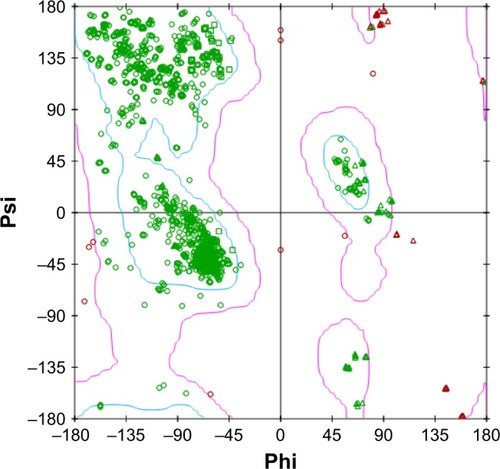
The in silico docking studies were performed to determine the binding affinity of the synthesized compounds with target protein. It has been reported that docking is an effective predictive tool regarding possible interactions of ligands with target protein.Citation55 It further enables illustration of the possible atomic interactions between the particular functional group of a ligand in the active binding site of protein. To date, the most successful strategy to control hyperlipidemic disorders has been to inhibit HMG CoA reductase. This enzyme catalyzes the conversion of HMG CoA to mevalonic acid, ultimately producing cholesterol.Citation56 HMG CoA reductase model characterized with X-ray diffraction at a resolution of 2.22 Å was downloaded from Protein Data Bank (). The structure was validated by using the Ramachandran plot, which demonstrated that >95.7% residues were lying in favorable region and 99.4% residues were lying in allowed region of plot (). The experimental compounds (phenolic derivatives) tightly entrapped in active binding sites of HMG CoA reductase enzyme were visualized through Discovery Studio 4.1 (). In the present study, we compared docking outcomes of our experimental phenolic compounds with atorvastatin (most widely used US Food and Drug Administration-approved statin), 4a and 6a showed even better binding affinity (−8.3 and −7.9 kcal/mol, respectively) energy values compared with atorvastatin (−7.8 kcal/mol) (). Analysis of residual interaction between inhibitor and target enzyme in active binding site revealed that the compound 4a interacted with Val805 (at a distance of 1.89 Å), Met656 (at a distance of 2.96 Å), Thr558 (at a distance of 2.70 Å), and Glu559 (at a distance of 2.20 Å) via hydrogen bonding in the active binding site of HMG CoA reductase (). In contrast, 6a interacts with Asn658 via hydrogen bond at a distance of 3.06 Å and with aspartate 767 at a distance of 4.33 Å (). These interaction patterns give a strong impression that synthesized phenolic derivatives possess good inhibitory potential against HMG CoA reductase.
Figure 4 Three dimensional structure of homosapien HMG CoA reductase downloaded from Protein Data Bank with PDB ID 1HWK bound to ligands.
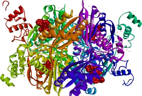
Figure 5 The phenolic derivatives 4a, 6a, and 6b tightly entrapped in active binding sites of HMG COA reductase enzyme visualized through Discovery Studio 4.1.
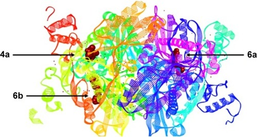
Figure 6 Residual interaction of 4a in binding pocket of HMG CO A reductase.
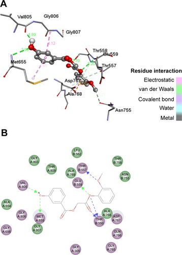
Figure 7 Residual interaction of 6a in binding pocket of HMG CO A reductase.
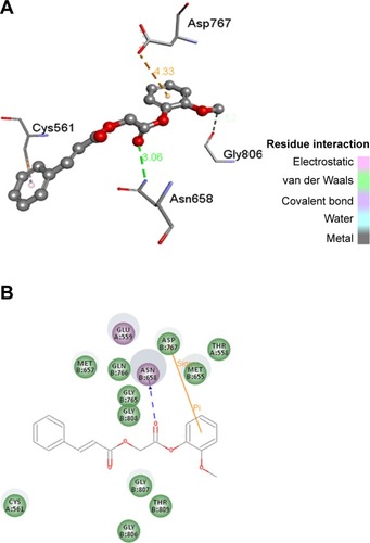
Figure 8 Ramachandran plot of protein structure of target protein (HMG COA reductase).
Scheme 1 Synthesis of phenolic derivatives (4a–4e).
Scheme 2 Synthesis of phenolic derivatives (6a–6c).
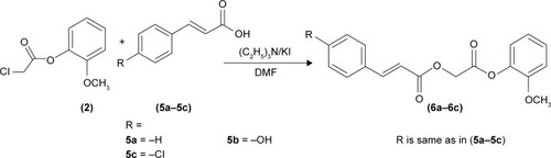
Table 4 Binding affinity/energy values against HMG CoA reductase (PDB ID: 1HWK)
Conclusion
The synthesis of some phenolic derivatives 4a–e and 6a–c was successfully accomplished starting with simplest precursors. The results of in silico docking studies showed that compounds 4a and 6a exhibited maximum binding affinity with target protein having binding energies −8.3 and −7.9 kcal, respectively. Compound 4a interacts with amino acids Val805 having distance 1.89 Å and Met656, Thr558, and Glu559 having bonding distances 2.96, 2.70, and 2.20 Å, respectively. Compound 4a exhibited excellent in vivo antihyperlipidemic activity with minimum weight increment (20%) compared to standard atorvastatin (35% weight increment). The synthesized compounds were nontoxic and well tolerated because none of the mice were found to suffer from any kind of morbidity and death during 6 weeks of dosing. Based on our pharmacological evaluation, we may propose that compound 4a may act as a lead structure for the design and development of more potent antihyperlipidemic drugs.
Disclosure
The authors report no conflicts of interest in this work.
References
- WaniFAAlbahrawyAZRahimanSHypolipidemic activity of olive oil (oleaeuropaea) against high fat diet-induced nonalcoholic fatty liver disease (NAFLD) in miceOpen J Path20155373
- MengXYZhangHXMezeiMCuiMMolecular docking: a powerful approach for structure-based drug discoveryCurr Comput Aided Drug Des20117214615721534921
- KapetanovicIMComputer-aided drug discovery and development (CADDD): in silico-chemico-biological approachChem Biol Interact2008171216517617229415
- ChiavariGVittorioCGuidoCGElectrochemical detection in the high-performance liquid chromatographic analysis of plant phenolicsAnalyst1988113191943358526
- RehmanMRSyedMASMaisamASComprehensive review on anti-oxidant activity of plantsPak J Med Biol Sci2017114151
- GanRYXiangRXFengLSAntioxidant activity and total phenolic content of medicinal plants associated with prevention and treatment of cardiovascular and cerebrovascular diseasesJ Med Plant Res201042224382444
- RiceECatherineANicholasJMStructure-antioxidant activity relationships of flavonoids and phenolic acidsFree Radical Bio Med19962079339568743980
- SkergetMKotnikPHadolinMPhenols, proanthocyanidins, flavones and flavonols in some plant materials and their antioxidant activitiesFood Chem2005892191198
- TripoliEGiammancoMTabacchiGDi MajoDGiammancoSLa GuardiaMThe phenolic compounds of olive oil: structure, biological activity and beneficial effects on human healthNutr Res Rev20051819811219079898
- LiuLZubikLCollinsFWMarkoMMeydaniMThe antiatherogenic potential of oat phenolic compoundsAtherosclerosis20041751394915186945
- TurnerRNicolasEMariaGAAntioxidant and anti-atherogenic activities of olive oil phenolicsInt J Vitam Nutr Res2005751617015830923
- NegroCTommasiLMiceliAPhenolic compounds and antioxidant activity from red grape marc extractsBioresour Technol2003871414412733573
- KelmMANairMGStrasburgGMDeWittDLAntioxidant and cyclooxygenase inhibitory phenolic compounds from Ocimum sanctum LinnPhytomedicine20007171310782484
- ZhangLRavipatiASKoyyalamudiSRAntioxidant and anti-inflammatory activities of selected medicinal plants containing phenolic and flavonoid compoundsJ Agric Food Chem20115923123611236722023309
- ConfortiFSosaSMarrelliMIn vivo anti-inflammatory and in vitro antioxidant activities of Mediterranean dietary plantsJ Ethnopharmacol2008116114415118164564
- KassimMAchouiMMustafaMRMohdMAYusoffKMEllagic acid, phenolic acids, and flavonoids in Malaysian honey extracts demonstrate in vitro anti-inflammatory activityNutr Res201030965065920934607
- HanNGuYYeCCaoYLiuZYinJAntithrombotic activity of fractions and components obtained from raspberry leaves (Rubuschingii)Food Chem2012132118118526434278
- KimMSKyungALAntithrombotic activity of methanolic extract of Umbilicaria esculentaJ Ethnopharmacol2006105334234516384677
- CarrieriCRosa AnnaMFrancescaIAntithrombotic activity of 12 table grape varieties. Relationship with polyphenolic profileFood Chem2013140464765323692748
- HsuCLWuCHHuangSLYenGCPhenolic compounds rutin and o-coumaric acid ameliorate obesity induced by high-fat diet in ratsJ Agric Food Chem200957242543119119847
- KaurGMeenaCEvaluation of anti-hyperlipidemic potential of combinatorial extract of curcumin, piperine and quercetin in Triton induced hyperlipidemia in ratsSci Int2013135763
- ChenCYMilburyPEKwakHKCollinsFWSamuelPBlumbergJBAvenanthramides and phenolic acids from oats are bioavailable and act synergistically with vitamin C to enhance hamster and human LDL resistance to oxidationJ Nutr200413461459146615173412
- MauryaDKDevasagayamTPAntioxidant and prooxidant nature of hydroxycinnamic acid derivatives ferulic and caffeic acidsFood Chem Toxicol201048123369337320837085
- ChengJCFangDBoZAntioxidant activity of hydroxycinnamic acid derivatives in human low-density lipoprotein: mechanism and structure–activity relationshipFood Chem20071041132139
- TrombinoSSeriniSDiNFAntioxidant effect of ferulic acid in isolated membranes and intact cells: synergistic interactions with α-tocopherol, β-carotene, and ascorbic acidJ Agric Food Chem20045282411242015080655
- RehmanMRFarhanSUmbreenNMedicinal plants having wound healing activity; a reviewPak J Med Biol Sci2017116369
- OuSKinchorKFerulic acid: pharmaceutical functions, preparation and applications in foodsJ Sci Food Agric2004841112611269
- ChengJCDaiFZhouBAntioxidant activity of hydroxycinnamic acid derivatives in human low-density lipoprotein: mechanism and structure–activity relationshipFood Chem20071041132139
- ChenJHChi-TangHAntioxidant activities of caffeic acid and its related hydroxycinnamic acid compoundsJ Agric Food Chem199745723742378
- GülçinİAntioxidant activity of caffeic acid (3, 4-dihydroxycinnamic acid)Toxicol20062172213220
- LeeYJLiaoPHChenWKYangCYPreferential cytotoxicity of caffeic acid phenethyl ester analogues on oral cancer cellsCancer Lett20001531515610779629
- FrescoPBorgesFMarquesMPDinizCThe anticancer properties of dietary polyphenols and its relation with apoptosisCurr Pharm Des201016111413420214622
- HuangMTSmartRCWongCQConneyAHInhibitory effect of curcumin, chlorogenic acid, caffeic acid, and ferulic acid on tumor promotion in mouse skin by 12-O-tetradecanoylphorbol-13-acetateCancer Res19884821594159463139287
- IslamBSharmaCAdemAAburawiEOjhaSInsight into the mechanism of polyphenols on the activity of HMGR by molecular dockingDrug des Devel Ther201594943
- EkinsSJordiMBernardTIn silico pharmacology for drug discovery: methods for virtual ligand screening and profilingBr J Pharmacol2007152192017549047
- BenderAJosefSMeirGAnalysis of pharmacology data and the prediction of adverse drug reactions and off-target effects from chemical structureChemMedChem20072686187317477341
- BowesJBrownAJHamonJReducing safety-related drug attrition: the use of in vitro pharmacological profilingNat Rev Drug Discov2012111290992223197038
- KitchenDBDecornezHFurrJRBajorathJDocking and scoring in virtual screening for drug discovery: methods and applicationsNat Rev Drug Discov200431193594915520816
- SashidharaKVKumarAKumarMSrivastavaAPuriASynthesis and antihyperlipidemic activity of novel coumarin bisindole derivativesBioorg Med Chem Lett201020226504650720932744
- JangadAPeriasamySNaYLComparison of hypolipidemic activity of synthetic gallic acid–linoleic acid ester with mixture of gallic acid and linoleic acid, gallic acid, and linoleic acid on high-fat diet induced obesity in C57BL/6 Cr Slc miceChem Biol Interact2008174210911718571153
- RosePWBeranBBiCThe RCSB protein data bank: redesigned web site and web servicesNucleic Acids Res2010391S392401
- DavisIWAndrewLFVincentBCMol Probity: all-atom contacts and structure validation for proteins and nucleic acidsNucleic Acids Res2007352SW375W38317452350
- GoddardTDConradCHThomasEFVisualizing density maps with UCSF ChimeraJ Struct Biol2007157128128716963278
- MorrisGMHueyRLindstromWAutoDock4 and auto dock tools 4: automated docking with selective receptor flexibilityJ Comput Chem200930162785279119399780
- WorachartcheewanANantasenamatCNaennaTIsarankura-Na- AyudhyaCPrachayasittikulVModeling the activity of furin inhibitors using artificial neural networkEur J Med Chem20094441664167318977558
- ForliSHueyRPiqueMESannerMFGoodsellDSOlsonAJComputational protein-ligand docking and virtual drug screening with the auto dock suiteNat Protoc201611590591927077332
- DallakyanSOlsonAJSmall-molecule library screening by docking with PyRxMethods Mol Biol2015126324325025618350
- AshrafZAlamgeerKanwalMFlurbiprofen–antioxidant mutual pro drugs as safer nonsteroidal anti-inflammatory drugs: synthesis, pharmacological investigation, and computational molecular modelingDrug Des Dev Ther20161024012419
- BrayGAFislerJYorkDANeuroendocrine control of the development of obesity: understanding gained from studies of experimental animal modelsFront Neuroendocrinol1990112128181
- LangerTHoffmannRDVirtual screening an effective tool for lead structure discoveryCurr Pharm Des20017750952711375766
- FrostegardJSLE, atherosclerosis and cardiovascular diseaseJ Intern Med2005257648549515910552
- LawMRWaldNJRudnickaARQuantifying effect of statins on low density lipoprotein cholesterol, ischaemic heart disease, and stroke: systematic review and meta-analysisBr Med J20033267404142312829554
- FestiDColecchiaASaccoTBondiMRodaEMarchesiniGHepatic steatosis in obese patients: clinical aspects and prognostic significanceObes Rev200451274214969505
- WanlessIRLentzJSFatty liver hepatitis (steatohepatitis) and obesity: an autopsy study with analysis of risk factorsHepatology1990125110611102227807
- IstvanESDeisenhoferJStructural mechanism for statin inhibition of HMG-CoA reductaseScience200129255191160116411349148
- MovahedianASadeghiHGhannadiAGharaviMAzarpajoohSHypolipidemic activity of Allium porrum L. in cholesterol-fed rabbitsJ Med Food2006919810116579735