
Abstract
Objective
Lobaplatin shows antitumor activity against a wide range of tumors, including metastatic breast cancer (BCa). The overexpression of metadherin (MTDH) is associated with poor prognosis of BCa patients. This study was designed to investigate the effect of lobaplatin on MCF-7 cell proliferation and its association with MTDH expression.
Patients and methods
Clinical treatment for BCa using lobaplatin, in combination with other general chemotherapy drugs, was administered to 32 BCa patients. The safety, effectiveness, and prognosis in lobaplatin-treated BCa patients were compared with those in controls (n=32). In vitro experiments were performed in MCF-7 cells to investigate the effect of lobaplatin on cell proliferation, apoptosis, and MTDH expression.
Results
We found the intraoperative local chemotherapy using lobaplatin was safe and effective for BCa treatment, in comparison with the patients administered general chemotherapy drugs. Treatment of MCF-7 cell cultures with lobaplatin significantly reduced cell proliferation and increased cell apoptotic percentage. The expression of MTDH and Bcl-2 was inhibited by lobaplatin and that of Bax was increased by lobaplatin. Moreover, we observed the inhibition of MTDH by shRNA reduced cell proliferation and enhanced cell apoptosis.
Conclusion
Lobaplatin was a safe and effective adjuvant chemotherapy for BCa. The effect of lobaplatin on inhibiting MCF-7 cell proliferation and inducing cell apoptosis might be, as least in part, mediated by suppressing the expression of oncogene MTDH.
Introduction
Breast cancer (BCa) is a common malignancy among women, with an increasing prevalence worldwide.Citation1,Citation2 BCa-related death is the second cause of cancer death among women worldwide.Citation1 The drug and chemoradiotherapy resistance, higher recurrence during follow-up, and higher rates of genetic mutations in BCa patients make BCa treatment challenging.Citation1,Citation3,Citation4 It is well known that the rate of BCa cells’ resistance to chemoradiotherapy is high.Citation5,Citation6 These obstacles make it an urgent need to find new agents or neoadjuvant chemotherapy for treatment of BCa.
Lobaplatin is a representative of the third-generation platinum antineoplastic agents, which has wide-range activities of overcoming tumor resistance to chemoradiotherapy drugs, including cisplatin and carboplatin.Citation1,Citation7,Citation8 Studies have shown the antitumor activity of lobaplatin in cancers, including human cholangiocarcinoma,Citation9,Citation10 lung cancer,Citation11 human cervical cancer,Citation12 melanoma,Citation13 gastric cancer,Citation7,Citation14 esophageal squamous cell carcinoma,Citation15 and BCa.Citation16–Citation18 Some clinical studies reported that the intraoperative local chemotherapy using lobaplatin for BCa was safe and effective,Citation17 while others reported that administration of lobaplatin as a neoadjuvant chemotherapy to docetaxel and epirubicin regimen for triple-negative BCa (TNBC) showed increased side effects.Citation15–Citation17,Citation19 Regimen using lobaplatin for TNBC, primary and metastatic BC had been reported.Citation16–Citation18 It has been reported that lobaplatin inhibited cancer cell proliferation and induced cancer cell apoptosis by arresting cell cycle progression, thus leading to the suppression of cancer metastasis and development of antitumor activity.Citation11–Citation13,Citation15
Metadherin (MTDH) is an oncogenic protein and functions by promoting cancer cell proliferation, invasion, and drug resistance.Citation20,Citation21 The expression of MTDH was associated with various signaling pathways, including AKT signaling pathway, and miRNAs which were involved in cell proliferation and tumorigenesis.Citation22–Citation26 The downregulation of MTDH, however, could induce the apoptosis of BCa MCF-7 cells,Citation1 prostate cancer DU145 cells,Citation26 and lung cancer A549 cells.Citation23 Wang showed that cell proliferation and the expression of MTDH in lobaplatin-treated MCF-7 cells were inhibited, with increased cell apoptosis (in Chinese).Citation27 Similarly, Chen showed that intraoperative local chemotherapy using lobaplatin in radical mastectomy for BCa resulted in reduced exfoliated cancer cells.Citation17 Engel et al reported that the administration of lobaplatin inhibited BCa cell proliferation.Citation28 In addition, the downregulation of MTDH in MCF-7 cells was related to cell apoptosis.Citation1 These studies might suggest that lobaplatin treatment for cancer cells and inhibition of MTDH were, respectively, associated with the inhibition of cancer cell proliferation. However, little information is available on MTDH expression in response to lobaplatin treatment for BCa.
To investigate the effect of lobaplatin on BCa and to explore the association of MTDH expression with lobaplatin-induced cell apoptosis, we performed the clinical case– control study using lobaplatin as an intraoperative local chemotherapy for BCa. Cellular experiments were performed to detect the influence of lobaplatin on MCF-7 cell proliferation and MTDH expression. The association between lobaplatin and MTDH expression would be discussed. This study would provide us with more basic information on the relation of MTDH expression with lobaplatin in MCF-7 cells in vitro.
Patients and methods
Subjects, treatments, and surgical procedure
Female patients with primary diagnosis of BCa were enrolled in this study from Daping Hospital, Army Military Medical University, Chongqing, China, between March 2009 and September 2012. Patients were diagnosed with BCa by imaging (magnetic resonance imaging) and pathology. All BCa patients had Karnofsky Performance Score ≥80. Subjects participating in this study met the following criteria: 1) no obvious chemotherapy taboo; 2) no obvious dysfunction in heart, lung, liver, and kidney; 3) no significant difference in basic information between patients when randomly assigned; and 4) no history of malignancy and diabetes. Patients were assigned to control or lobaplatin-treated (experimental) group according to individual willingness. A total of 32 patients were assigned to the experimental group (n=32) and the other 32 age-matched patients were assigned to the control group (n=32). Experimental protocols were approved by the Ethics Committee of Daping Hospital. Informed written consents were obtained from these 64 patients with BCa.
All patients were subjected to radical mastectomies. After cleaning, the wounds were washed with normal saline (NS) supplemented with and without lobaplatin (50 mg in 100 mL NS). Before closing them with stitches, 50 mg lobaplatin (in 100 mL NS) was sprayed onto the surface of tumor bed, axillary wound cavity, and other areas suspicious for having residual cancer cells.Citation17 The total volume of drainage fluids in 72 hours, wound healing time, complications (local pain, nausea, vomiting, diarrhea), myelosuppression (white cell count and platelet count), and liver and renal functions (creatinine and alanine aminotransferase) were recorded and compared. All patients received normal chemoradiotherapy plus lobaplatin as intraoperative local chemotherapy.
At 72 hours post-surgery, drainage fluid was collected, centrifuged, and cell pellets were collected for smears, which were then subjected to the detection of exfoliated cancer cells.Citation17 Also, recurrences and other complications during the follow-up of 4 years were compared.
Cells, culture conditions, and lobaplatin treatment
Human BCa cell line MCF-7 was obtained from ATCC (Manassas, VA, USA) and cultured in DMEM (Thermo Fisher Scientific, Waltham, MA, USA) supplemented with 10% fetal bovine serum (Thermo Fisher Scientific) and 1% antibiotic–antimycotic (Thermo Fisher Scientific) at 37°C with 5% CO2. For lobaplatin treatment, MCF-7 cells were treated with 0, 5, 10, and 50 mg/L lobaplatin (Hainan Chang’an International Pharmaceutical Co., Ltd, Hainan, China) for 24 hours.Citation7 Each cellular experiment was performed in triplicate.
Cell transfection
To generate an MTDH-low-expressing cell model in vitro, we transfected MCF-7 cells with shRNA-MTDH plasmids (based on pSUPER-EGFP vector; OligoEngine, Seattle, WA, USA) using Lipofectamine 2000 (Thermo Fisher Scientific) according to the manufacturer’s instructions.Citation29 Cells were transfected with shRNA-MTDH and pSuper-EGFP vector containing control shRNAs (shRNA-NC) for 6 hours prior to lobaplatin treatment.
Cell proliferation assay
The effect of lobaplatin and MTDH inhibition on MCF-7 cells viability was examined using the MTT assay (Thermo Fisher Scientific). Briefly, cells were plated into 96-well plates (5×10⁴ cells/well) and treated with lobaplatin for 24, 48, and 72 hours. Then, the cell cultures were treated with MTT reagent for 4 hours and dimethyl sulfoxide (150 µL/well) for another 20 minutes, followed by detection at OD 570 nm.
Cell apoptosis analysis by flow cytometry
To confirm the effect of lobaplatin and MTDH expression on cell apoptosis, the MCF-7 cells (1×10⁵ cells/well in six-well plates) were treated with either shRNA-MTDH transfection or lobaplatin for 24 hours. Then, Annexin V-FITC Apoptosis Detection Kit (Sigma) was utilized to measure cell apoptosis. The percentage of apoptotic cells was examined using a FACS Canto™ II cytometry instrument and analyzed using CellQuest software (BD Diagnostics, Franklin lakes, NJ, USA).
RNA isolation and real-time polymerase chain reaction
Total RNA was extracted from MCF-7 cells using an RNeasy kit (Qiagen, Valencia, CA, USA) at 24 hours post-lobaplatin treatment, and cDNA was generated using the M-MLV1 reverse transcription kit (Takara, Tokyo, Japan). Amplification was conducted on PE-9600 thermal cycler (Thermo Fisher Scientific) using a SYBR Green PCR kit (Qiagen, Los Angeles, CA, USA) with the following reaction conditions: 95°C for 5 minutes, 40 cycles of 95°C for 30 seconds, and 60°C for 30 seconds. The 2−∆∆Ct methods were used for calculating the mRNA relative expression level of MTDH gene, with normalization to β-actin.
Western blot analysis
Protein was isolated from MCF-7 cells using RIPA lysis buffer (Thermo Fisher Scientific), followed by quantification using a BCA kit (Qiagen). Protein was separated onto sodium dodecyl sulfate-polyacrylamide gel electrophoresis and electroblotted onto polyvinylidene difluoride membranes (EMD Millipore, Billerica, MA, USA) according to standard methods. Membranes were then incubated with specific primary antibody anti-MTDH (Sigma), anti-Bax (Cell Signaling Technology [CST], Danvers, MA, USA), anti-Bcl-2 (CST), and anti-β-actin (CST) at 4°C overnight and related secondary antibodies at room temperature for 40 minutes. Bio-Rad Quantity One software (Bio-Rad) was used to analyze protein expression with the enhanced chemiluminescence system.
Statistical analysis
Continuous variables are expressed as mean ± SD. Statistical significances between groups were assessed using Student’s t-test, and those among groups were assessed using one-way analysis of variance followed by Dunnett’s post hoc test. Categorical variables are expressed as frequencies, and differences between groups were compared by using chi-squared statistics. P<0.05 was considered statistically significant.
Results
Multi-point spraying of lobaplatin promotes wound healing and reduces recurrence
As shown in , there was difference in the volume of drainage within 72 hours after operation between the two groups (P<0.05). The white cell count, platelet count, creatinine, alanine aminotransferase, wound healing time, and complications between groups were similar (P>0.05; ). The detection rate of exfoliated cancer cells in the control group was higher than that in the treatment group (P<0.05). Within 72 hours postoperations, exfoliated cancer cells were found in the drainage of four patients (4/32, 12.50%) treated with lobaplatin, which was obviously lower than that in the control group (10/32, 31.25%; P<0.05). No differences were found in the rates of adverse events (recurrence and metastasis) between the groups during a 4-year follow-up. These data suggested the safety of using lobaplatin as intraoperative local chemotherapy for BCa patients.
Table 1 The differences in general data and complications within 72 hours after operations, and the adverse events during a 4-year follow-up
Lobaplatin suppresses MCF-7 cells proliferation and promotes cell apoptosis
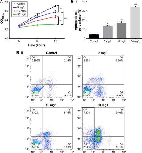
To investigate the effect of lobaplatin on BCa cell proliferation, we treated MCF-7 cells in vitro with lobaplatin and found inhibited cell viability (). In comparison with the control cells, lobaplatin-treated MCF-7 cells showed inhibited cell viability (P<0.05). MCF-7 cells treated with 5 mg/L lobaplatin presented with significantly reduced cell viability compared with controls (P<0.05), which was then enhanced with the increment of lobaplatin concentration. In addition, flow cytometry analysis showed that lobaplatin promoted cell apoptosis in a dose-dependent manner. After being treated with lobaplatin for 24 hours, 13.20%±1.22%, 16.73%±2.10%, and 34.0%±1.35% apoptotic cells were found in MCF-7 cells treated with 5 mg/L (P<0.05), 10 mg/L (P<0.01), and 50 mg/L lobaplatin (P<0.01), respectively, which were significantly higher than that of control cells (4.22%±0.12%; ). These data suggest that lobaplatin inhibited MCF-7 cell proliferation and induced cell apoptosis.
Figure 1 Lobaplatin induces MCF-7 cell apoptosis.
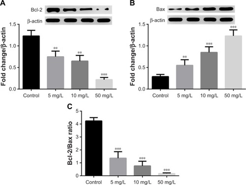
Lobaplatin decreases Bcl-2/Bax ratio
In addition, we found the expression of Bcl-2 and Bax proteins was inhibited and promoted by lobaplatin, respectively (). The Bcl-2/Bax ratio in MCF-7 cells was decreased by lobaplatin administration (<1.36±0.50) in a dose-dependent manner () compared with that in control cells (4.24±0.26, P<0.001). This fact suggested that lobaplatin inhibited the Bcl-2/Bax ratio in MCF-7 cells.
Figure 2 Lobaplatin affected the expression of MTDH and apoptosis-related protein in MCF-7 cells.
Abbreviation: MTDH, metadherin.
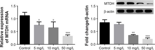
Lobaplatin suppresses MTDH expression in MCF-7 cells
It was reported previously that MTDH expression was associated with cancer cells’ proliferation and the latter could be dysregulated by lobaplatin.Citation1,Citation7,Citation13,Citation29 Accordingly, we thought there might be an association between MTDH and lobaplatin-mediated apoptosis. As expected, we found the expression of MTDH mRNA and protein was reduced in MCF-7 cells receiving lobaplatin treatments, in a dose-dependent manner (). This result revealed the expression of MTDH in MCF-7 cells was inhibited by lobaplatin administration.
Figure 3 The expression of MTDH in lobaplatin-treated cells.
Abbreviations: MTDH, metadherin; qRT-PCR, quantitative reverse transcriptase-polymerase chain reaction.
MTDH inhibition promotes MCF-7 cell apoptosis
Since the administration of lobaplatin suppressed MTDH expression and promoted the apoptosis of MCF-7 cells, we thought the inhibition of MTDH might be a crucial regulator for lobaplatin-mediated cell apoptosis. To investigate this, we knocked out MTDH expression in MCF-7 cells by shRNA technology (). We found the knockdown of MTDH expression significantly increased the percentage of apoptotic MCF-7 cells (45.20%±1.35%) compared with control (4.51%±0.12%, P<0.001; ), and was superior to 50 mg/L lobaplatin administration (28.00%±2.12%, P<0.001).
Figure 4 Effect of MTDH inhibition on MCF-7 cell apoptosis and proliferation.
Abbreviation: MTDH, metadherin.
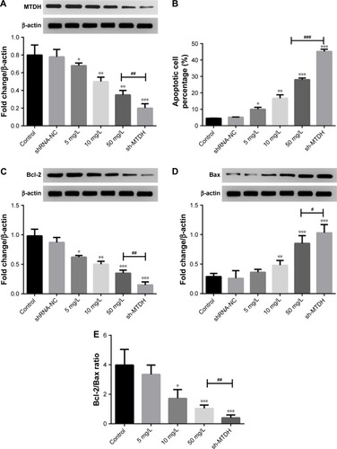
Moreover, we also found the knockdown of MTDH showed superior effect on suppressing Bcl-2 expression (P<0.01, ) and inducing Bax expression (P<0.05, ) to 50 mg/L lobaplatin administration in MCF-7 cells. Accordingly, prominent lower Bcl-2/Bax ratio (0.44±0.18) was observed in shRNA-MTDH groups compared with that in 50 mg/L lobaplatin-administered cells (1.04±0.23, P<0.01; ). These data demonstrate that lower MTDH expression correlated with higher apoptotic percentage in MCF-7 cells, and lobaplatin-mediated MCF-7 cell apoptosis was at least, in part, related with inhibition of MTDH and Bcl-2/Bax ratio.
Discussion
Lobaplatin is an effective chemotherapy agent with antitumor characteristics.Citation1,Citation13,Citation17 The overexpression and inhibition of MTDH promoted and inhibited cancer cell proliferation, respectively.Citation1,Citation29 There are plenty of in vitro and in vivo clinical studies showing the antitumor effect of lobaplatin.Citation12,Citation13,Citation18 Our current data suggested that lobaplatin-reduced cell proliferation of MCF-7 cells was related to the inhibition of an oncogene MTDH.
Using the case–control study, we demonstrated the safety and efficacy of using lobaplatin as one of the chemotherapy agents for BCa treatment after surgery. These results were in accordance with a previous clinical study by Chen.Citation17 BCa patients who were treated with general agents combined with lobaplatin after radical mastectomies showed higher amount of drainage and lower exfoliated cancer cells compared with patients receiving only general agents. In our study, the fact that no differences were observed in complications within 72 hours after operations and during a 4-year follow-up revealed the safety of using lobaplatin for BCa treatment. This was in consistent with the results from Chen et al. This suggests the effectiveness of using lobaplatin. However, Wu et al showed that although the addition of lobaplatin as neoadjuvant chemotherapy to TNBC treatments improved the pathologic complete response, the rate of side effects (anemia and thrombocytopenia) in BCa patients receiving lobaplatin was elevated.Citation16 All these studies suggested the effectiveness of using lobaplatin for BCa treatment. But the opposing point on side effects is worth pursuing further.
Yang et al showed that lobaplatin arrested B16-F10 melanoma cell cycle progression and induced apoptosis, thus inhibiting the migration and invasion of melanoma cells.Citation13 Similar results of using lobaplatin treatment for in vitro cancers had been reported on human cervical cancer cells,Citation14 gastric carcinoma cells,Citation12 and esophageal squamous cell carcinoma cells.Citation15 The antiapoptotic protein Bcl-2 and the proapoptotic protein Bax are associated with cell apoptosis and cancer cell resistance.Citation13,Citation15,Citation30 The Bcl-2/Bax ratio, particularly, could be used as a marker for therapeutic response to radiotherapy.Citation31,Citation32 Du et al showed that lobaplatin induced esophageal squamous cell carcinoma KYSE-410 and EC-109 cells’ apoptosis and decreased the Bcl-2/Bax ratio.Citation42 In our in vitro experiments, we demonstrated that the administration of lobaplatin to MCF-7 cells significantly reduced cell proliferation, Bcl-2/Bax ratio, and induced cell apoptosis. Collectively, these data suggest that lobaplatin might be an effective chemotherapeutic agent in BCa treatment.
Previous studies had shown that MTDH played important roles in tumorigenesis.Citation20,Citation29,Citation33 The evidence on the association of MTDH with microRNAs, AKT, and β-catenin signaling pathways reveals the crucial functional roles of MTDH overexpression in tumorigenesis.Citation21,Citation24,Citation25,Citation34,Citation35 Li et al showed that MTDH was a direct target of tumor suppressor miRNA miR-30a-5p and the overexpression of miR-30a-5p induced hepatocellular carcinoma cell apoptosis via inhibiting MTDH, upregulating PTEN protein, and suppressing Akt.Citation35 The expression of MTDH was regulated by various tumor suppressing and oncogenic miRNAs.Citation35–Citation37 The expression of MTDH in tumors was associated with tumor progression via regulating epithelial–mesenchymal transition and chemoresistance in cancer cells,Citation29,Citation38,Citation39 suggesting the crucial roles of MTDH expression in tumorigenesis and progression.
In BCa, the overexpression of MTDH was associated with an aggressive phenotype and short overall survival as well as short distant metastasis-free survival.Citation20 In addition, the downregulation of a tumor suppressing miRNA, miR-26a, was associated with the overexpression of MTDH in TNBC, and the ectopic expression of miR-26a suppressed MTDH and the subsequent cell proliferation and metastasis.Citation21 Our data suggest that lobaplatin induces MCF-7 cells proliferation as well as MTDH suppression. Also, the inhibition of MTDH by shRNA in MCF-7 cells showed similar results to lobaplatin treatment. Taken together, we thought there might be an association between MTDH inhibition and lobaplatin-induced cell apoptosis in BCa.
MTDH is involved in various pathwaysCitation21,Citation24,Citation25,Citation40. It has been reported that MTDH could promote the differentiation of cancer cell stemness in BCa cells by activating Twist1, thus promoting tumorigenicity.Citation41 A latest study suggested that the expression of MTDH in BCa cells could be decreased by the overexpression of tumor suppressor F-box and WD-40 domain protein 7 (FBXW7, hCDC4), thus inhibiting cancer cell proliferation and promoting apoptosis.Citation33 In our present study, we suggest that lobaplatin-induced cell apoptosis in BCa cells might be associated with MTDH inhibition. Also, MTDH-shRNA–induced apoptosis in MCF-7 cells was at least, in part, related to increase in Bcl-2/Bax ratio. These suggest that MTDH plays important roles in tumorigenicity, and the MTDH-mediated mechanism in BCa progression should be explored in future experiments.
Conclusion
In summary, we primarily found treatment of BCa patients with lobaplatin was a safe and effective strategy. In the in vitro experiments, we found lobaplatin promoted MCF-7 cell apoptosis, inhibited cell proliferation, Bcl-2/Bax ratio, as well as the expression of MTDH in BCa cells. We found that lobaplatin-mediated MCF-7 cell apoptosis was at least, in part, mediated by suppression of the oncogene MTDH.
Acknowledgments
This study was supported by the Chongqing Science and Technology Commission Huimin Plan-funded projects (cstc2016kjhmpt100-16).
Disclosure
The authors report no conflicts of interest in this work.
References
- LuoZHuXXiongHA polysaccharide from Huaier induced apoptosis in MCF-7 breast cancer cells via down-regulation of MTDH proteinCarbohydr Polym20161511027103327474651
- DesantisCEFedewaSAGoding SauerAKramerJLSmithRAJemalABreast cancer statistics, 2015: convergence of incidence rates between black and white womenCA Cancer J Clin2016661314226513636
- Garcia-MurillasISchiavonGWeigeltBMutation tracking in circulating tumor DNA predicts relapse in early breast cancerSci Transl Med20157302302ra133
- RheinbayEParasuramanPGrimsbyJRecurrent and functional regulatory mutations in breast cancerNature20175477661556028658208
- LiJLiuJGuoNZhangXReversal of multidrug resistance in breast cancer MCF-7/ADR cells by h-R3-siMDR1-PAMAM complexesInt J Pharm2016511143644527444552
- LiuJLiJLiuNIn vitro studies of phospholipid-modified PAMAM-siMDR1 complexes for the reversal of multidrug resistance in human breast cancer cellsInt J Pharm20175301–229129928619457
- YinCYLinXLTianLYeMYangXYXiaoXYLobaplatin inhibits growth of gastric cancer cells by inducing apoptosisWorld J Gastroenterol201420461742625516654
- DaiHYLiuLQinSKHeXMLiSYLobaplatin suppresses proliferation and induces apoptosis in the human colorectal carcinoma cell Line LOVO in vitroBiomed Pharmacother201165313714121612887
- WangZTangXZhangYLobaplatin induces apoptosis and arrests cell cycle progression in human cholangiocarcinoma cell line RBEBiomed Pharmacother201266316116622425181
- LiekeTRamackersWBergmannSKlempnauerJWinklerMKloseJImpact of salinomycin on human cholangiocarcinoma: induction of apoptosis and impairment of tumor cell proliferation in vitroBMC Cancer201212146623057720
- XieCYXuYPJinWLouLGAntitumor activity of lobaplatin alone or in combination with antitubulin agents in non-small-cell lung cancerAnticancer Drugs201223769870522441567
- LiXRanLFangWWangDLobaplatin arrests cell cycle progression, induces apoptosis and alters the proteome in human cervical cancer cell line CaSkiBiomed Pharmacother201468329129724239273
- YangFYuYLeiQLobaplatin arrests cell cycle progression, induces apoptosis and impairs migration and invasion in B16-F10 melanoma cell line in vitroBiomed Pharmacother20156940240825661389
- LiYLiuBYangFLobaplatin induces BGC-823 human gastric carcinoma cell apoptosis via ROS-mitochondrial apoptotic pathway and impairs cell migration and invasionBiomed Pharmacother2016831239124627565846
- DuLFeiZSongSWeiNAntitumor activity of lobaplatin against esophageal squamous cell carcinoma through caspase-dependent apoptosis and increasing the Bax/Bcl-2 ratioBiomed Pharmacother20179544745228865364
- WuXTangPLiSA randomized and open-label Phase II trial reports the efficacy of neoadjuvant lobaplatin in breast cancerNat Commun20189183229483583
- ChenWXClinical observation of intraoperative local chemotherapy with lobaplatin in breast cancer modified radical mastectomyInt J Clin Exp Med20171071083410839
- WangZXuLWangHLobaplatin-based regimens outperform cisplatin for metastatic breast cancer after anthracyclines and taxanes treatmentSaudi J Biol Sci201825590991630108440
- LiDA clinical study of combination regimen containing lobaplatin in treating advanced metastatic breast cancerJournal of Shanxi Medical College for Continuing Education20164004
- TokunagaENakashimaYYamashitaNOverexpression of metadherin/MTDH is associated with an aggressive phenotype and a poor prognosis in invasive breast cancerBreast Cancer201421334134922903204
- LiuPTangHChenBmiR-26a suppresses tumour proliferation and metastasis by targeting metadherin in triple negative breast cancerCancer Lett2015357138439225434799
- LiPPYaoQMZhouHMetadherin contribute to BCR signaling in chronic lymphocytic leukemiaInt J Clin Exp Pathol201474158824817955
- ZouYQinXXiongHZhuFChenTWuHApoptosis of human non-small-cell lung cancer A549 cells triggered by evodiamine through MTDH-dependent signaling pathwayTumour Biol20153675187519325652471
- LiangCDingJYangYDengLLiXMicroRNA-433 inhibits cervical cancer progression by directly targeting metadherin to regulate the AKT and β-catenin signalling pathwaysOncol Rep20173863639364929130111
- YuCLiuYTanHMetadherin regulates metastasis of squamous cell carcinoma of the head and neck via AKT signalling pathway-mediated epithelial-mesenchymal transitionCancer Lett2014343225826724099913
- QianBYaoYLiuCZhangJChenHLiHSU6668 modulates prostate cancer progression by downregulating MTDH/AKT signaling pathwayInt J Oncol20175051601161128339027
- WangQEffect of Lobaplatin on Apoptosis of Breast Cancer MCF-7 Cells and Expression of MTDH Gene [master’s thesis]The Fourth Military Medical University (in Chinese)2010
- EngelJBMartensTHahneJCEffects of lobaplatin as a single agent and in combination with TRAIL on the growth of triple-negative p53-mutated breast cancers in vitroAnticancer Drugs201223442643622314264
- MengXBrachovaPYangSKnockdown of MTDH sensitizes endometrial cancer cells to cell death induction by death receptor ligand TRAIL and HDAC inhibitor LBH589 co-treatmentPLoS One201166e2092021687633
- SharifiSBararJHejaziMSSamadiNRoles of the Bcl-2/Bax ratio, caspase-8 and 9 in resistance of breast cancer cells to paclitaxelAsian Pac J Cancer Prev201415208617862225374178
- MackeyTJBorkowskiAAminPJacobsSCKyprianouNBcl-2/Bax ratio as a predictive marker for therapeutic response to radiotherapy in patients with prostate cancerUrology1998526108510909836559
- KhodapasandEJafarzadehNFarrokhiFKamalidehghanBHoushmandMIs Bax/Bcl-2 ratio considered as a prognostic marker with age and tumor location in colorectal cancer?Iran Biomed J2015192697525864810
- ChenXLiXYLongMThe FBXW7 tumor suppressor inhibits breast cancer proliferation and promotes apoptosis by targeting MTDH for degradationNeoplasma201865220120929534580
- ZhuLZhangPYangYAbstract A53: metadherin functions as a laminin receptor that is essential for metastasis and is associated with poor survival in osteosarcomaCancer Res20147420 SupplementA53
- LiWFDaiHOuQZuoGQLiuCAOverexpression of microRNA-30a-5p inhibits liver cancer cell proliferation and induces apoptosis by targeting MTDH/PTEN/AKT pathwayTumour Biol20163755885589526589417
- TangYLiuXSuBMicroRNA-22 acts as a metastasis suppressor by targeting metadherin in gastric cancerMol Med Rep201511145446025323629
- WangYWeiYTongHMiR-302c-3p suppresses invasion and proliferation of glioma cells via down-regulating metadherin (MTDH) expressionCancer Biol Ther20151691308131526176806
- WangZTangZYYinZMetadherin regulates epithelial–mesenchymal transition in carcinomaOnco Targets Ther20169Issue 12429243627143938
- LiWZhaiLZhaoCLvSMiR-153 inhibits epithelial–mesenchymal transition by targeting metadherin in human breast cancerBreast Cancer Res Treat2015150350150925794773
- LiPPFengLLChenNMetadherin contributes to the pathogenesis of chronic lymphocytic leukemia partially through Wnt/β-catenin pathwayMed Oncol201532221
- LiangYHuJLiJEpigenetic activation of TWIST1 by MTDH promotes cancer stem-like cell traits in breast cancerCancer Res201575173672368026141861
- DuLFeiZWeiNAntitumor activity of Lobaplatin against esophageal squamous cell carcinoma through caspase-dependent apoptosis and increasing the Bax/Bcl-2 ratioBiomed Pharmacother20179544745228865364