
 ?Mathematical formulae have been encoded as MathML and are displayed in this HTML version using MathJax in order to improve their display. Uncheck the box to turn MathJax off. This feature requires Javascript. Click on a formula to zoom.
Abstract
Background
For stroke patients, stimulating neurorepair mechanisms is necessary to reduce morbidity and disability. Our previous studies on brain and spinal cord trauma show that exogenous treatment with the S-nitrosylating agent S-nitrosoglutathione (GSNO) – a nitric oxide and glutathione metabolite of the human body – stimulates neurorepair and aids functional recovery. Using a rat model of cerebral ischemia and reperfusion (IR) in this study, we tested the hypothesis that GSNO invokes the neurorepair process and improves neurobehavioral functions through the angiogenic HIF-1α/VEGF pathway.
Methods
Stroke was induced by middle cerebral artery occlusion for 60 minutes followed by reperfusion in adult male rats. The injured animals were treated with saline (IR group, n=7), GSNO (0.25 mg/kg, GSNO group, n=7), and GSNO plus the HIF-1α inhibitor 2-methoxyestra-diol (2-ME) (0.25 mg/kg GSNO + 5.0 mg/kg 2-ME, GSNO + 2-ME group, n=7). The groups were studied for either 7 or 14 days to determine neurorepair mediators and functional recovery. Brain capillary endothelial cells were used to show that GSNO promotes angiogenesis and that GSNO-mediated induction of VEGF and the stimulation of angiogenesis are dependent on HIF-1α activity.
Results
IR injury increased the expression of neurorepair mediators HIF-1α, VEGF, and PECAM-1 and vessel markers to a limited degree that correlate well with significantly compromised neurobehavioral functions compared with sham animals. GSNO treatment of IR not only remarkably enhanced further the expression of HIF-1α, VEGF, and PECAM-1 but also improved functioning compared with IR. The GSNO group also had a higher degree of vessel density than the IR group. Increased expression of VEGF and the degree of tube formation (angiogenesis) by GSNO were reduced after the inhibition of HIF-1α by 2-ME in an endothelial cell culture model. 2-ME treatment of the GSNO group also blocked not only GSNO’s effect of reduced infarct volume, decreased neuronal loss, and enhanced expression of PECAM-1 (P<0.001), but also its improvement of motor and neurological functions (P<0.001).
Conclusion
GSNO stimulates the process of neurorepair, promotes angiogenesis, and aids functional recovery through the HIF-1α-dependent pathway, showing therapeutic and translational promise for stroke.
Introduction
Stroke is a leading cause of serious long-term disability and ranks fourth among all causes of death in the United States.Citation1 Following stroke, direct trauma causes necrotic neuronal death; however, even greater apoptotic neuronal damage occurs hours and days later, caused by secondary injury due to inflammation and oxidative stress.Citation2 These secondary injury mechanisms hamper the neurorepair process, leading to long-term chronic disabilities.Citation3 Unfortunately, proven effective therapy for neuroprotection/neurorepair following stroke is presently not available, mainly because of the biphasic (acute and chronic) nature of the disease and limited understanding of the differential mechanisms involved in these phases.Citation4 For functional recovery, clinical trials in human stroke show that neuroprotective drugs failed due to the lack of efficacy in the chronic phase.Citation5,Citation6 Therefore, an ideal therapy must ameliorate acute as well as chronic phases of the injury by well-understood mechanisms.
Recently, we and others identified an S-nitrosylating agent, S-nitrosoglutathione (GSNO),Citation7,Citation8 that provided robust neurovascular protection and blood–brain barrier (BBB) repair in a rat model of experimental stroke.Citation7,Citation9,Citation10 This neurovascular protection was not associated with free nitric oxide (NO) donors.Citation11 Later, we observed that GSNO was equally effective in aiding functional recovery in a rat model of ischemia and reperfusion (IR),Citation12 spinal cord injury,Citation13 and traumatic brain injury,Citation14,Citation15 indicating that GSNO could improve functional recovery following central nervous system injury. GSNO also improved learning and memory in a rat model of cerebral hypoperfusion, a vascular dementia model for experimental Alzheimer disease,Citation16 suggesting that GSNO’s activities are associated with motor as well as cognitive function recovery. However, GSNO’s neurorepair mechanisms following central nervous system injury are not understood. The term “neurorepair” refers to the combination of neurochemical and cellular events, such as stimulation of angiogenic mediators (eg, PECAM-1 and vascular endothelial growth factor [VEGF]) and formation of vessels, leading to regenerative repair, neurorestoration, and functional recovery. In this study, we investigated the mechanisms of GSNO-mediated stimulation of the neurorepair process with a focus on the hypoxia-inducible factor-1 alpha (HIF-1α)/VEGF/angiogenesis pathway using a rat model of experimental stroke.
GSNO directly regulates several angiogenic and neurorepair mediators, including stabilization of HIF-1αCitation17 and induction of neurorepair mechanisms.Citation12 S-nitrosylation-mediated stabilization of HIF-1α has also been shown to protect against myocardial injury via the VEGF/angiogenesis pathway in GSNO reductase (GSNOR) knockout mice,Citation18 indicating that HIF-1 is a key player in the repair processes. These studies provide a rationale for investigating S-nitrosylation-mediated modulation of HIF-1α for neurorepair in stroke.
HIF-1 is a nuclear transcription factor characterized as the master regulator of cellular oxygen homeostasis. HIF-1α is rapidly upregulated in response to hypoxia and is rapidly degraded upon reoxygenation/reperfusion.Citation19 It activates the tissue survival pathways by inducing several key enzymes involved in cell metabolism (GLUT), angiogenesis (VEGF, VEGFR1, angiopoietin), and free radical scavenging (heme oxygenase-1 [HO-1]). HIF-1α knockout mice show impaired vascular development and embryonic lethality, indicating HIF-1’s protective role in vascular diseases.Citation20 The HIF-1α pathway is deeply involved in both pathological (hypoxia) and neurorepair (normoxia) pathways following stroke.Citation21 The HIF-1α stabilizers/inducers, such as desferrioxamine (an iron chelator approved for hemochromatosis treatment), promote a number of survival pathways, including neuroprotection, angiogenesis, and neurotrophins. These HIF-1α stabilizers also reduce brain infarctions when administered pre- or post-stroke.Citation21 Prolyl hydroxylase (PHD) inhibitors, such as FG-4539, are presently in a Phase II anemia trial because of their activity to stabilize HIF-1α.Citation22 However, inhibition of HIF-1α under hypoxic/ischemic conditions in the acute phase of IR injury has also been reported to be neuroprotective.Citation22
Under normoxic conditions, studies are lacking on direct stabilization of HIF-1α by secondary modification and the induction of consequent protective genes. However, the S-nitrosylation reaction has been shown to stabilize HIF-1 protein expression and activity in normoxic endothelial cells.Citation23 It was also confirmed that, while GSNO stabilizes HIF-1α by S-nitrosylation, reactive oxygen species (peroxynitrite, superoxide) destabilize it.Citation24 The GSNO/S-nitrosylation-mediated stabilization of HIF-1α has been shown to involve upstream PI3K/Akt activity.Citation25 GSNO also attenuates PHD activity during normoxia and inhibits proteasomal degradation of HIF-1α.Citation26
GSNO is a natural component of the human body produced by the reaction of NO with glutathione (GSH) in the presence of oxygen.Citation27 GSNO executes its action mainly via S-nitrosylation of target proteins.Citation28 Under physiological conditions, GSNO and S-nitrosylated proteins are present in blood and brain.Citation29–Citation32 The concentration of GSNO in adult rat brain tissue is estimated to be 6–8 μM.Citation30 A study on GSNO metabolism and its membrane crossing ability has been reported.Citation33 Using an in vitro BBB model, we have also reported GSNO crossing the cellular membrane.Citation7 Studies have also reported that GSNO inhibits platelet activationCitation34 and protects both BBB integrity and epithelial permeability.Citation15,Citation35 Similarly, exogenous administration of GSNOCitation36 also protects against cardiac ischemic injury,Citation18,Citation37 supporting the therapeutic potential of GSNO. Pharmacological inhibition of GSNOR has also been shown to improve endothelial functions,Citation38 indicating a protective role of GSNO in neurovascular dysfunction-related diseases.
In this study, we demonstrate that GSNO-mediated HIF-1α/VEGF/PECAM-1-dependent mechanisms stimulate activation of angiogenesis-associated mediators, leading to increased vessel density and improved neurobehavioral functions during the delayed phases after IR. GSNO, via the stabilization of HIF-1α, thereby confers improved long-term outcomes with reduced IR injury and better functional recovery after stroke. The beneficial effects of GSNO are blocked by the HIF-1α inhibitor 2-methoxyestradiol (2-ME), thus supporting the conclusion that GSNO-stimulated beneficial effects are dependent on the activity of HIF-1α.
Methods
Reagents
GSNO was purchased from World Precision Instruments (Sarasota, FL, USA). 2-ME and all other chemicals and reagents were purchased from Sigma-Aldrich Co. (St Louis, MO, USA), unless stated otherwise.
Animals and experimental design
Animals were male Sprague Dawley rats (n=121) weighing 250–290 g at the time of surgery. All animals received humane care in compliance with the Medical University of South Carolina (MUSC)’s guidelines and the National Research Council’s criteria for humane care. Animal procedures were approved by the MUSC Institutional Animal Care and Use Committee. The animals were allowed to acclimatize for 3–5 days before the experiments. They were randomly divided into four groups: 1) IR for 7 and 14 days (IR group); 2) IR + GSNO treatment for 7 and 14 days (GSNO group);3) IR + GSNO + 2-ME (GSNO + 2-ME group) for 7 days; and 4) sham-operated control without treatment (Sham group) for 14 days. The number of animals used in each experiment is indicated in –.
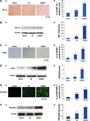
Figure 1 Photomicrographs of immunoreactivity (immunohistochemistry) and Western blots of HIF-1α, VEGF, and PECAM-1 in the cortical peri-infarct area at 14 days after IR.
Abbreviations: GSNO, S-nitrosoglutathione; IR, ischemia–reperfusion; Sham, sham-operated animals; VEGF, vascular endothelial growth factor; HIF-1α, hypoxia-inducible factor-1 alpha.
Middle cerebral artery occlusion (MCAO) rat model and GSNO treatment
Rats were anesthetized by ketamine hydrochloride (90 mg/kg body weight) and xylazine (10 mg/kg body weight) administered intraperitoneally. Stroke was induced by 60-minute left middle cerebral artery occlusion (MCAO) using an intraluminal filament as previously described.Citation11 Regional cerebral blood flow was monitored during the occlusion and early reperfusion to ensure the obstruction of blood flow, as previously described.Citation11,Citation12 Reperfusion was established by withdrawal of the filament. After 7 or 14 days, animals were sacrificed with an overdose of Nembutal. The brain, including the peri-infarct region, was sliced for histological and immunohistochemical analyses, and 2,3,5-triphenyltetrazolium chloride (TTC) staining.
In the GSNO group, GSNO (0.25 mg/kg body weight) in saline (~250 μL) was slowly infused by jugular vein cannulation at 1 hour after reperfusion. From 1 day after MCAO and thereafter, the same dose of GSNO was gavage fed until 7 or 14 days following IR. Physiological parameters did not alter after GSNO treatment. Details of the study on physiologic parameters in IR and GSNO-treated rats have been reported earlier.Citation11 In GSNO + 2-ME group, GSNO was treated for 7 days as described above. 2-ME (5 mg/kg, intraperitoneally) administration to the GSNO group was initiated 24 hours after the first dose of GSNO treatment and continued until the 7th day after IR.
Evaluation of ischemic infarct
Coronal sections (2 mm) were immersed in 1% solution of TTC in phosphate-buffered saline (pH 7.4) at 37°C for 15 minutes, as described previously.Citation12 After staining, infarctions were measured using Scion Image software (Scion Corp., Frederick, MD, USA). Total infarct area was multiplied by the thickness of the brain sections to obtain infarct volume as described previously.Citation7 To minimize the error introduced by edema and liquefaction after infarction, an indirect method for calculating infarct volume was also used.Citation39,Citation40 The non-infarcted area in the ipsilateral hemisphere was subtracted from that in the contralateral hemisphere, and infarct volume was calculated using the following formula:
Evaluation of neurological score and motor behavior
Animals in each group were evaluated for neurological scores, subtle behavior (using body swing test), and motor coordination and balance (using rotarod task) before and at the 7th and 14th days after reperfusion. The body weight of each animal was determined before surgery and at days 7 and 14 after IR.
Neurological examination was performed by an observer masked from the identity of the groups. A neurological grading system with a 4-point scale (0–3), as described previously,Citation7 was used: 0, no observable neurological deficit (normal); 1, failure to extend right forepaw on lifting the whole body by tail (mild); 2, circling to the contralateral side (moderate); and 3, leaning to the contralateral side at rest or no spontaneous motor activity (severe). The animals not showing paralysis at 1 hour after MCAO were excluded from the study because the reduction of blood flow may not have produced an infarction of adequate size to cause quantifiable neurological deficits in those animals.
Modified neurological severity score (mNSS) measurement was performed as described previously.Citation41 The test is sensitive to unilateral cortical injury because it reflects multiple asymmetries, including postural, sensory, and forelimb and hind limb use. A detailed description of this functional test has been previously reported.Citation42 In our studies, mNSS was scaled from 0 to 12, with 0 as normal and 12 as the maximal deficit.Citation15
The body swing test reflects symmetry of striatal function.Citation43 A normal rat typically has an equal number of swings to both sides. Each rat was held along the vertical axis (defined as no more than 10° to either the left or the right side) approximately 2.5 centimeters from the base of its tail and elevated 2.5 centimeters above a table surface. A swing was recorded whenever the rat moved its head out of the vertical axis to either side. The rats have to return to the vertical position for the next swing to be counted. Thirty total swings (score 3) were counted.
In the motor coordination and balance test, rats were examined on an accelerating rotarod task by trained personnel blinded to animal groups as described previously.Citation44 Walking time and speed on rotarod was recorded as previously described from our laboratory.Citation12 Speed was increased from 0 rpm to 30 rpm, with an increment of 2 rpm every 5 seconds. Each rat was placed on the rotarod cylinder, and the speed (rpm) at which the animal fell off the drum was recorded. The trial ended if the animal fell off the drum. Each animal was given three trials, and the mean speed (rpm) of three trials was calculated for each animal.
Immunohistochemistry and histology
Paraffin-embedded sections from the formalin-fixed brain tissues, processed at 7 or 14 days after reperfusion, were stained using antibodies against angiogenesis markers/mediators HIF-1α, VEGF, and PECAM-1 (Santa Cruz Biotechnology Inc., Dallas, TX, USA) and vessel markers laminin (Sigma-Aldrich Co.) and GSL-1 (Griffonia simplicifolia lectin 1) (Vector Lab, Burlingame, CA, USA).
Cell proliferation was assessed using Ki67 (Abcam, Cambridge, MA, USA). Secondary anti-rabbit and mouse IgG, conjugated with Alexa Fluor 488, were incubated on slides for 60 minutes. HIF-1α and VEGF sections were allowed to react with anti-mouse IgG conjugated to a peroxidase-labeled dextran polymer and then developed with diaminobenzidine substrate. All sections were examined for immunoreactivity in the peri-infarct area using an Olympus microscope as previously described.Citation9,Citation14 Three areas in the peri-infarct area of each immunostained section were digitized by a 40× microscope objective with microscope and camera without visual field overlap. Semiquantitative cell counting was performed using Scion Image software as described previously.Citation12
The degree of neuronal damage (loss of viable neurons) was evaluated by cresyl violet (Nissl) staining. The staining highlights important structural features of neurons. The brain sections described above were used to stain with Nissl, and the staining was performed according to classical histology methods.Citation12 Cells that contained Nissl substance were considered to be viable neurons. Condensed fragmented staining shows neuronal degeneration. The viable neurons were quantified by manually counting cells with visible nucleoli, as described previously,Citation45 in the peri-infarct area using an Olympus microscope.
Western blot analysis
In the peri-infarct area from the ipsilateral injured brain tissue, Western blot was performed using antibodies against HIF-1α and PECAM-1 (Abcam), VEGF (Santa Cruz Biotechnology Inc.), and β-actin, as described earlier.Citation9 Protein concentrations were determined using protein assay dye from Bio-Rad Laboratories Inc. (Hercules, CA, USA). Densitometry of protein expression was performed using a GS800 calibrated densitometer from Bio-Rad Laboratories Inc.
siRNA silencing of HIF-1α expression and treatment with GSNO in a mouse brain capillary endothelial cell line (bEnd3)
bEnd3, an immortalized mouse brain endothelial cell line originally generated and characterized,Citation46 and lately used in our studies,Citation47 is commercially available at American Type Culture Collection ([ATCC] CRL-2299; Manassas, VA, USA). It is a well-characterized endothelial cell line that shows the expression of ICAM-1 and VCAM-1.Citation47 Cells were grown according to the supplier’s instructions and were allowed to grow to ~70%–80% confluence. HIF-1α expression was silenced for 72 hours by two different predesigned siRNA sequence primers from Santa Cruz Biotechnology Inc. (A: SC-44308 and B: SC-35562) and the corresponding scrambled siRNA (negative control) into the cells using the bEnd3 standard transfection reagent (CRL-2299; Alto-gen Biosystems, Las Vegas, ND, USA) according to the manufacturer’s instructions and as elsewhere described.Citation48 Transfected cells were treated with freshly prepared GSNO (100 μM) for 24 hours. Western blots for HIF-1α (ab65979; Abcam), VEGF (SC507; Santa Cruz Biotechnology Inc.), and β-actin (4967; Cell Signaling, Danvers, MA, USA) were performed using 20 μg protein samples.
Matrigel analysis
Matrigel assay was performed as described previously.Citation49 Briefly, bEnd3 endothelial cells were plated in a six-well Matrigel plate (BD Biocoat Matrigel Matrix, 354432). The cells were seeded at a density of 105 cells per well in 2 mL medium. At 2 hours after seeding in Matrigel, the cells were treated with GSNO (100 μM), 2-ME (5 μM), or GSNO followed by 2-ME (30 minutes later). The cells were observed under a microscope for formation of tube-like structures at 3, 18, and 48 hours. Tubule length ratio, branch number per field, and aggregation area were analyzed using ImageJ software.
Statistical evaluation
Statistical analysis was performed as described previouslyCitation50 using GraphPad Prism 5.01 software. Unless otherwise stated, all values are expressed as mean ± standard deviation of n determinations or as mentioned. The results were examined by unpaired Student’s t-test. Multiple comparisons were performed using the Kruskal–Wallis test, or using analysis of variance followed by the Bonferroni test as appropriate. A P-value less than 0.05 was considered significant.
Results
GSNO treatment of IR for 2 weeks enhanced the expression of angiogenic mediators
Essential factors for the neurorepair process following stroke include the transcriptional activity of HIF-1α and enhanced expression of the angiogenic factor VEGF along with the endothelial cell proliferation mediator PECAM-1 (angiogenesis). S-Nitrosylation/GSNO has been shown to stabilize HIF-1α, leading to increased VEGF expression, angiogenesis, and protection against myocardial injury.Citation18 In this study, we observed that immunohistochemical staining 14 days after IR shows an increased expression of HIF-1α (), VEGF (), and PECAM-1 () in the peri-infarct area. GSNO treatment of IR significantly increased the expression further of HIF-1α, VEGF, and PECAM-1. Similar results of increased expression in the GSNO group from the peri-infract area at the 14th day after IR were confirmed for HIF-1α (), VEGF (), and PECAM-1 () using Western blot. In summary, GSNO treatment of IR increased the expression of angiogenic mediators.
GSNO treatment of IR for 2 weeks increased the number of blood vessels and enhanced the expression of a cell proliferation marker
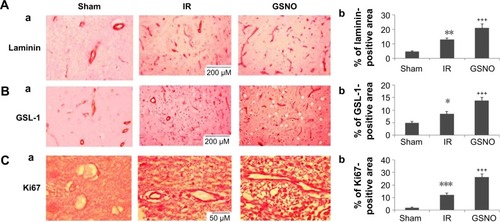
Quantitatively comparing vessel density using markers for vessels and a cell proliferation marker provides a measure of angiogenic vessel formation.Citation51 GSNO treatment of IR for 2 weeks increased the expression of markers of vessel density, including laminin () and GSL-1 (), indicating that GSNO may have promoted new vessel formation. Supporting the notion that the increased number of vessels present in the peri-infarct area resulted from angiogenic processes, we determined the expression of Ki67, a marker of cell proliferation. The expression of Ki67 () was significantly increased in GSNO-treated animals compared with IR animals. We anticipate that proliferating cells include endothelial cells also because vessel density is increased; however, we did not identify Ki67-positive cell types in this study. Sham animals had significantly lower expressions of angiogenic markers ().
Figure 2 Photomicrographs of immunohistochemistry of blood vessel markers and cell proliferation marker at 14 days after IR.
Abbreviations: GSNO, S-nitrosoglutathione; IR, ischemia–reperfusion; Sham, sham-operated animals; GSL-1, Griffonia simplicifolia lectin 1.
GSNO treatment of IR for 2 weeks improved motor function and accelerated the recovery of subtle behavior
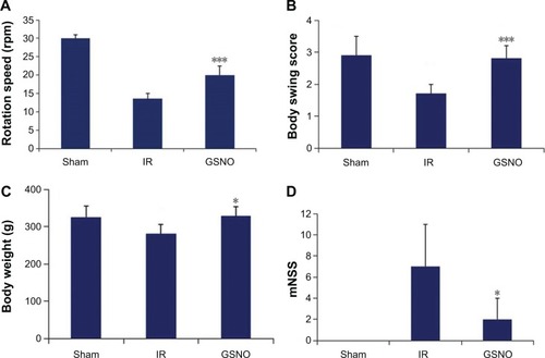
Compromised neurobehavioral functions, especially motor coordination and balance, and neurological functions are the leading causes of disability and morbidity following stroke. We determined the efficacy of GSNO in improving motor and subtle behaviors. GSNO treatment significantly improved tolerated rotation speed (rotarod task) () compared with the IR group measured at the 14th day after IR. The treatment with GSNO also improved body swing behavior () and mNSS measured at day 14 after IR (). The body weight of animals in the GSNO group was also significantly improved compared with animals in the IR group ().
Figure 3 Effect of GSNO on improvement of neurobehavioral functions at 14 days after IR.
Abbreviations: GSNO, S-nitrosoglutathione; IR, ischemia–reperfusion; mNSS, modified neurological severity score; Sham, sham-operated animals.
Beneficial effects of GSNO on neuroprotection and functional recovery were blocked by HIF-1α inhibitor 2-ME
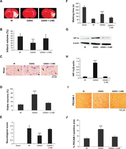
2-ME is a natural metabolite of estradiol and has been documented to inhibit HIF-1α stabilization and thus its transcriptional activity.Citation52 Therefore, we investigated whether 2-ME reverses GSNO’s HIF-1α-dependent activities (). Administration of 2-ME to GSNO-treated animals for 6 days (first dose of 2-ME was administered 24 hours after IR to investigate its effect after the acute phase) blocked GSNO’s protective effect on brain infarctions and functional recovery as evaluated by TTC staining (), Nissl staining (), and neurological score (). Furthermore, 2-ME treatment completely blocked the beneficial effect of GSNO on motor function recovery evaluated by rotarod performance (). As expected, 2-ME treatment robustly inhibited HIF-1α protein expression () as well as significantly reversed GSNO-mediated enhanced expression of PECAM-1 (). Reversal of GSNO’s effect by 2-ME was remarkably significant at day 7 after IR; therefore, the experiment was stopped at the 7th day after the injury.
Figure 4 Effect of inhibition of HIF-1α by 2-ME on GSNO-mediated protective effects at 7 days after IR.
Abbreviations: 2-ME, 2-methoxyestradiol; GSNO, S-nitrosoglutathione; IR, ischemia–reperfusion; Sham, sham-operated animals; TTC, 2,3,5-triphenyltetrazolium chloride; HIF-1α, hypoxia-inducible factor-1 alpha.
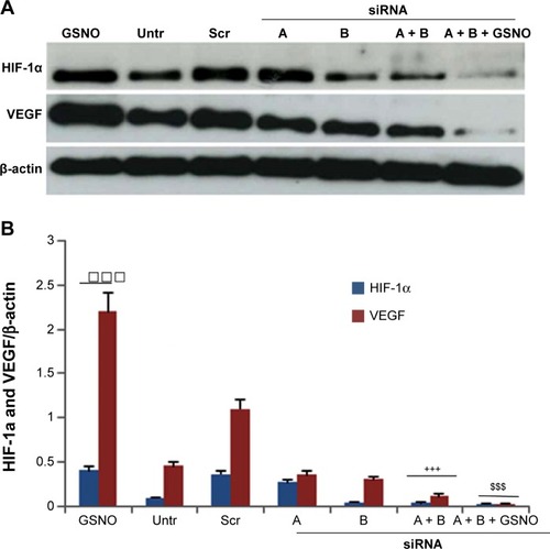
GSNO-mediated enhanced protein expression of VEGF was reversed by silencing HIF-1α in endothelial cells
Stabilization of HIF-1α results in increased HIF-1 transcriptional activity, leading to induction of HIF-1-dependent genes, including angiogenic VEGF. GSNO treatment resulted in greater protein expression of both HIF-1α and VEGF compared with untreated cells (). Single siRNA (A or B) silencing for HIF-1α reduced the expression of HIF-1α as well as VEGF; however, silencing with both A and B siRNA remarkably reduced the expression of HIF-1α and VEGF. Moreover, cell transfection with two HIF-1α-targeted siRNAs markedly reduced GSNO-mediated HIF-1α stabilization and prevented the resultant expression of VEGF (), consistent with a role of GSNO in HIF-1α stabilization and the stimulation of angiogenesis. However, a reduced level of both HIF-1α and VEGF in A + B + GSNO compared with A + B set was unanticipated and may be attributed to the stability of GSNO under bEnd3 cell transfection conditions.
Figure 5 Effects of GSNO on expression of HIF-1α and VEGF in HIF-1α-silenced endothelial (bEnd3) cells.
Abbreviations: GSNO, S-nitrosoglutathione; Scr, scrambled; Untr, untreated; VEGF, vascular endothelial growth factor; HIF-1α, hypoxia-inducible factor-1 alpha; RIPA, radioimmunoprecipitation assay.
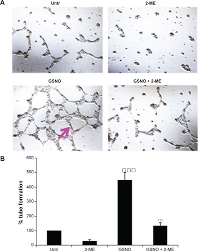
GSNO-stimulated formation of capillary-like structures on endothelial cells was blunted by the inhibition of HIF-1α
We next examined the ability of GSNO to stimulate formation and stabilization of tube/capillary-like proangiogenic structures on endothelial cultured cells using Matrigel (in vitro angiogenesis). Tube/capillary-like proangiogenic structures were significantly increased after 48-hour GSNO treatment (). Further, we assessed the effect of HIF-1α inhibition using 2-ME on GSNO-stimulated capillary-like structure formation. 2-ME is a specific HIF-1α inhibitor and has been widely used for the inhibition of angiogenesis.Citation52 We observed that GSNO’s capillary-like structure formation ability was prevented by 2-ME, indicating that the GSNO-stimulated proangiogenic process was mediated by HIF-1α activity ().
Figure 6 Effect of GSNO on formation of capillary-like proangiogenic structures on endothelial cells.
Abbreviations: 2-ME, 2-methoxyestradiol; GSNO, S-nitrosoglutathione; Untr, untreated.
Discussion
To facilitate functional recovery, an ideal stroke therapy must ameliorate acute as well as chronic phases of the injury by well-understood mechanisms. Our previous studies showed that GSNO treatment of IR provided neuroprotection in the acute phaseCitation7,Citation9 and improved neurobehavioral function in the chronic phase.Citation12 However, the mechanisms of GSNO-mediated stimulation of the neurorepair process require elucidation in order to determine and then to widen its therapeutic potential.
Functional recovery in stroke is associated with vascular integrity and the stimulation of both angiogenesis and neurogenesis.Citation53 Stroke patients who have higher cerebral blood vessel density recover better and survive longer than patients who have lower vascular density.Citation54 A low degree of restorative angiogenesis after stroke results in limited and insufficient functional recovery. However, angiogenesis-promoting therapeutics confer not only a greater degree but also a faster rate of functional improvement.Citation55,Citation56 Because GSNO also produces effects that increase vessel density () and enhance functional recovery (), GSNO likely promotes angiogenesis.Citation54 Findings from other studies that observed GSNO-induced angiogenesis in ischemic heartCitation18 and in a cell culture modelCitation57 support our observation of GSNO-mediated increased expression of angiogenic markers () and a cell proliferation marker (), as well as an increased number of vessels ().
The angiogenic marker VEGF is closely related to both angiogenesis and neurogenesis.Citation53 Increased neurogenesis is accompanied by increased angiogenesis, whereas angio-genesis upregulates neurogenesis and NO-based drugs upregulate both.Citation58 Angiogenesis itself is regulated by VEGF, mainly via HIF-1-based transcription. VEGF has been shown to modulate coupling of angiogenesis and neurogenesis; hence, it is an essential factor for regeneration.Citation53,Citation59 Therefore, we investigated the effect of GSNO on VEGF as well as PECAM-1 (markers of angiogenesis), finding that GSNO increased the expression of both following IR (). This observation indicates that GSNO might be acting via the upregulation of VEGF. VEGF is transcriptionally regulated by HIF-1, and HIF-1α is stabilized by GSNO.Citation25,Citation26 shows that GSNO increased the expression of and thereby stabilized HIF-1α. Consistent with our findings, endogenous GSNO-induced stabilization of HIF-1α via S-nitrosylation was shown to induce the expression of VEGF, resulting in angiogenesis and myocardial protection.Citation18
A potential concern of GSNO’s impact on VEGF is that HIF-1α-dependent upregulation of VEGF has been implicated in edema and BBB leakage in the acute phase of IR and cerebral hemorrhage.Citation60,Citation61 For VEGF-mediated BBB disruption to occur, MMP-9 and endothelial dysfunction are required.Citation9,Citation62 However, in addition to upregulating VEGF, GSNO transcriptionally downregulated MMP-9 as well as ICAM-1 and E-selectin, leading to reduced edema and increased BBB protection in the acute phase of IR and traumatic brain injury.Citation9,Citation15 Similarly, MMP-9-associated BBB disruption in experimental diabetes has been prevented by an exogenous treatment of GSNO.Citation63 Exogenous inhibition of GSNOR and thus increased levels of GSNO have also been reported to improve endothelial function.Citation38 Decreased levels of HIF-1α in cerebrospinal fluid from hypoxic/ischemic humansCitation64 support the notion that HIF-1α-mediated mechanisms may ameliorate stroke injury. These observations indicate that HIF-1α is likely an important beneficial component of neuroprotection and the neurovascular repair process.
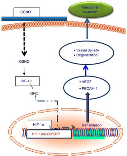
To show that GSNO invokes its neurorestorative/neurorepair effects via the HIF-1α/VEGF pathway, as depicted in , we inhibited HIF-1α, anticipating that it would block the effect of GSNO on angiogenic mediators and functional recovery. The inhibition of HIF-1α in GSNO-treated animals reduced the expression of HIF-1α () as well as PECAM-1 (), indicating that the angiogenic effect of GSNO was dependent on the activity of HIF-1α. 2-ME treatment of GSNO animals also blunted GSNO-mediated neuroprotection (), decreased neurological score (), and reduced motor function recovery (). These findings support the protective role of HIF-1α. Furthermore, endothelial cell culture studies showing reduced tube formation (Matrigel assay for angiogenesis) by 2-ME in GSNO-treated cells () provide evidence that GSNO’s regenerative repair effects are mediated by HIF-1α/VEGF pathway-induced angiogen-esis. Reduced VEGF expression in GSNO-treated HIF-1α-silenced cells () confirms that the GSNO-stimulated VEGF/angiogenesis pathway is dependent on the transcriptional activity of HIF-1α. Recently, stabilization of HIF-1α by N-acetylcysteine has been documented to provide neu-roprotection in stroke,Citation65 and its inhibition by 2-ME reduces neuroprotection and downregulates neurorepair processes in cerebral IR.Citation65,Citation66 2-ME has also been reported to worsen outcomes after global ischemia.Citation67 These observations further suggest that GSNO-induced angiogenic and neurorestorative processes are mediated through the stabilization of HIF-1α. The documented HIF-1α stabilization by GSNO-mediated S-nitrosylation (Cys-800) leading to stimulation of its transcriptional activityCitation68 supports our finding of GSNO-mediated enhanced transcriptional activity of HIF-1α and the expression of VEGF (). An enhanced level of GSNO has been shown to stimulate the HIF-1α-dependent angiogenic process, protecting against cardiac ischemic injury.Citation18,Citation37 In view of the above observations, we hypothesized that GSNO-mediated S-nitrosylation and stabilization of HIF-1α will enhance VEGF and PECAM-1, leading to increased vessel density and functional recovery ().
Figure 7 Schematic showing GSNO-mediated events leading to neuroprotection and neurorepair as well as functional recovery.
Both deficient S-nitrosylation and a reduction in NO signaling pathways are observed in several neurodegenerative disease processes, including stroke.Citation9,Citation69–Citation72 These pathologies occur due to decreased bioavailability of NO and, hence, reduced biosynthesis of GSNO. We have previously documented reduced levels of NO and decreased expression of S-nitrosocysteine proteins (-SNO) in the peri-infarct area. This decreased -SNO was normalized by an exogenous GSNO treatment.Citation9 In short, reduced NO and -SNO are associated with IR pathophysiology. These observations are further supported by a report showing decreased levels of plasma NO and associated poor outcomes in stroke patients.Citation73 Plasma S-nitrosothiols are also decreased in patients with endothelial dysfunction.Citation74 These results indicate that S-nitrosylation is reduced in IR; therefore, exogenous GSNO supplementation in stroke therapy is a promising strategy. Although HIF-1α stabilization-based therapy using chemical inhibitors of PHDs is under investigation in chronic ischemic diseases, they also invoke HIF-1α-independent effects.Citation22 Therefore, a direct stabilization by S-nitrosylation of HIF-1α through the endogenous signaling molecule GSNO is mechanistically a more specific therapeutic approach especially for IR stroke therapy. The role of the GSNO/HIF-1α/VEGF pathway in permanent ischemic stroke is less clear. Nonetheless, evidence from clinical studies suggests that administration of NO-based therapeutics in humans is safe and may be an effective treatment for stroke injury in human subjects.Citation75,Citation76
Conclusion
GSNO-mediated HIF-1α stabilization for neuroprotection and the stimulation of the neurorepair process provides a novel target mechanism for a therapy that could confer benefits in both the acute and chronic phases of stroke. The translational potential of GSNO is high because it occurs naturally in the human body and its exogenous administration to humans is not associated with any known side effects or toxicity. This study shows the feasibility of a new treatment paradigm for stroke, introducing the concept that stabilization of HIF-1α using GSNO may reduce disabilities in stroke survivors.
Author contributions
This study is based on an original idea of M Khan and AK Singh. All authors have contributed to conception and design of the study, analysis and interpretation of data and drafting the manuscript. Drs. TS Dhammu, F Matsuda, M Baarine, M Khan and Mr. TS Dhindsa are responsible for acquisition of data. All authors hereby approve the content of the article and agree to be accountable for all aspects of the work.
Acknowledgments
This work was supported by grants from NIH (NS-72511) and VA merit award (BX001062). This work was also supported by the NIH, Grants C06 RR018823 and No C06 RR015455 from the Extramural Research Facilities Program of the National Center for Research Resources. We thank Ms Joyce Bryan and Ms Terry Hope for their technical help and secretarial assistance. We are grateful to Dr Harutoshi Sakakima and Dr Yoshihiro Yoshida for their valuable input. We also acknowledge Dr Tom Smith and Dr Lisa Kerr from the MUSC Writing Center for their valuable editing of the manuscript.
Disclosure
The authors report no conflicts of interest in this work.
Notes
M Khan (PhD) is a research associate professor at the MUSC with a strong background in neuroprotection and functional recovery in animal models of stroke, traumatic brain injury, and spinal cord injury. He has published more than 80 articles in the field of neuroprotection and neuropharmacology. TS Dhammu (PhD) and M Baarine (PhD) are postdoctoral fellows with expertise in cell culture and biochemical techniques. TS Dhammu has expertise in creating MCAO animal model of stroke and evaluating neurobehavioral functions of rodents. F Matsuda (PhD) is an assistant professor at the Kagoshima University, Kagoshima, Japan, and she has a strong background in evaluation of animals’ neurobehavioral functions. TS Dhindsa is a medical student at the MUSC and works in our group on projects related to neurodegeneration. He is an enthusiastic scientist with a special interest in neuropharmacology of stroke and traumatic brain injury. I Singh is a PhD and a distinguished university professor and Scientific Director of the Children’s Research Institute at the MUSC. He has published more than 280 articles in the field of neuroprotection and functional recovery. AK Singh is an MD and a practicing pathologist. She is professor at the MUSC and Director of the Pathology Laboratory at the VA Medical Center, Charleston, SC, USA. Her major research interest is traumatic brain injury and neurodegenerative diseases including stroke and Alzheimer’s. She has published more than 130 articles related to neurodegenerative diseases in peer-reviewed journals.
References
- GoASMozaffarianDRogerVLAmerican Heart Association Statistics Committee and Stroke Statistics SubcommitteeHeart disease and stroke statistics – 2014 update: a report from the American Heart AssociationCirculation20141293e28e29224352519
- RosamondWFlegalKFurieKAmerican Heart Association Statistics Committee and Stroke Statistics SubcommitteeHeart disease and stroke statistics – 2008 update: a report from the American Heart Association Statistics Committee and Stroke Statistics SubcommitteeCirculation20081174e25e14618086926
- MoskowitzMALoEHIadecolaCThe science of stroke: mechanisms in search of treatmentsNeuron201067218119820670828
- MakiTHayakawaKPhamLDXingCLoEHAraiKBiphasic mechanisms of neurovascular unit injury and protection in CNS diseasesCNS Neurol Disord Drug Targets201312330231523469847
- CramerSCRepairing the human brain after stroke. II. Restorative therapiesAnn Neurol200863554956018481291
- CramerSCRepairing the human brain after stroke: I. Mechanisms of spontaneous recoveryAnn Neurol200863327228718383072
- KhanMSekhonBGiriSS-Nitrosoglutathione reduces inflammation and protects brain against focal cerebral ischemia in a rat model of experimental strokeJ Cereb Blood Flow Metab200525217719215647746
- LiuDHYuanFGHuSQEndogenous nitric oxide induces activation of apoptosis signal-regulating kinase 1 via S-nitrosylation in rat hippocampus during cerebral ischemia-reperfusionNeuroscience2013229364823137546
- KhanMDhammuTSSakakimaHThe inhibitory effect of S-nitrosoglutathione on blood-brain barrier disruption and peroxynitrite formation in a rat model of experimental strokeJ Neurochem2012123Suppl 2869723050646
- YinXHYanJZHouXYWuSLZhangGYNeuroprotection of S-nitrosoglutathione against ischemic injury by down-regulating Fas S-nitrosylation and downstream signalingNeuroscience201324829029823792322
- KhanMJatanaMElangoCPaintliaASSinghAKSinghICerebrovascular protection by various nitric oxide donors in rats after experimental strokeNitric Oxide200615211412416524750
- SakakimaHKhanMDhammuTSStimulation of functional recovery via the mechanisms of neurorepair by S-nitrosoglutathione and motor exercise in a rat model of transient cerebral ischemia and reperfusionRestor Neurol Neurosci201230538339622717646
- ChouPCShunmugavelAEl SayedHPreclinical use of longitudinal MRI for screening the efficacy of S-nitrosoglutathione in treating spinal cord injuryJ Magn Reson Imaging20113361301131121590998
- KhanMSakakimaHDhammuTSS-nitrosoglutathione reduces oxidative injury and promotes mechanisms of neurorepair following traumatic brain injury in ratsJ Neuroinflammation201187821733162
- KhanMImYBShunmugavelAAdministration of S-nitrosoglutathione after traumatic brain injury protects the neuro-vascular unit and reduces secondary injury in a rat model of controlled cortical impactJ Neuroinflammation200963219889224
- WonJSKimJAnnamalaiBShunmugavelASinghISinghAKProtective role of S-nitrosoglutathione (GSNO) against cognitive impairment in rat model of chronic cerebral hypoperfusionJ Alzheimers Dis201334362163523254638
- KimuraHOguraTKurashimaYWeiszAEsumiHEffects of nitric oxide donors on vascular endothelial growth factor gene inductionBiochem Biophys Res Commun2002296497698212200144
- LimaBLamGKXieLEndogenous S-nitrosothiols protect against myocardial injuryProc Natl Acad Sci U S A2009106156297630219325130
- KeQCostaMHypoxia-inducible factor-1 (HIF-1)Mol Pharmacol20067051469148016887934
- IyerNVKotchLEAganiFCellular and developmental control of O2 homeostasis by hypoxia-inducible factor 1 alphaGenes Dev19981221491629436976
- KasivisvanathanVShalhoubJLimCSShepherdACThaparADaviesAHHypoxia-inducible factor-1 in arterial disease: a putative therapeutic targetCurr Vasc Pharmacol20119333334920807188
- HartenSKAshcroftMMaxwellPHProlyl hydroxylase domain inhibitors: a route to HIF activation and neuroprotectionAntioxid Redox Signal201012445948019737089
- PalmerLAGastonBJohnsRANormoxic stabilization of hypoxia-inducible factor-1 expression and activity: redox-dependent effect of nitrogen oxidesMol Pharmacol20005861197120311093754
- WellmanTLJenkinsJPenarPLTranmerBZahrRLounsburyKMNitric oxide and reactive oxygen species exert opposing effects on the stability of hypoxia-inducible factor-1alpha (HIF-1alpha) in explants of human pial arteriesFASEB J200418237938114657004
- CarverDJGastonBDerondeKPalmerLAAkt-mediated activation of HIF-1 in pulmonary vascular endothelial cells by S-nitrosoglutathioneAm J Respir Cell Mol Biol200737325526317541013
- MetzenEZhouJJelkmannWFandreyJBruneBNitric oxide impairs normoxic degradation of HIF-1alpha by inhibition of prolyl hydroxylasesMol Biol Cell20031483470348112925778
- SinghSPWishnokJSKeshiveMDeenWMTannenbaumSRThe chemistry of the S-nitrosoglutathione/glutathione systemProc Natl Acad Sci U S A1996932514428144338962068
- HessDTStamlerJSRegulation by S-nitrosylation of protein post-translational modificationJ Biol Chem201228774411441822147701
- StamlerJSJarakiOOsborneJNitric oxide circulates in mammalian plasma primarily as an S-nitroso adduct of serum albuminProc Natl Acad Sci U S A19928916767476771502182
- KlugeIGutteck-AmslerUZollingerMDoKQS-nitrosoglutathione in rat cerebellum: identification and quantification by liquid chromatography-mass spectrometryJ Neurochem1997696259926079375694
- BryanNSRassafTMaloneyRECellular targets and mechanisms of nitros(yl)ation: an insight into their nature and kinetics in vivoProc Natl Acad Sci U S A2004101124308431315014175
- TsikasDSandmannJHolzbergDPantazisPRaidaMFrölichJCDetermination of S-nitrosoglutathione in human and rat plasma by high-performance liquid chromatography with fluorescence and ultraviolet absorbance detection after precolumn derivatization with o-phthalaldehydeAnal Biochem19992731324010452796
- MatsumotoAGowAJMembrane transfer of S-nitrosothiolsNitric Oxide201125210210721377531
- RadomskiMWReesDDDutraAMoncadaSS-nitroso-glutathione inhibits platelet activation in vitro and in vivoBr J Pharmacol199210737457491335336
- SavidgeTCNewmanPPothoulakisCEnteric glia regulate intestinal barrier function and inflammation via release of S-nitroso-glutathioneGastroenterology200713241344135817408650
- RassafTPollLWBrouzosPPositive effects of nitric oxide on left ventricular function in humansEur Heart J200627141699170516782717
- KonorevEATarpeyMMJosephJBakerJEKalyanaramanBS-nitrosoglutathione improves functional recovery in the isolated rat heart after cardioplegic ischemic arrest-evidence for a cardioprotective effect of nitric oxideJ Pharmacol Exp Ther199527412002067616400
- ChenQSieversREVargaMPharmacological inhibition of S-nitrosoglutathione reductase improves endothelial vasodilatory function in rats in vivoJ Appl Physiol (1985)2013114675276023349456
- MatsudaFSakakimaHYoshidaYThe effects of early exercise on brain damage and recovery after focal cerebral infarction in ratsActa Physiol (Oxf)2011201227528720726846
- SwansonRAMortonMTTsao-WuGSavalosRADavidsonCSharpFRA semiautomated method for measuring brain infarct volumeJ Cereb Blood Flow Metab19901022902931689322
- MahmoodAGoussevALuDLong-lasting benefits after treatment of traumatic brain injury (TBI) in rats with combination therapy of marrow stromal cells (MSCs) and simvastatinJ Neurotrauma200825121441144719072586
- LiYChenJChenXGHuman marrow stromal cell therapy for stroke in rat: neurotrophins and functional recoveryNeurology200259451452312196642
- BorlonganCVSanbergPRElevated body swing test: a new behavioral parameter for rats with 6-hydroxydopamine-induced hemiparkinsonismJ Neurosci1995157 Pt 2537253787623159
- MonvilleCTorresEMDunnettSBComparison of incremental and accelerating protocols of the rotarod test for the assessment of motor deficits in the 6-OHDA modelJ Neurosci Methods2006158221922316837051
- ZhuYBuQLiuXHuWWangYNeuroprotective effect of TAT-14-3-3ε fusion protein against cerebral ischemia/reperfusion injury in ratsPLoS One201493e9333424671253
- MontesanoRPepperMSMöhle-SteinleinURisauWWagnerEFOrciLIncreased proteolytic activity is responsible for the aberrant morphogenetic behavior of endothelial cells expressing the middle T oncogeneCell19906234354452379237
- PrasadRGiriSNathNSinghISinghAKGSNO attenuates EAE disease by S-nitrosylation-mediated modulation of endothelial-monocyte interactionsGlia2007551657717019693
- BovePFHristovaMWesleyUVOlsonNLounsburyKMvan der VlietAInflammatory levels of nitric oxide inhibit airway epithelial cell migration by inhibition of the kinase ERK1/2 and activation of hypoxia-inducible factor-1 alphaJ Biol Chem200828326179191792818424783
- HePPhilbrickMJAnXWuJMessmer-BlustAFLiJEndothe-lial differentiation gene-1, a new downstream gene is involved in RTEF-1 induced angiogenesis in endothelial cellsPLoS One201492e8814324520353
- JatanaMGiriSAnsariMAInhibition of NF-kappaB activation by 5-lipoxygenase inhibitors protects brain against injury in a rat model of focal cerebral ischemiaJ Neuroinflammation200631216689995
- ValableSMontanerJBellailAVEGF-induced BBB permeability is associated with an MMP-9 activity increase in cerebral isch-emia: both effects decreased by Ang-1J Cereb Blood Flow Metab200525111491150415902195
- MooberrySLMechanism of action of 2-methoxyestradiol: new developmentsDrug Resist Updat20036635536114744499
- LokJGuptaPGuoSCell-cell signaling in the neurovascular unitNeurochem Res200732122032204517457674
- WeiLErinjeriJPRovainenCMWoolseyTACollateral growth and angiogenesis around cortical strokeStroke20013292179218411546914
- ChenJZhangZGLiYStatins induce angiogenesis, neurogenesis, and synaptogenesis after strokeAnn Neurol200353674375112783420
- ChenJCuiXZacharekANiaspan increases angiogenesis and improves functional recovery after strokeAnn Neurol2007621495817557352
- KawasakiKSmithRSJrHsiehCMSunJChaoJLiaoJKActivation of the phosphatidylinositol 3-kinase/protein kinase Akt pathway mediates nitric oxide-induced endothelial cell migration and angiogenesisMol Cell Biol200323165726573712897144
- ZhangRLZhangZGChoppMTargeting nitric oxide in the subacute restorative treatment of ischemic strokeExpert Opin Investig Drugs2013227843851
- TengHZhangZGWangLCoupling of angiogenesis and neu-rogenesis in cultured endothelial cells and neural progenitor cells after strokeJ Cereb Blood Flow Metab200828476477117971789
- LeeCZXueZZhuYYangGYYoungWLMatrix metalloproteinase-9 inhibition attenuates vascular endothelial growth factor-induced intracerebral hemorrhageStroke20073892563256817673717
- BauerATBürgersHFRabieTMartiHHMatrix metalloproteinase-9 mediates hypoxia-induced vascular leakage in the brain via tight junction rearrangementJ Cereb Blood Flow Metab201030483784819997118
- LeeCZXueZHaoQYangGYYoungWLNitric oxide in vascular endothelial growth factor-induced focal angiogenesis and matrix metalloproteinase-9 activity in the mouse brainStroke20094082879288119498186
- AggarwalAKheraASinghISandhirRS-nitrosoglutathione prevents blood–brain barrier disruption associated with increased matrix metalloproteinase-9 activity in experimental diabetesJ Neurochem Epub201494
- KeXJZhangJJChanges in HIF-1α, VEGF, NGF and BDNF levels in cerebrospinal fluid and their relationship with cognitive impairment in patients with cerebral infarctionJ Huazhong Univ Sci Technolog Med Sci201333343343723771673
- ZhangZYanJTaheriSLiuKJShiHHypoxia-inducible factor 1 contributes to N-acetylcysteine’s protection in strokeFree Radic Biol Med20146882124296245
- WangZTsaiLKMunasingheJChronic valproate treatment enhances postischemic angiogenesis and promotes functional recovery in a rat model of ischemic strokeStroke20124392430243622811460
- ZhouDMatchettGAJadhavVDachNZhangJHThe effect of 2-methoxyestradiol, a HIF-1 alpha inhibitor, in global cerebral ischemia in ratsNeurol Res200830326827117716391
- YasinskaIMSumbayevVVS-nitrosation of Cys-800 of HIF-1alpha protein activates its interaction with p300 and stimulates its transcriptional activityFEBS Lett20035491–310510912914934
- SchonhoffCMMatsuokaMTummalaHS-nitrosothiol depletion in amyotrophic lateral sclerosisProc Natl Acad Sci U S A200610372404240916461917
- JuTCChenSDLiuCCYangDIProtective effects of S-nitrosoglutathione against amyloid beta-peptide neurotoxicityFree Radic Biol Med200538793894915749390
- RauhalaPMohanakumarKPSzirakiILinAMChiuehCCS-nitrosothiols and nitric oxide, but not sodium nitroprusside, protect nigrostriatal dopamine neurons against iron-induced oxidative stress in vivoSynapse199623158608723136
- TerpolilliNAMoskowitzMAPlesnilaNNitric oxide: considerations for the treatment of ischemic strokeJ Cereb Blood Flow Metab20123271332134622333622
- RashidPAWhitehurstALawsonNBathPMPlasma nitric oxide (nitrate/nitrite) levels in acute stroke and their relationship with severity and outcomeJ Stroke Cerebrovasc Dis2003122828717903909
- HeissCLauerTDejamAPlasma nitroso compounds are decreased in patients with endothelial dysfunctionJ Am Coll Cardiol200647357357916458138
- RobertsBWMitchellJKilgannonJHChanskyMETrzeciakSNitric oxide donor agents for the treatment of ischemia/reperfusion injury in human subjects: a systematic reviewShock201339322923923358103
- PlutaRMOldfieldEHBakhtianKDSafety and feasibility of long-term intravenous sodium nitrite infusion in healthy volunteersPLoS One201161e1450421249218