
 ?Mathematical formulae have been encoded as MathML and are displayed in this HTML version using MathJax in order to improve their display. Uncheck the box to turn MathJax off. This feature requires Javascript. Click on a formula to zoom.
Abstract
The aim of the study was to improve corneal penetration and bioavailability of ofloxacin (OFX) eye preparations. OFX was incorporated in poly (lactide-co-glycolide) as biodegradable microspheres using oil in oil emulsion solvent evaporation technique. The prepared OFX microspheres were then incorporated in Gelrite® in situ gel preparation. In addition, OFX Gelrite-based in situ gel formulations were prepared. OFX formulations were characterized for gelling capacity, viscosity, and rheological properties. Release studies for OFX microspheres, OFX in situ gel, and OFX-loaded microspheres in situ gel formulations were carried out to investigate release characteristics of the drug. The prepared OFX formulations were then investigated in vivo compared with commercially available OFX eyedrops. Results showed that the optimum Gelrite concentration was at 0.4%–0.7% w/v; the prepared formulations were viscous liquid transformed into a pourable gel immediately after the addition of simulated tear fluid with a gelling factor of 27–35. Incorporation of OFX-loaded microspheres in Gelrite solution (0.4% w/v) significantly altered the release profiles of OFX-loaded microspheres in situ gel formula compared with the corresponding OFX gels and OFX microspheres. In vivo results in rabbits showed that OFX-loaded microspheres in situ gel formula improved the relative bioavailability by 11.7-fold compared with the commercially available OFX eyedrops. In addition, the longer duration of action of OFX-loaded microspheres in situ gel formula preparations is thought to avoid frequent instillations, which improves patient tolerability and compliance.
Introduction
Ofloxacin (OFX) is a second-generation fluoroquinolone with a broad spectrum of action against gram-positive and gram-negative bacteria.Citation1,Citation2 It is a potent agent against various ocular species, including Staphylococcus aureus, Staphylococcus epidermidis, Streptococcus pneumonia, chlamydial species, and various anaerobes.Citation3 Reports showed that the mean concentration of OFX in the aqueous humor exceeded the minimum inhibitory concentration for 90% (MIC90) of the frequently occurring gram-positive and gram-negative bacteria after topical application of 0.3% OFX eye drops.Citation4,Citation5 Of the topically applied fluoroquinolones, OFX achieved the highest aqueous humor concentration.Citation4 Corneal precipitations of topically applied fluoroquinolones frequently occur due to the formation of the zwitterionic form near the physiologic pH. This lowers their solubility and hence precipitation can occur at this pH. Although precipitation has not yet been reported for OFX eye drops, deposits may occur.Citation6
Topical drug delivery is the most common method of administration for treatment of diseases of the anterior segment of the eye as it is simple, convenient, and painless.Citation7–Citation9 However, less than 10% bioavailability is achieved for topical ocular drug application. The low bioavailability is attributed to the ocular protecting biological barriers such as the impermeable corneal epithelium, which represents a barrier for drugs to penetrate into the eye.Citation7–Citation9 Moreover, rapid elimination of instilled ophthalmic drug solution occurs from the precorneal area as a result of the lacrimal secretion and nasolacrimal drainage. Consequently, frequent instillation of ophthalmic solutions is required to achieve the required therapeutic effects.Citation10 Accordingly, efficient topical delivery of drugs to ocular tissues is a challenge to formulation scientists.Citation9
Novel technologies were developed to improve the bioavailability of ophthalmic drugs. Mucoadhesive systems, in situ gelling systems, microparticulate systems, microemulsions, vesicular systems, implants, prodrugs, penetration enhancers, and dendrimers are examples of the applied technologies.Citation11–Citation21 Pharmaceutical particulate technology for the delivery of ophthalmic drugs is used to extend drug release and improve patient compliance.Citation22 The technology can also improve the residence time of the encapsulated drug.Citation14 Ophthalmic microparticles should not exceed 10 μm in size, otherwise, a scratching feeling in the eye can occur that could lead to corneal abrasion and injury.Citation15 Bioadhesive ocular microspheres are novel delivery systems that remain adherent to the ocular surface for a prolonged time. As a result, the bioavailability of the encapsulated drug is improved.Citation12,Citation23,Citation24
In situ-forming hydrogels are liquids which undergo phase transition upon instillation to a viscoelastic gel in response to physiological conditions in the cul-de-sac.Citation12,Citation14 Three types of physiological changes can induce phase transition; they are: change in temperature, change in pH, or change in electrolyte composition.Citation14 The increase in the viscosity upon instillation results in prolonged ocular residence time.Citation14,Citation25 In situ gels are preferred because they are liquids that can be easily dropped in the eye. In addition, they allow for reproducible instillation of accurate doses in comparison with instillation of preformed gels.Citation12 Low acylated gellan gum (commercially available as Gelrite®) is a polysaccharide that undergoes phase transition to clear gel in the presence of mono- or divalent cations.Citation26 Gelrite-based formulations have been tested for different types of drugs such as timolol maleate, ciprofloxacin hydrochloride, indomethacin, pefloxacin mesylate, carteolol, and gatifloxacin.Citation26–Citation31
In the present study, OFX was incorporated in poly (lactide-co-glycolide) (PLGA) biodegradable microspheres, and then loaded into in situ gel formula. The incorporation of OFX microspheres in the in situ gel preparation aimed to improve OFX corneal penetration and consequently enhance its efficacy against ocular infections. In addition, the longer duration of action of this formula preparations is thought to avoid frequent instillations, which improves patient tolerability and compliance.
Materials and methods
Materials
Poly (DL-lactide-co-glycolide), Gelrite (deacylated gellan gum), and cellophane membrane were purchased from Sigma-Aldrich Co. (St Louis, MO, USA). OFX was kindly gifted by Al-Kahira Pharmaceutical Company (Cairo, Egypt). Polyvinyl alcohol was purchased from Fluka Chemie GmbH (Steinheim, Germany). Dichloromethane (DCM), acetonitrile (ACN), methanol (MeOH), petroleum ether, liquid paraffin, sodium bicarbonate, calcium chloride dihydrate, sodium chloride, polyethylene glycol 6,000, and isopropyl myristate were purchased from El-Nasr Pharmaceutical Co. (Cairo, Egypt). Ketamine 50 mg/mL was supplied by Sigma-Tec Pharmaceutical Industries (Cairo, Egypt), and ACN and MeOH for high performance liquid chromatography (HPLC) was supplied by EMD Millipore (Billerica, MA, USA). All other solvents and chemicals were of analytical grade.
Methods
Preparation of OFX microspheres using oil in oil emulsion technique
OFX microspheres were prepared using oil in oil (O/O) emulsion solvent evaporation technique. Briefly, poly (DL-lactide-co-glycolide) was dissolved in a mixture of ACN, DCM, and MeOH in a ratio of 6:4:1, in screw-capped test tubes, to make 4% w/v solutions. OFX was added to the organic phase and then sonicated for 10 seconds using a probe sonicator under cooling (Heidolph MR 3001; Heidolph, Schwabach, Germany). The organic phase was then added dropwise to 50 mL of liquid paraffin containing lecithin (2% w/v) and was homogenized at 20,000 rpm (T10 basic ULTRA-TURRAX, IKA, Staufen, Germany) for 2 minutes to obtain the O/O emulsion. The formed emulsion was then stirred under a slower speed (500 rpm) overnight to allow slow evaporation of organic solvent. Microspheres were then harvested by centrifugation at 4,000 rpm for 30 minutes (Fischer Centrific® Centrifuge; Thermo Fisher Scientific, Waltham, MA, USA), washed three times with petroleum ether, and air-dried.
Encapsulation efficiency
Weighed amounts of the prepared microspheres were added to 50 mL DCM and sonicated for 30 minutes. OFX concentration was measured after appropriate dilution at wavelength of maximum absorption (λmax) 294 nm using a ultraviolet/visible spectrophotometer (Spectronic Genesys® with Winspec Software; Thermo Fisher Scientific). The percentage encapsulation efficiency (EE %) was calculated according to EquationEquation 1(1) :
Particle size analysis
Particle size and size distribution of the prepared OFX microspheres were measured using a laser scattering particle size distribution analyzer (HORIBA-LA300; Retsch Technology GmbH, Düsseldorf, Germany). Five analyses were performed for each microsphere sample to determine the mean diameter.
Morphology
Morphology and the surface characteristics of OFX microspheres were examined by scanning electron microscope (Jeol Fine-Coat JFC 1100E; Jeol JSM-5400 LV; JEOL, Tokyo, Japan).
Preparation of OFX Gelrite based in situ gel formulations
Solutions of Gelrite (0.1%–0.9% w/v) in boric–borax buffer (pH 7.4) were prepared by heating OFX dispersions to 90°C for 20 minutes while stirring. Solutions were then cooled at room temperature while stirring. The prepared formulations were stored in amber colored glass vials and were sterilized by autoclaving.
Characterization of in situ gel formulations
Gelling capacity and flow behavior
Gelling capacity was determined by addition of 7 mL of simulated tear fluid (STF) to a vial containing 25 mL of Gelrite-based formulation at 37°C, and then visually evaluating the gel formation and observing the flow behavior of the formulations at each concentration, before and after addition of STF. The gelling factor was calculated according to EquationEquation 2(2) :
Viscosity and rheological properties
Viscosity and rheological properties of the prepared formulae were carried out using a Brookfield rheometer (DV-III Ultra; Brookfield Engineering Laboratories, Inc., Middleboro, MA, USA). STF of pH 7.4 was added in increments of 25 mL to 200 mL to the formulations, and the viscosities at which gelation occurred were recorded. The angular velocity increased gradually from 0.01 to 100 rpm. The average of the two readings was used to calculate the viscosity.
Effect of autoclaving
Characteristics of the prepared in situ Gelrite-based gels were investigated before and after sterilization by autoclaving. Briefly, the formulations were packaged in amber-colored glass vials and were sterilized by terminal autoclaving at 121°C and 204, 774.292 pascals (15 psig) for 20 minutes.
Release study of OFX-loaded in situ gel formulations
OFX in vitro release from the prepared in situ gel formulations was carried out using a small cylindrical glass tube with a surface area of 1.77 cm2. Cellophane membrane was soaked overnight in STF, and then was clamped into one end of the tube. The in situ gelling formulation (1 mL) was placed over the cellophane membrane. The end of the tube was immersed in 100 mL of STF, kept at 37°C, and stirred at 40 rpm. The release experiments were designed to maintain sink conditions. Two-milliliter samples were withdrawn at the different time intervals. Each sample was replaced by fresh STF. The concentration of OFX was analyzed at λmax 294 nm. The release of OFX from the combined microsphere–gel formulations was compared to the release of OFX from corresponding microsphere formulations and in situ gels, prepared separately.
Inclusion of microspheres into the in situ gel
OFX microspheres were homogeneously dispersed into the preformed Gelrite solution in order to produce the final OFX concentration of 0.3% w/v in the microsphere-loaded in situ gel formula. The formula was prepared by dispersing OFX microspheres in 0.4% w/v Gelrite. It is important to mention that 0.4% w/v Gelrite was used to produce the in situ gelling system. The effect of OFX microspheres inclusion on gelling capacity, rheology, and in vitro release was investigated.
In vivo studies
Animals
Adult male rabbits (2.5–3.0 kg, free of ocular defects) were used. The animals were obtained from the National Research Centre, Dokky, Giza, Egypt. Animal use was approved by the Review Board for Preclinical and Clinical Research Minia University who ensured the care and use of animals conformed to the Declaration of Helsinki and the Guiding Principle in Care and Use of Animals (DHEW publication NIH 80-23). Rabbits were classified into five groups. Each group contained four rabbits. The first group served as an untreated control. The second group (positive control) received suspension of OFX microspheres in isotonic boric–borax buffer (pH 6.8). The third group received OFX in situ gel. The fourth group received OFX microspheres in 0.4% Gelrite in situ gel. The last group received OFX marketed product. All the aforementioned preparations contained 0.3% w/v OFX. The test was carried out by dropping 100 μL (corresponding to 0.3 mg OFX) of each preparation into the lower conjunctival sac of the right eye of the rabbit. The rabbits were anaesthetized by intramuscular injection of 30 mg/kg ketamine, and then 80 μL of aqueous humor were withdrawn from the anterior chamber of the eye at different time intervals. The aqueous humor samples were stored at −18°C. The relative area under the curve compared with commercial product (AUCrel), maximum OFX concentration in aqueous humor (Cmax), and the time to reach this concentration (Tmax) were calculated.
HPLC analysis of aqueous humor samples
Aqueous humor samples, after suitable dilutions, were analysed for OFX content using the previously reported HPLC method.Citation32 An HPLC system (Agilent Technologies, Santa Clara, CA, USA) with a fluorescence detector (Model FP-2020; Jasco International Co., Ltd., Hachioji, Tokyo, Japan) was used. The analytical column was a Venusil XBP C18 (L) (250 mm ×4.6 mm, 5 μm) (Agilent Technologies) in conjunction with a pre-column module (Agilent Technologies) with a Venusil Cl8 insert. Data acquisition was performed on Young Lin Autochro-3000 software (YL Instrument Co., Ltd., Anyang-si, Gyeonggi-do, The Republic of Korea). The mobile phase was MeOH, CAN, and 0.4 M citric acid (3:1:10, v/v/v). The flow rate was 0.8 mL/min at ambient temperature. The excitation and emission wavelengths were set at 290 nm and 500 nm, respectively.
Results and discussion
Preparation and characterization of OFX-loaded PLGA microspheres
O/O emulsion solvent evaporation method was adopted for OFX incorporation in microspheres to improve the entrapment efficiency. A disadvantage for the use of microspheres suspensions in the eye is the possible drainage of the particles that leads to an incomplete drug release. The major advantage of using in situ gel formulation containing microspheres is the improved precorneal retention of the administered microspheres, provided that the gel would remain in the eye during the time for drug release for a longer period of time compared with the microspheres-alone formula.
The composition of the liquid that is dispersed in liquid paraffin consisted of ACN, DCM, and MeOH. DCM was included to lower the polarity of ACN and to ensure formation of stable emulsion.Citation33 OFX showed poor solubility in ACN (2.5 mg/mL) as measured practically in our lab. Therefore, MeOH and DCM were used to optimize OFX encapsulation within the microspheres by increasing the solubility of OFX in the dispersed phase. The solubility of OFX in the tertiary mixture of ACN–MeOH–DCM (6:4:1 v/v/v) solvent system was 15 mg/mL. It is worth mentioning that DCM was found to be miscible with liquid paraffin in all proportions while MeOH is immiscible with liquid paraffin. OFX formula prepared using O/O method utilized 4% PLGA with an OFX:PLGA ratio of 1:3 and lecithin (emulsifier) concentration of 2% w/v.
PLGA is a synthetic, biocompatible, and biodegradable copolymer of polylactic acid and polyglycolic acid.Citation12 PLGA is well tolerated by the human body and was previously investigated for ocular drug delivery.Citation34 Burst release may result with the use of PLGA particles. This could be considered disadvantageous in ocular formulations and the utilization of in situ gelling systems could reduce the burst-release effect of PLGA microspheres.Citation12
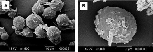
Scanning electron microscope images of OFX microspheres () showed rough surfaces due to presence of needle-shaped drug crystals interspersed within the polymer surface. This could have resulted from the extraction of DCM from the internal phase to the liquid paraffin when the polymer–drug solution was introduced into the external oily phase.Citation35 DCM migration draws the drug towards the surface of the half-formed microspheres. Therefore, the drug was deposited close to the surface of the microspheres during solidification.Citation36 Microspheres prepared using the solvent system ACN–MeOH–DCM in a volume ratio of 6:4:1 are spherical in shape with well-defined boundaries as shown in . The particle size of the prepared microspheres was 8.5 μm with 100 % entrapment efficiency.
Figure 1 SEM images of OFX microspheres formulation at different magnifications.
Abbreviations: OFX, ofloxacin; SEM, scanning electron microscope.
Characterization of OFX Gelrite in situ gel
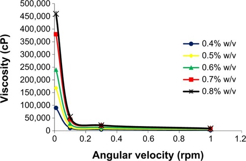
shows flow behavior of the prepared in situ gel formulations. All formulations exhibited an increase in viscosity after addition of STF even at the lowest Gelrite concentration of 0.1% w/v, but with different gelling factors. The gelling factor was increased by the increase in the Gelrite concentration. Gelling factor values were two to three for formulations prepared at concentrations up to 0.3% w/v, after addition of STF. However, a significant increase in gelling factor resulted when the Gelrite concentration was increased above 0.3%. The gelling factor increased from three to 27 when the Gelrite concentration was increased from 0.3% to 0.4% w/v. At 0.4%–0.7% w/v Gelrite concentration, the formulations were viscous liquid transformed into a pourable gel immediately after the addition of STF with a gelling factor of 27–35. Using Gelrite in concentrations greater than 0.7% w/v, the formulations were gel before addition of STF. The gelling capacity is determined in order to identify Gelrite concentrations that would have an optimum viscosity to facilitate instillation into the eye as a liquid, and then undergo rapid gelation.Citation28 The rapid gelation in the eye improves drug residence time.
Table 1 Gelling capacity and viscosity of OFX in situ gel formulations
The gelation of Gelrite after addition of STF is attributed to the cross-linking of Gelrite helices with STF solution cations. Similarly, gelation of Gelrite happened when it was administered to ocular mucosa due to contact with tear fluid cations, resulting in the formation of a clear gel.Citation14,Citation37,Citation38 The sharp increase in gelling factor with the increase in Gelrite concentration from 0.3% to 0.4 % could be attributed to the significant increase in Gelrite cross-linking with STF cations and the interaction between polymer helices that leads to the formation of a denser structure.
Gelrite-based formulations exhibited pseudoplastic flow. The prepared gels, after addition of STF, showed thixotropic behavior where the gel was thinned when shearing force was increased, and then thickened when the stress was removed. For example, the viscosity of 0.4% Gelrite was decreased from 90,000 cP to 10,622 cP and finally to 5,000 cP when the shear rate increased from 0.01 rpm to 0.3 rpm then to 1.0 rpm, respectively (). Similar results were reported previously.Citation27–Citation30 Accordingly, the increased viscosity of the formulation at low shear rates will maintain long contact between the corneal surface and the gel. During blinking, when shear rate is increased, the gel will be thinned and will be well distributed over the eye surface.Citation39
Figure 2 Effect of different Gelrite concentrations on rheology behavior of Gelrite-based in situ gels.
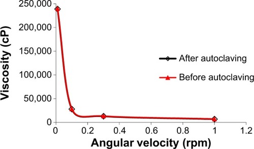
The effect of autoclaving on the properties of polymer solutions is considered an important parameter for ocular preparations. shows the viscosity profiles of in situ gel before and after autoclaving. The results showed no significant change of Gelrite-based formulation properties before and after autoclaving. This finding is in accordance with previously published reports.Citation29,Citation31 Gelrite is able to better withstand the elevated temperatures of autoclaving compared to other polymers.Citation31 So, autoclaving was used to sterilize Gelrite-based formulations in order to avoid microbial contamination.
Figure 3 Effect of autoclaving on rheology of Gelrite-based in situ gel formula.
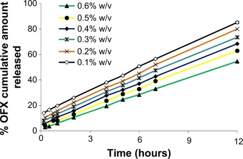
shows the cumulative percent of OFX released as a function of time for various Gelrite formulations. All the investigated formulations showed a linear release profile for the first 12 hours of the study. The in situ gel formula with Gelrite concentration of 0.1% w/v released more than 80% of the drug after 12 hours of the experiment. The release of OFX, after 12 hours, from Gelrite in situ gel formulation was decreased with the increase in Gelrite concentration; the Gelrite in situ gel formula with 0.6% w/v Gelrite showed about 54% cumulative OFX release after 12 hours (). The decrease in the release of OFX from in situ gel formulations is attributed to the increase of the polymer concentration, as by increasing the polymer concentration, a denser polymeric chain structure is produced. Thus, the diffusion of OFX through the denser formulations is reduced. In addition, the entrance of water into these formulations was reduced; thus, the rates of both dissolution and erosion were reduced.
Figure 4 Effect of different Gelrite concentrations on the release of OFX from in situ gels.
Abbreviation: OFX, ofloxacin.
In vitro release study of OFX microspheres loaded in situ gels
Hydrogel is used to incorporate microspheres in order to prolong the precorneal residence time of OFX microparticulate formula, and hence improve the bioavailability of the ocular drug.Citation40 It is worth reporting that inclusion of microspheres in in situ gels did not change the viscosity, gelling capacity, or rheogram of the prepared Gelrite-based in situ gel formulations (data not shown). Incorporation of OFX-loaded microspheres in Gelrite solution (0.4% w/v) significantly altered the release profile of OFX-loaded microspheres in situ gel formula compared with the corresponding OFX gels and OFX microspheres ().
Figure 5 Release profile of OFX from microspheres, Gelrite in situ gel, and microsphere-loaded Gelrite in situ gel.

The prepared OFX microspheres showed a 40% burst release after 1 hour that was reduced by the incorporation of the microspheres in 0.4% w/v Gelrite to 8.4%. On the other hand, microspheres released 80% of OFX content within the first 12 hours (). While OFX-loaded microspheres in situ gel formula released 54.4% of OFX content after 24 hours. These results showed that incorporation of microspheres in the in situ gel successfully prevented the burst release and resulted in an improved sustained-release OFX profile. A disadvantage of the use of microsphere suspensions in the eye might be drainage of the particles, leading to an incomplete drug release. The major advantage of using in situ gel formulations loaded with OFX microspheres is the improved precorneal residence time compared with the corresponding OFX microspheres.Citation35
In vivo study
Aqueous humor drug concentrations following various times of instillation of 0.3 mg of OFX as microspheres, Gelrite-loaded microspheres in situ gel, and the commercially available eye drops are shown in . Compared with the other investigated formulations, OFX microspheres showed the highest Cmax and shortest Tmax values (, inset table). This indicates the highest rate of absorption. Commercial OFX eye drops showed the lowest Cmax, which could possibly be attributed to the rapid drainage, and hence reduced residence time, of the applied OFX dose from the commercial eye drops. OFX microspheres in Gelrite in situ gel formula showed improved Cmax data compared with the commercial product and delayed action compared to all investigated formulations. This result indicates a prolonged pattern of release and absorption from OFX microspheres in Gelrite in situ gel formula.
Figure 6 Concentration time profiles in aqueous humor of rabbits, following topical administration of different OFX formulations.
Abbreviations: AUC, area under the curve maximum; AUCrel, the relative area under the curve compared with commercial product; Cmax, maximum OFX concentration in aqueous humor; OFX, ofloxacin; SD, standard deviation; Tmax, the time to reach this concentration.
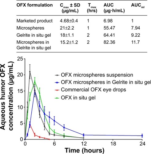
Regarding the area under the curve value for the first 24 hours after OFX administration, microspheres in Gelrite in situ gel showed the highest value of area under the curve, with relative bioavailability (AUCrel) of 11.7 compared to commercial OFX product (, inset table). This result indicates the greatest extent of drug absorption as a result of the improved residence time of OFX microspheres in Gelrite in situ gel formula.
The reported MIC90 of test organisms of OFX against 190 gram-negative and 229 gram-positive organisms from ocular sources is 2 μg/mL.Citation5 shows that the commercial OFX eye drops only achieve a concentration equal to or higher than the MIC90 for the first hour after instillation. However, OFX microspheres sustained this level for approximately 4 hours. On the other hand, OFX microspheres in the in situ gel formula sustained concentrations higher than the MIC90 for approximately 8 hours (). These results indicate that formulation of OFX as microspheres in the in situ gel of Gelrite successfully achieve the sought objectives of the present study. The formulation effectively maintains the drug concentration in the aqueous humor of rabbits above MIC90 for up to 8 hours. This will eliminate the need for frequent instillation of the drug and enhance patient compliance.
Conclusion
In this study, in situ gel formula loaded with OFX microspheres showed a prolonged pattern of release and absorption compared with OFX microspheres and with OFX in situ gel formulations. OFX microspheres were prepared by O/O emulsion solvent evaporation method utilizing PLGA as a biodegradable polymer. The prepared microspheres were added to Gelrite in situ gelling system. The improved release pattern and bioavailability of the prepared OFX-loaded microspheres in situ gel formula is attributed to improved precorneal residence time. These results support the potential use of in situ gel formulation loaded with OFX microspheres as a novel ocular drug delivery system.
Disclosure
The authors report no conflicts of interest in this work.
References
- NagayamaANakaoTTaenHIn vitro activities of ofloxacin and four other new quinoline-carboxylic acids against Chlamydia trachomatisAntimicrob Agents Chemother198832173517373150916
- ChanKPChuKOLaiWWDetermination of ofloxacin and moxifloxacin and their penetration in human aqueous and vitreous humor by using high-performance liquid chromatography fluorescence detectionAnal Biochem2006353303616620758
- GwonATopical ofloxacin compared with gentamicin in the treatment of external ocular infection. Ofloxacin Study GroupBrit J Ophthalmol1992767147181486071
- BeckRvan KeyserlingkJFischerUGuthoffRDrewelowBPenetration of ciprofloxacin, norfloxacin and ofloxacin into the aqueous humor using different topical application modesGraefes Arch Clin Exp Ophthalmol199923789929987622
- RichmanJZolezioHTang-LiuDComparison of ofloxacin, gentamicin, and tobramycin concentrations in tears and in vitro MICs for 90% of test organismsAntimicrob Agents Chemother199034160216042221871
- SinnaeveBADecaesteckerTNClaerhoutIJKestelynPRemonJPVan BocxlaerJFConfirmation of ofloxacin precipitation in corneal deposits by microbore liquid chromatography-quadrupole time-of-flight tandem mass spectrometryJ Chromatogr B Analyt Technol Biomed Life Sci2003785193196
- JärvinenKJärvinenTUrttiAOcular absorption following topical deliveryAdv Drug Deliv Rev199516319
- ZhangWPrausnitzMREdwardsAModel of transient drug diffusion across corneaJ Control Release20049924125815380634
- DaviesNMBiopharmaceutical considerations in topical ocular drug deliveryClin Exp Pharmacol Physiol20002755856210874518
- LinHRSungKCCarbopol/pluronic phase change solutions for ophthalmic drug deliveryJ Control Release20006937938811102678
- SouzaJGDiasKPereiraTABernardiDSLopezRFTopical delivery of ocular therapeutics: carrier systems and physical methodsJ Pharm Pharmacol20146650753024635555
- LudwigAThe use of mucoadhesive polymers in ocular drug deliveryAdv Drug Deliv Rev2005571595163916198021
- HornofMWeyenbergWLudwigABernkop-SchnürchAMucoadhesive ocular insert based on thiolated poly(acrylic acid): development and in vivo evaluation in humansJ Control Release20038941942812737844
- NanjawadeBKManviFVManjappaASIn situ-forming hydrogels for sustained ophthalmic drug deliveryJ Control Release200712211913417719120
- ZimmerAKreuterJMicrospheres and nanoparticles used in ocular delivery systemsAdv Drug Deliv Rev1995166173
- Radomska-SoukharevAWojciechowskaJMicroemulsions as potential ocular drug delivery systems: phase diagrams and physical properties depending on ingredientsActa Pol Pharm20056246547116583987
- CarvalhoELDe la FuenteMSeijo ReyBLiposomes as ocular drug delivery systemsArch Soc Esp Oftalmol200479151152 Spannish15124069
- AlmeidaHAmaralMHLobãoPLoboJMIn situ gelling systems: a strategy to improve the bioavailability of ophthalmic pharmaceutical formulationsDrug Discov Today20141940041224120893
- JärvinenTJärvinenKProdrugs for improved ocular drug deliveryAdv Drug Deliv Rev199619203224
- SultanaYAqilMAliAZafarSEvaluation of carbopol-methyl cellulose based sustained-release ocular delivery system for pefloxacin mesylate using rabbit eye modelPharm Dev Technol20061131331916895842
- SvensonSDendrimers as versatile platform in drug delivery applicationsEur J Pharm Biopharm20097144546218976707
- SultanaYAqilMAliASamadAAdvances in the topical ocular drug deliveryExpert Rev Ophthalmol20072309323
- Bin ChoyYParkJHPrausnitzMRMucoadhesive Microparticles Engineered for Ophthalmic Drug DeliveryJ Phys Chem Solids2008691533153620657721
- PatilSBSawantKKMucoadhesive microspheres: a promising tool in drug deliveryCurr Drug Deliv2008531231818855602
- LiuZLiJNieSLiuHDingPPanWStudy of an alginate/HPMC-based in situ gelling ophthalmic delivery system for gatifloxacinInt J Pharm2006315121716616442
- RozierAMazuelCGroveJPlazonnetBGelrite®: A novel, ion-activated, in-situ gelling polymer for ophthalmic vehicles. Effect on bioavailability of timololInt J Pharm198957163168
- BalasubramaniamJPanditJKIon-activated in situ gelling systems for sustained ophthalmic delivery of ciprofloxacin hydrochlorideDrug Deliv20031018519112944139
- BalasubramaniamJKantSPanditJKIn vitro and in vivo evaluation of the Gelrite gellan gum-based ocular delivery system for IndomethacinActa Pharm20035325126114769232
- SultanaYAqilMAliAIon-activated, Gelrite®-based in situ ophthalmic gels of pefloxacin mesylate: comparison with conventional eye dropsDrug Deliv20061321521916556574
- El-KamelAAl-DosariHAl-JenoobiFEnvironmentally responsive ophthalmic gel formulation of carteolol hydrochlorideDrug Deliv200613555916401594
- KalamMASultanaYSamadAGelrite-Based In Vitro Gelation Ophthalmic Drug Delivery System of GatifloxacinJ Dispers Sci Technol2008298996
- BasciNEHanioglu-KargiSSoysalHBozkurtAKayaalpSODetermination of ofloxacin in human aqueous humour by high-performance liquid chromatography with fluorescence detectionJ Pharm Biomed Anal1997156636669127278
- KimBKHwangSJParkJBParkHJPreparation and characterization of drug-loaded polymethacrylate microspheres by an emulsion solvent evaporation methodJ Microencapsul20021981182212569029
- GaviniEChetoniPCossuMAlvarezMGSaettoneMFGiunchediPPLGA microspheres for the ocular delivery of a peptide drug, vancomycin using emulsification/spray-drying as the preparation method: in vitro/in vivo studiesEur J Pharm Biopharm20045720721215018976
- AlbertssonACCarlforsJSturessonCPreparation and characterisation of poly(adipic anhydride) microspheres for ocular drug deliveryJ Appl Polym Sci199662695705
- NafeaEHEl-MassikMAEl-KhordaguiLKMareiMKhalafallahNMAlendronate PLGA microspheres with high loading efficiency for dental applicationsJ Microencapsul20072452553817654173
- DaiLLiuXTongZCritical behavior at sol–gel transition in gellan gum aqueous solutions with KCl and CaCl2 of different concentrationsCarbohydr Polym201081207212
- OgawaEMatsuzawaHIwahashiMConformational transition of gellan gum of sodium, lithium, and potassium types in aqueous solutionsFood Hydrocoll20021619
- RupenthalIDGreenCRAlanyRGComparison of ion-activated in situ gelling systems for ocular drug delivery. Part 1: physicochemical characterisation and in vitro releaseInt J pharm2011411697721453762
- Ravi KumarMNNano and microparticles as controlled drug delivery devicesJ Pharm Pharm Sci2000323425810994037