Abstract
Purpose
The triglyceride-glucose (TyG) index is a new index of insulin resistance (IR), and its association with hyperuricemia (HUA) is unclear. The aim of this study was to investigate whether TyG is an independent risk factor for hyperuricemia (HUA) in patients with nonalcoholic fatty liver disease (NAFLD).
Patients and Methods
We retrospectively analyzed 461 patients with ultrasound-confirmed NAFLD and calculated the TyG index. Multivariate logistic regression was used to analyze the relationship between the TyG index and HUA in NAFLD patients. The correlation between the TyG index and HUA was further confirmed by a restricted cubic spline. Furthermore, the stability of the association between TyG index and HUA was examined using subgroup analysis. Receiver operating characteristic (ROC) curves were constructed to evaluate the predictive value of the TyG index on HUA. Multivariate linear regression was used to analyze the linear relationship between the TyG index and serum uric acid.
Results
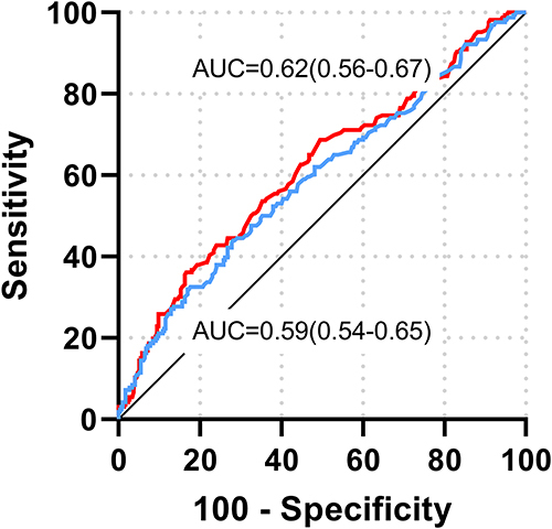
A total of 166 HUA patients and 295 non-HUA patients were included in the study. The results of multivariate logistic regression analysis showed that after controlling the confounding risk factors, TyG was still an independent risk factor for HUA (OR = 2.00, 95% CI: 1.38 −2.91, p < 0.001). Restricted cubic splines showed that HUA risk increased linearly with TyG across the entire TyG range. The ROC curve showed that TyG index was better than triglyceride in predicting HUA in NAFLD patients, with AUC values of 0.62 and 0.59, respectively. Multiple linear regression analysis showed that TyG index was significantly positively correlated with blood uric acid (B = 1.37, 95% CI: 0.67–2.08, p < 0.001).
Conclusion
TyG index is an independent risk factor for HUA in patients with NAFLD. The increase of the TyG index level is closely related to the occurrence and development of HUA in patients with NAFLD.
Introduction
In recent years, with changes in people’s lifestyles and eating habits, the prevalence of hyperuricemia (HUA) has been increasing worldwide year by year.Citation1 HUA not only causes inflammatory joint disease (gout), but also increases the risk of cardiometabolic and renal diseases, and even causes multi-target organ damage.Citation2,Citation3 It is worth noting that although the age of onset of HUA tends to be younger, most patients with early HUA have no obvious symptoms and are often ignored.Citation4 Therefore, early detection of high-risk groups with HUA and early intervention are crucial to improving the quality of life of patients and reducing the pressure on the medical system.
Non-alcoholic fatty liver disease (NAFLD) is one of the most common chronic metabolic diseases worldwide.Citation5 The disease burden caused by NAFLD comes not only from the disease itself but also from other disease risks associated with NAFLD (including HUA).Citation6 Both serum uric acid (SUA) and NAFLD are important risk factors for vascular diseases.Citation7,Citation8 The coexistence of the two will bring higher cardiovascular and cerebrovascular risks, which lead to a significant increase in cardiovascular mortality in NAFLD patients.Citation9
NAFLD is considered a hepatic manifestation of metabolic syndrome (Mets).Citation10 SUA levels have been found to be bidirectional in relation to MetS/NAFLD risk, with higher SUA levels associated with an increased risk of MetS/NAFLD.Citation11,Citation12 High levels of circulating SUA are always comorbidities with MetS and several of its components, including NAFLD.Citation13 Secondly, several clinical studies have found that SUA and uric-acid-based markers are associated with inflammatory and metabolic diseases including hypertension (HTN), such as MetS, type 2 diabetes, NAFLD, and thyroiditis.Citation14–18
Insulin resistance (IR) is one of the most important pathogeneses in NAFLD patients.Citation19 It has also been confirmed to be closely related to the onset of HUA. Studies have shown that the hyper insulinemic environment caused by IR can lead to decreased uric acid excretion and increased production, which in turn leads to uric acid accumulation.Citation20 As a novel indicator to measure the degree of IR in the body, TyG is composed of triglyceride and fasting glucose level. Substantial evidence currently suggests that triglyceride-based inflammatory markers are highly associated with hypertension, metabolic syndrome, and type 2 diabetes, all of which are characterized by a high inflammatory burden. This indicated that TyG index may reflect the inflammatory load of the body to a certain extent. TyG has been confirmed to be related to the occurrence of NAFLD and the severity of liver lesions.Citation21–23 A recent study revealed a correlation between the TyG index and HUA.Citation24 However, the relationship between TyG and HUA in fatty liver populations has not been proven. The aim of this study was to determine the correlation between TyG index and the occurrence of HUA in NAFLD patients.
Materials and Methods
Research Population
This study was a retrospective case-control study. Patients with NAFLD diagnosed by abdominal ultrasound (US) admitted to the Department of Endocrinology of the Second Hospital of Shanxi Medical University from 2020 to 2022 were selected. Finally, a total of 461 NAFLD patients were included in this study, including 271 females and 190 males. The study was approved by the Committee of the Second Hospital of Shanxi Medical University. Conforms to the Declaration of Helsinki. As this was a retrospective analysis, the requirement for informed consent was waived.
The diagnostic criteria for HUA adopt international standards, which are defined as SUA >360 μmol/L for females and SUA >420 μmol/L for males under a normal purine diet.Citation25
The diagnosis of NAFLDCitation26 was based on the criteria recommended by the Chinese Society of Liver Diseases, based on the ultrasound findings of fatty liver, and excluding other causes of chronic liver disease. At least two of the following three findings are defined as fatty liver: 1. Diffuse enhancement of liver near-field echo, which is stronger than that of the kidney; 2. Intrahepatic bile duct structure is unclear; 3. Liver far-field echo gradually attenuates.
According to the criteria of the Diabetes Society of the Chinese Medical Association,Citation27 people with three or more of the following criteria can be diagnosed with MetS: overweight or obese (BMI ≥25 kg/m2); Fasting glucose≥ 6.1mmol /L; SBP/DBP≥140/90 mmHg (1 mmHg = 0.133kPa) or taking anti-hypertensive drugs; Dyslipidemia: fasting TG ≥1.7 mmol/L and/or fasting HDL-C <0.9 mmol/L (male) or <1.0 mmol/L (female).
Clinical and Laboratory Data
Anthropometric data and anthropometric parameters were collected through electronic medical records, such as gender, age, height, weight, systolic blood pressure (SBP), diastolic blood pressure (DBP), hypertension (HTN), diabetes mellitus (DM), smoking history, and other general clinical data; Overnight fasting blood samples for analysis of biochemical variables including alanine aminotransferase (ALT), aspartate aminotransferase (AST), serum creatinine (Scr), serum uric acid (SUA), total cholesterol (TC), triglycerides (TG), high-density lipoprotein cholesterol (HDL-C), low-density lipoprotein cholesterol (LDL-C), fasting blood glucose (FBG), and other biochemical indicators. Additionally, we calculated body mass (BMI) index, TyG index and TyG-BMI index. BMI was calculated by weight (in kilograms)/height square (in meters), the TyG index was calculated by the formula Ln (triglyceride [mg/dL] × fasting blood glucose [mg/dL]/2), and the triglyceride glucose-body mass (TyG-BMI) index was calculated as BMI multiplied by TyG index.Citation23
Statistical Analysis
Statistical analysis was performed using SPSS version 26.0 software and R version 4.0.1 software. Shapiro–Wilk test was used to test the normality of the data for continuous variables, with P > 0.05 considered to be consistent with a normal distribution. The normal distribution data is represented by mean and standard deviation, while the non-normal distribution data is represented by median and quartile range. Categorical variables are described as frequencies and percentages (%). The independent sample t-test or Mann–Whitney test was used for the comparison of continuous variables, and the Chi-square test or Fisher’s exact test was used for the comparison of categorical variables. A restricted cubic spline with 3 nodes (10th, 50th, and 90th percentiles) was used to evaluate the non-linear correlation between the TyG index and HUA, and a multivariate logistic regression analysis was used to study the relationship between the TyG index and HUA. Subgroup analysis was performed according to gender, age, BMI, smoking, HTN, and diabetes to detect the stability of the correlation between the TyG index and HUA. Categorical variables between different TyG levels were analyzed using the chi-square test for trend. ROC curves were constructed to evaluate the predictive value of the TyG index on HUA. A linear relationship between the TyG index and SUA was confirmed by univariate and multiple linear regression analysis. P < 0.05 (two-sided) was considered statistically significant.
Results
Baseline Data
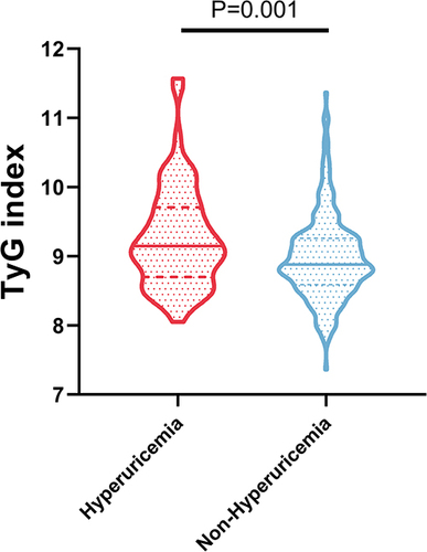
lists the clinical characteristics and laboratory parameters of 166 HUA patients and 295 non-HUA patients. Compared with non-HUA patients, HUA patients had significantly higher BMI, ALT, AST, Scr, TC, TG, and LDL-C values than non-HUA patients. However, the HUA group was younger and had lower HDL-C levels. On the other hand, the TyG level of HUA patients was significantly higher than that of non-HUA patients (P = 0.001) (). There were no significant differences in diabetes, systolic blood pressure, diastolic blood pressure, MetS and FPG between the two groups (P > 0.05).
Table 1 Demographic and Clinical Characteristics of Participants by the Presence of HUA
Multivariate Logistic Regression Analysis
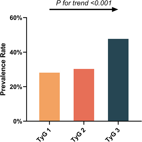
As shown in , in the unadjusted model, model 1, and model 2, the risk of HUA increased significantly (p < 0.001) with an increasing TyG index. TyG levels were divided into tertiles, and the first tertile (TyG 1) was used as the control group. Logistic regression analysis was performed on the HUA risk of TyG 2 and TyG 3, adjusting age, gender, BMI, and HTN, after diabetes, smoking, ALT, AST, and Scr, there was no significant difference in TyG 2 group (OR, 1.13; 95% CI, 0.67–1.92; P = 0.650), while TyG 3 increased the risk of HUA by 2.11 times compared with TyG 1 group (OR, 2.11; 95% CI, 1.21–3.68; P = 0.008) (). Secondly, the trend test showed that there was a trend in the prevalence of HUA among the TyG tertiles (P for trend <0.001) (). In short, the higher the TyG level, the higher the risk of HUA.
Table 2 Univariate and Multivariate Logistic Regression Assesses the Correlation Between TyG and the Prevalence of HUA
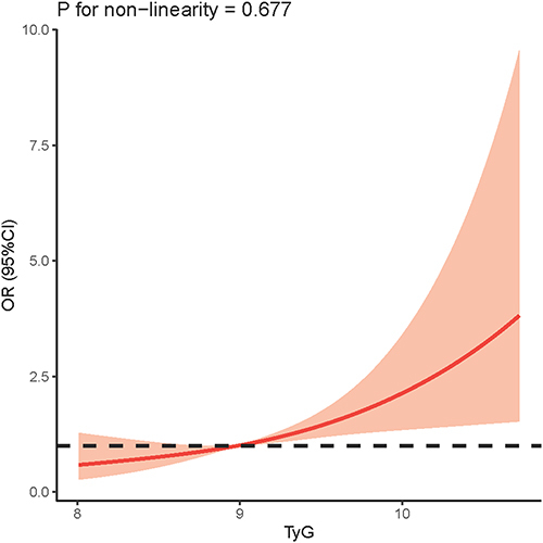
Our study further used restricted cubic splines to prove the correlation between TyG and HUA, and the results are shown in . The risk of HUA increased linearly with increasing TyG across the entire range of TyG. Furthermore, the log-likelihood test demonstrated significant linearity (P nonlinear = 0.677).
Subgroup Analysis of the Correlation Between the TyG Index and HUA
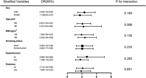
We performed several stratified analyzes to further assess the robustness of the relationship between the TyG index and HUA. As shown in , after grouping according to sex, age, BMI, smoking, HTN, and diabetes, TyG was associated with a high risk of HUA among all subgroups, and no interaction was found in all subgroup analyses (P > 0.05).
ROC Analysis of the TyG Index
We performed ROC analysis between TyG and HUA occurrence to evaluate the usefulness of TyG in predicting HUA in NAFLD. The AUC of TyG and TG were 0.62 and 0.59 (95% CI: 0.56 ~ 0.67, P < 0.001), respectively (95% CI: 0.54 ~ 0.65, P = 0.001). This indicated that TyG had sufficient accuracy for HUA, but TG had poor accuracy for HUA. The critical value of the TyG index for HUA was 8.894 (sensitivity, 68.7%; specificity, 51.2%) ().
Univariate and Multiple Linear Regression of SUA Levels
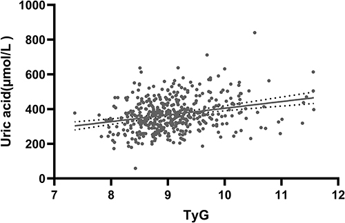
As shown in , we performed linear regression analysis on TyG and SUA levels, and the results showed that SUA levels were significantly positively correlated with the TyG index (p < 0.05). For every 0.1 unit increase in TyG, SUA levels increased by 1.53 units (β=1.53; 95% CI, 0.67–2.08; P < 0.001). The scatter plot of TyG and SUA levels also showed a positive correlation ().
Table 3 Univariate and Multivariate Linear Regression of Uric Acid
Discussion
HUA is a disorder of purine metabolism, which is associated with excessive secretion or decreased clearance of uric acid in the body.Citation28 The burden of HUA increases with socio-economic development, especially in high-income and economically developing countries that adopt Western lifestyles.Citation29 Although HUA is not used as one of the diagnostic criteria for MetS, high levels of SUA can induce oxidative stress in vascular endothelial cells, produce lipid peroxidation, activate inflammatory pathways, stimulate the expression of C-reactive protein, interleukin, tumor necrosis factor α and other inflammatory transmitters, lead to long-term chronic inflammation, aggravate lipid metabolism disorder and IR.Citation30 In addition, Chen et alCitation31 found bidirectional regulating relationship between SUA and MetS through CHARLS longitudinal study. Therefore, most scholars believe that high uric acid is a precipitating factor for Mets.
Meanwhile, HUA has been associated with adverse outcomes in Mets, namely cardiometabolic and renal disease. Uric acid has been proven to be an independent risk factor for high-risk cardiovascular and cerebrovascular diseases. A meta-analysisCitation32 confirmed that for every 1 mg/dL increase in SUA levels, the combined relative risk of coronary heart disease death was 1.13. Another meta-analysisCitation33 showed that HUA was associated with a 1.22-fold increased risk of stroke and a 1.33-fold increased risk of death.
NAFLD is a clinical syndrome with lesions concentrated in the hepatic lobules and no history of excessive alcohol consumption, characterized by hepatic steatosis and fat accumulation.Citation34 MetS is a group of clinical syndromes characterized by the co-occurrence of multiple metabolic diseases. Its pathogenesis is complex, involving not only IR, chronic inflammation and oxidative stress imbalance, but also genetic and environmental factors.Citation35 It is worth noting that the occurrence and development of MetS in individuals are often the result of the combined action of multiple pathogenic mechanisms.
Epidemiological findingsCitation36 show that about 70% to 90% of MetS patients suffer from NAFLD, which is a type of metabolic stress liver injury closely related to IR and genetic susceptibility. Metabolic risk factors of MetS can amplify the effect of genetic polymorphic mutations leading to NAFLD, and MetS is an independent predictor of progression from NAFLD to liver fibrosis.Citation37 Traditionally, NAFLD is the hepatic manifestation of MetS. However, a series of longitudinal studies have reportedCitation38 that NAFLD may be a precursor to MetS, suggesting that NAFLD is a risk factor for MetS and not just its liver presentation. Another studyCitation39 from China also proved that NAFLD and MetS are mutually causal.
IR and oxidative stress play a very important role in the occurrence and development of NAFLD.Citation40 The burden of disease caused by NAFLD comes not only from the disease itself, but also from other disease risks associated with NAFLD. Studies have shownCitation41–43 that chronic kidney disease, liver cancer, and cardiovascular disease may be associated with NAFLD. Among the many diseases associated with NAFLD, cardiovascular disease is currently considered to be the leading cause of death in patients with NAFLD. SUA is also an important risk factor for cardiovascular disease. The coexistence of both causes higher cardiovascular and cerebrovascular risks. Kim et alCitation44 found that SUA was significantly associated with a higher risk of CVD in patients with non-alcoholic fatty liver disease.
Current studies have shown that NAFLD may have a causal effect on cardiovascular disease through IR or systemic inflammation, and the progression of NAFLD may increase the risk of cardiovascular disease in patients with HUA.Citation45 On the other hand, IR is also a common driver of NAFLD and HUA. Although NAFLD is highly associated with hepatic IR, NAFLD is highly associated with systemic metabolic dysfunction and can directly lead to HUA, and IR may amplify this interaction.Citation46 Meanwhile, a large number of cross-sectional and longitudinal studies have confirmed the correlation between SUA level and NAFLD. A 7-year prospective studyCitation47 showed that NAFLD significantly increased the risk of subsequent HUA in 5541 people without baseline HUA. Therefore, elevated uric acid may be one of the important therapeutic risk factors for NAFLD disease, and lowering SUA may be a promising potential therapy for NAFLD patients.
On the other hand, IR has been found to be central to the pathogenesis of many metabolic diseases, and SUA levels are also closely associated with IR.Citation48 Many clinical articles and basic studies have revealed this correlation. In 2021, a bidirectional Mendelian randomized studyCitation49 found a causal relationship between elevated fasting insulin (a measure of IR and a precursor of cardiometabolic disease) and clinical endpoints for HUA and gout. In addition, prospective clinical studiesCitation50 have found that homeostasis model of insulin resistance (HOMA-IR) can independently predict HUA, suggesting that IR itself or compensatory hyperinsulinemia may contribute to HUA. It is currently believed that IR may lead to increased SUA levels through the following mechanisms: First, IR can transfer glycolysis intermediates to 5-phosphoribose and phosphoribose pyrophosphate, which will further lead to increased SUA production.Citation51 Second, high insulin levels due to IR led to a lower renal glucose threshold, which stimulates Na-H exchange in renal tubules and increases uric acid reabsorptionCitation52 In addition, xanthine oxidoreductase is an enzyme closely related to the metabolism of purine synthesized by the liver. IR-activated inflammatory cascade can lead to liver dysfunction, thus increasing the activity of xanthine oxidoreductase and catalyzing the increase of uric acid synthesis.Citation53 Moreover, hyperglycemia and hyperlipidemia associated with IR reduce glyceraldehyde-3-phosphate dehydrogenase activity, which also leads to increased uric acid synthesis.Citation54 Therefore, systemic IR associated with NAFLD may be an important pathophysiological mechanism of elevated uric acid, which further aggravates IR and inflammatory disorders, leading to the progression of NAFLD.
The TyG index is a novel, easy-to-use clinical measure that is considered a simple and reliable alternative clinical marker of IR.Citation55 Previously, a large number of literaturesCitation55–57 reported that TyG index was associated with a variety of diseases, including coronary atherosclerotic heart disease, cerebrovascular disease, DM, HTN, MetS, kidney disease, etc. In recent years, the relationship between TyG index and NAFLD has also been gradually discovered.Citation58 Meanwhile, some studies also revealed the correlation between TyG index and SUA. Luo et alCitation59 found that TyG index was positively correlated with SUA in non-obese T2DM patients, and TyG might be better than HOMA-IR in predicting HUA in non-obese T2DM patients. In a studyCitation60 of physical examination population in Xinjiang, China, it was found that TyG index was significantly correlated with HUA, which was superior to obesity index in identifying HUA. NAFLD is a disease highly associated with IR, and TyG, as an indirect marker of IR, may be associated with uric acid level in patients with NAFLD. Our findings further revealed the relationship between TyG index and the occurrence of HUA in patients with NAFLD.
This study revealed the correlation between TyG and HUA risk in NAFLD patients, further expanding the application range of TyG. These findings suggest that IR caused by NAFLD is not limited to the liver but is systemic, leading to a higher risk of other metabolic diseases. Secondly, NAFLD is the most common chronic liver disease, and the presence of HUA undoubtedly greatly increases the cardiovascular risk. Therefore, early identification and intervention of HUA high-risk groups is very necessary. TyG index is different from previous evaluation criteria. Because of its simple, cheap and reliable characteristics, it can be widely used in primary hospitals and communities. It can be used as a supplement to classic risk. In addition, although HUA is an indicator of morbidity and mortality in MetS, elevated uric acid involves multiple mechanisms and is also associated with MetS/NAFLD, and SUA level is greatly influenced by dietary habits. Therefore, the prediction of long-term prognosis in patients with metabolic disease NAFLD with a single uric acid level is limited. TyG has been found to be highly associated with the risk of cardiovascular and other complications in MetS/NAFLD patients. Therefore, compared with uric acid, TyG will play a more comprehensive and stable role in predicting long-term prognosis in this population and may be a potential long-term prognostic indicator. In addition, higher TyG levels indicate more severe IR, so the use of insulin sensitizers may reduce the risk of HUA and cardiovascular disease in NAFLD patients. This could lead to new therapeutic strategies for lowering uric acid in Mets patients; however, these hypotheses still need to be confirmed in ground-breaking studies with large samples.
This study also has its limitations. First, this study was a cross-sectional study with a small sample size; a single center and small sample size may have introduced bias; second, although our model was adjusted for many covariates, there were no data on dietary habits and physical activity, which factors known to affect uric acid levels. Third, SUA is derived from a single blood sample and only reflects the individual’s uric acid level at a certain point in time. Fourth, because this is a cross-sectional study, the results can show that the TyG index is positively associated with HUA in NAFLD patients, but cannot claim predictive value. Future large-scale multicenter prospective studies are needed to verify the predictive power of the TyG index on the risk of HUA in NAFLD patients.
Conclusion
The results of this study revealed a significant independent association between the TyG index and the risk of HUA in NAFLD patients. As a simple and easy-to-obtain parameter, TyG can distinguish high-risk groups of NAFLD and help reduce the occurrence of HUA and related diseases.
Abbreviations
ALT, Alanine aminotransferase; AST, Aspartate aminotransferase; SBP, Systolic blood pressure; DBP, Diastolic blood pressure; DM, Diabetes mellitus; FBG, fasting blood glucose; HDL-C, High-density lipoprotein cholesterol; HUA, hyperuricemia; HTN, Hypertension; IR, Insulin resistance; LDL-C, Low-density lipoprotein cholesterol; NAFLD, Non-alcoholic fatty liver disease; Scr, Serum creatinine; SUA, Serum uric acid; TC, Total cholesterol; TG, Triglyceride; TyG, Triglycerides-glucose index.
Data Sharing Statement
The data are available from the corresponding author on request.
Ethics Approval and Consent to Participants
The study was carried out in accordance with the principles of the Declaration of Helsinki. The study was approved by the Committee of the Second Hospital of Shanxi Medical University (Approval No. (2023) YX No. (145)). Patient informed consent was waived because all medical data had been retrospectively reviewed and analyzed anonymously.
Author Contributions
All authors have made significant contributions to this study, whether in data collection, study design, execution, or in data analysis and interpretation. They took part in drafting, revising, or reviewing the article; gave final approval of the version to be published. All authors have agreed on the journal to which the article will be submitted; and agree to be accountable for all aspects of the work.
Disclosure
The authors declare that they have no competing interests.
Additional information
Funding
References
- Chen‐Xu M, Yokose C, Rai SK, Pillinger MH, Choi HK. Contemporary prevalence of gout and hyperuricemia in the United States and decadal trends: the national health and nutrition examination survey, 2007–2016. Arthritis Rheumatol. 2019;71(6):991–999. doi:10.1002/art.40807
- Borghi C, Rosei EA, Bardin T, et al. Serum uric acid and the risk of cardiovascular and renal disease. J Hypertens. 2015;33(9):1729–1741. doi:10.1097/HJH.0000000000000701
- Targher G, Byrne CD, Nafld TH. and increased risk of cardiovascular disease: clinical associations, pathophysiological mechanisms and pharmacological implications. Gut. 2020;69(9):1691–1705. doi:10.1136/gutjnl-2020-320622
- She D, Wang Y, Liu J, et al. Changes in the prevalence of hyperuricemia in clients of health examination in Eastern China, 2009 to 2019. BMC Endocr Disord. 2022;22(1):202. doi:10.1186/s12902-022-01118-z
- Younossi ZM. Non-alcoholic fatty liver disease – a global public health perspective. J Hepatol. 2019;70(3):531–544. doi:10.1016/j.jhep.2018.10.033
- Byrne CD, Targher G. NAFLD: a multisystem disease. J Hepatol. 2015;62(1):S47–S64. doi:10.1016/j.jhep.2014.12.012
- Doehner W, Landmesser U. Xanthine oxidase and uric acid in cardiovascular disease: clinical impact and therapeutic options. Semin Nephrol. 2011;31(5):433–440. doi:10.1016/j.semnephrol.2011.08.007
- Stahl EP, Dhindsa DS, Lee SK, Sandesara PB, Chalasani NP, Sperling LS. Nonalcoholic fatty liver disease and the heart. J Am Coll Cardiol. 2019;73(8):948–963. doi:10.1016/j.jacc.2018.11.050
- Katsiki N G, Athyros V, Karagiannis A P, Mikhailidis D. Hyperuricaemia and Non-Alcoholic Fatty Liver Disease (NAFLD): a relationship with implications for vascular risk? CVP. 2011;9(6):698–705. doi:10.2174/157016111797484152
- Yki-Järvinen H. Non-alcoholic fatty liver disease as a cause and a consequence of metabolic syndrome. Lancet Diabetes Endocrinol. 2014;2(11):901–910. doi:10.1016/S2213-8587(14)70032-4
- Li C, Hsieh MC, Chang SJ. Metabolic syndrome, diabetes, and hyperuricemia. Curr Opin Rheumatol. 2013;25(2):210–216. doi:10.1097/BOR.0b013e32835d951e
- Li S, Fu Y, Liu Y, et al. Serum uric acid levels and nonalcoholic fatty liver disease: a 2-sample bidirectional Mendelian randomization study. J Clin Endocrinol Metab. 2022;107(8):e3497–e3503. doi:10.1210/clinem/dgac190
- Liu Z, Que S, Zhou L, Zheng S. Dose-response relationship of serum uric acid with metabolic syndrome and non-alcoholic fatty liver disease incidence: a meta-analysis of prospective studies. Sci Rep. 2015;5(1):14325. doi:10.1038/srep14325
- Aktas G, Khalid A, Kurtkulagi O, et al. Poorly controlled hypertension is associated with elevated serum uric acid to HDL-cholesterol ratio: a cross-sectional cohort study. Postgrad Med. 2022;134(3):297–302. doi:10.1080/00325481.2022.2039007
- Kocak MZ, Aktas G, Erkus E, Sincer I, Atak B, Duman T. Serum uric acid to HDL-cholesterol ratio is a strong predictor of metabolic syndrome in type 2 diabetes mellitus. Rev Assoc Med Bras. 2019;65(1):9–15. doi:10.1590/1806-9282.65.1.9
- Aktas G, Kocak MZ, Bilgin S, Atak BM, Duman TT, Kurtkulagi O. Uric acid to HDL cholesterol ratio is a strong predictor of diabetic control in men with type 2 diabetes mellitus. Aging Male. 2020;23(5):1098–1102. doi:10.1080/13685538.2019.1678126
- Kosekli MA, Kurtkulagii O, Kahveci G, et al. The association between serum uric acid to high density lipoprotein-cholesterol ratio and non-alcoholic fatty liver disease: the abund study. Rev Assoc Med Bras. 2021;67(4):549–554. doi:10.1590/1806-9282.20201005
- Kurtkulagi O, Tel BMA, Kahveci G, et al. Hashimoto’s thyroiditis is associated with elevated serum uric acid to high density lipoprotein-cholesterol ratio. Rom J Intern Med. 2021;59(4):403–408. doi:10.2478/rjim-2021-0023
- Buzzetti E, Pinzani M, Tsochatzis EA. The multiple-hit pathogenesis of non-alcoholic fatty liver disease (NAFLD). Metabolism. 2016;65(8):1038–1048. doi:10.1016/j.metabol.2015.12.012
- Yanai H, Adachi H, Hakoshima M, Katsuyama H. Molecular biological and clinical understanding of the pathophysiology and treatments of hyperuricemia and its association with metabolic syndrome, cardiovascular diseases and chronic kidney disease. IJMS. 2021;22(17):9221. doi:10.3390/ijms22179221
- Wang J, Yan S, Cui Y, Chen F, Piao M, Cui W. The diagnostic and prognostic value of the triglyceride-glucose index in metabolic dysfunction-associated fatty liver disease (MAFLD): a systematic review and meta-analysis. Nutrients. 2022;14(23):4969. doi:10.3390/nu14234969
- Zheng R, Du Z, Wang M, Mao Y, Mao W. A longitudinal epidemiological study on the triglyceride and glucose index and the incident nonalcoholic fatty liver disease. Lipids Health Dis. 2018;17(1):262. doi:10.1186/s12944-018-0913-3
- Guo W, Lu J, Qin P, et al. The triglyceride-glucose index is associated with the severity of hepatic steatosis and the presence of liver fibrosis in non-alcoholic fatty liver disease: a cross-sectional study in Chinese adults. Lipids Health Dis. 2020;19(1):218. doi:10.1186/s12944-020-01393-6
- Liu XZ, Xu X, Zhu JQ, Zhao DB. Association between three non-insulin-based indexes of insulin resistance and hyperuricemia. Clin Rheumatol. 2019;38(11):3227–3233. doi:10.1007/s10067-019-04671-6
- Wei CY, Sun CC, Wei JCC, et al. Association between hyperuricemia and metabolic syndrome: an epidemiological study of a labor force population in Taiwan. Biomed Res Int. 2015;2015:1–7.
- Jian-gao F; Chinese Liver Disease Association. Guidelines for management of nonalcoholic fatty liver disease: an updated and revised edition. Zhonghua Gan Zang Bing Za Zhi. 2010;18(3):163–166.
- Xi B, He D, Hu Y, Zhou D. Prevalence of metabolic syndrome and its influencing factors among the Chinese adults: the China Health and Nutrition Survey in 2009. Prev Med. 2013;57(6):867–871. doi:10.1016/j.ypmed.2013.09.023
- Wang H, Zhang J, Pu Y, et al. Comparison of different insulin resistance surrogates to predict hyperuricemia among U.S. non-diabetic adults. Front Endocrinol. 2022;13:1028167. doi:10.3389/fendo.2022.1028167
- Zhang S, Wang Y, Cheng J, et al. Hyperuricemia and cardiovascular disease. CPD. 2019;25(6):700–709. doi:10.2174/1381612825666190408122557
- Kimura Y, Tsukui D, Kono H. Uric acid in inflammation and the pathogenesis of atherosclerosis. IJMS. 2021;22(22):12394. doi:10.3390/ijms222212394
- Chen WY, Fu YP, Zhou M. The bidirectional relationship between metabolic syndrome and hyperuricemia in China: a longitudinal study from CHARLS. Endocrine. 2022;76(1):62–69. doi:10.1007/s12020-022-02979-z
- Li M, Hu X, Fan Y, et al. Hyperuricemia and the risk for coronary heart disease morbidity and mortality a systematic review and dose-response meta-analysis. Sci Rep. 2016;6(1):19520. doi:10.1038/srep19520
- Li M, Hou W, Zhang X, Hu L, Tang Z. Hyperuricemia and risk of stroke: a systematic review and meta-analysis of prospective studies. Atherosclerosis. 2014;232(2):265–270. doi:10.1016/j.atherosclerosis.2013.11.051
- Friedman SL, Neuschwander-Tetri BA, Rinella M, Sanyal AJ. Mechanisms of NAFLD development and therapeutic strategies. Nat Med. 2018;24(7):908–922. doi:10.1038/s41591-018-0104-9
- Fahed G, Aoun L, Bou Zerdan M, et al. Metabolic syndrome: updates on pathophysiology and management in 2021. IJMS. 2022;23(2):786. doi:10.3390/ijms23020786
- Fan JG, Zhu J, Li XJ, et al. Fatty liver and the metabolic syndrome among Shanghai adults. J Gastroenterol Hepatol. 2005;20(12):1825–1832. doi:10.1111/j.1440-1746.2005.04058.x
- Bence KK, Birnbaum MJ. Metabolic drivers of non-alcoholic fatty liver disease. Molcul Metabol. 2021;50:101143. doi:10.1016/j.molmet.2020.101143
- Yoo JJ, Cho EJ, Chung GE, et al. Nonalcoholic fatty liver disease is a precursor of new-onset metabolic syndrome in metabolically healthy young adults. JCM. 2022;11(4):935. doi:10.3390/jcm11040935
- Guo Z, Zhang T, Yun Z, et al. Assessing the causal relationships between human blood metabolites and the risk of NAFLD: a comprehensive Mendelian randomization study. Front Genet. 2023;28(14):1108086. doi:10.3389/fgene.2023.1108086
- Khan RS, Bril F, Cusi K, Newsome PN. Modulation of insulin resistance in nonalcoholic fatty liver disease. Hepatology. 2019;70(2):711–724. doi:10.1002/hep.30429
- Sun DQ, Jin Y, Wang TY, et al. MAFLD and risk of CKD. Metabolism. 2021;115:154433. doi:10.1016/j.metabol.2020.154433
- Ioannou GN. Epidemiology and risk-stratification of NAFLD-associated HCC. J Hepatol. 2021;75(6):1476–1484. doi:10.1016/j.jhep.2021.08.012
- Johnson RJ, Bakris GL, Borghi C, et al. Hyperuricemia, acute and chronic kidney disease, hypertension, and cardiovascular disease: report of a scientific workshop organized by the national kidney foundation. Am J Kidney Dis. 2018;71(6):851–865. doi:10.1053/j.ajkd.2017.12.009
- Kim K, Kang K, Sheol H, et al. The association between serum uric acid levels and 10-year cardiovascular disease risk in non-alcoholic fatty liver disease patients. IJERPH. 2022;19(3):1042. doi:10.3390/ijerph19031042
- Xu C. Hyperuricemia and nonalcoholic fatty liver disease: from bedside to bench and back. Hepatol Int. 2016;10(2):286–293. doi:10.1007/s12072-015-9682-5
- Sun DQ, Wu SJ, Liu WY, et al. Serum uric acid: a new therapeutic target for nonalcoholic fatty liver disease. Expert Opin Ther Targets. 2016;20(3):375–387. doi:10.1517/14728222.2016.1096930
- Xu C, Wan X, Xu L, et al. Xanthine oxidase in non-alcoholic fatty liver disease and hyperuricemia: one stone hits two birds. J Hepatol. 2015;62(6):1412–1419. doi:10.1016/j.jhep.2015.01.019
- Fernández‐Chirino L, Antonio‐Villa NE, Fermín‐Martínez CA, et al. Elevated serum uric acid is a facilitating mechanism for insulin resistance mediated accumulation of visceral adipose tissue. Clin Endocrinol. 2022;96(5):707–718. doi:10.1111/cen.14673
- McCormick N, O’Connor MJ, Yokose C, et al. Assessing the causal relationships between insulin resistance and hyperuricemia and gout using bidirectional Mendelian randomization. Arthritis Rheumatol. 2021;73(11):2096–2104. doi:10.1002/art.41779
- Nakamura K, Sakurai M, Miura K, et al. HOMA-IR and the risk of hyperuricemia: a prospective study in non-diabetic Japanese men. Diabetes Res Clin Pract. 2014;106(1):154–160. doi:10.1016/j.diabres.2014.07.006
- Li Q, Shao X, Zhou S, et al. Triglyceride-glucose index is significantly associated with the risk of hyperuricemia in patients with diabetic kidney disease. Sci Rep. 2022;12(1):19988. doi:10.1038/s41598-022-23478-1
- Liu H, Song X, Zhu J, et al. The elevated visceral adiposity index increases the risk of hyperuricemia in Chinese hypertensive patients: a cross-sectional study. Front Endocrinol. 2022;15(13):18971.
- Yang B, Xin M, Liang S, et al. New insight into the management of renal excretion and hyperuricemia: potential therapeutic strategies with natural bioactive compounds. Front Pharmacol. 2022;22(13):1026246. doi:10.3389/fphar.2022.1026246
- Gao Z, Zuo M, Han F, et al. Renal impairment markers in type 2 diabetes patients with different types of hyperuricemia. J Diabetes Investig. 2019;10(1):118–123. doi:10.1111/jdi.12850
- Yan F, Yan S, Wang J, et al. Association between triglyceride glucose index and risk of cerebrovascular disease: systematic review and meta-analysis. Cardiovasc Diabetol. 2022;21(1):226. doi:10.1186/s12933-022-01664-9
- Tao LC, Xu J, Wang T, Hua F, Li JJ. Triglyceride-glucose index as a marker in cardiovascular diseases: landscape and limitations. Cardiovasc Diabetol. 2022;21(1):68. doi:10.1186/s12933-022-01511-x
- Zhu Q, Chen Y, Cai X, et al. The non-linear relationship between triglyceride-glucose index and risk of chronic kidney disease in hypertensive patients with abnormal glucose metabolism: a cohort study. Front Med. 2022;20(9):1018083. doi:10.3389/fmed.2022.1018083
- Tutunchi H, Naeini F, Mobasseri M, Ostadrahimi A. Triglyceride glucose (TyG) index and the progression of liver fibrosis: a cross-sectional study. Clin Nutr ESPEN. 2021;44:483–487. doi:10.1016/j.clnesp.2021.04.025
- Luo Y, Hao J, He X, et al. Association between triglyceride-glucose index and serum uric acid levels: a biochemical study on anthropometry in non-obese type 2 diabetes mellitus patients. DMSO. 2022;15:3447–3458. doi:10.2147/DMSO.S387961
- Kahaer M, Zhang B, Chen W, et al. Triglyceride glucose index is more closely related to hyperuricemia than obesity indices in the medical checkup population in Xinjiang, China. Front Endocrinol. 2022;2(13):861760. doi:10.3389/fendo.2022.861760

