Abstract
Background
The degree of hippocampal magnetic resonance imaging (MRI) volume loss in Alzheimer’s disease (AD) is commonly accepted as a marker of disease severity, yet remains expensive, unavailable, or not tolerated by many patients.
Aim
To examine whether the presence of one or more apolipoprotein E (ApoE) e4 alleles is associated with smaller hippocampal MRI volumes in a population of early AD patients.
Methods
A total of 88 consecutive patients attending a community-based memory disorders clinic who had both mild dementia on the Clinical Dementia Rating scale and Diagnostic and Statistical Manual of Mental Disorders criteria for probable AD were recruited. We examined the relationship between ApoE e4 allele load and hippocampal atrophy on MRI volumes.
Results
There was no association between the ApoE e4 load and hippocampal volume in this cohort.
Conclusion
This study suggests that the presence of one or more ApoE e4 alleles cannot be used to estimate pathological disease load in early AD.
Introduction
There is a lack of consensus regarding the utility of the apolipoprotein E (ApoE) genotype in the clinical management and prognostication of individuals with Alzheimer’s disease (AD), where the ApoE e4 allele is reported to have multiple negative outcomes. As a cumulative and progressive neurodegenerative process, therapeutic intervention in the earliest stages of AD offers the most potential for preventing or slowing progression of the disease. The Clinical Dementia Rating (CDR) scaleCitation1 is both a useful and common tool for classification of dementia severity. It is used by many AD clinicians, but lacks the ability to predict the prognosis, rate of decline, response to drug therapy, or the onset of behavioral and psychotic symptoms. ApoE e4 has the potential to aid markedly in these clinical settings, but its optimal use has not been well defined. The degree of hippocampal magnetic resonance imaging (MRI) volume loss in AD is commonly accepted as a marker of disease severity, yet remains expensive, unavailable, or not tolerated by many patients. Examining the potential of the ApoE e4 allele as a simple, readily available substitute to MRI to infer the extent of hippocampal shrinkage has significant appeal, especially in the early stages of the disease.
ApoE is the major apolipoprotein species in the brain, and is involved in the catabolism of triglyceride-containing lipoproteins and the transport of these, fat-soluble vitamins and cholesterol. It is a 34 kDa protein mediating the binding of lipoproteins to the low-density lipoprotein receptor (mainly on neuronal membranes), and it plays a key role in the mobilization of cholesterol and phospholipids during neuronal membrane remodeling associated with synaptic plasticity and neuronal repair. Also, there is evidence that ApoE isoforms alter the transport and metabolism of Aβ amyloid in the brain.Citation2
The possibility of a link between the ApoE genotype and the cross-sectional degree of medial temporal lobe atrophy on MRI in AD has been examined in relatively small studies in the last decade, with sample sizes varying between 15 and 49,Citation3–Citation9 and the largest study having 138 AD patients.Citation10 The majority (six of eight) of these studies suggested a positive correlation between ApoE e4 and medial temporal lobe atrophy. One study suggested no difference across genotype in hippocampal volumes but significant atrophy in the adjoining amygdalae in the presence of an e4 allele.Citation11 However in this study subjects appear to have been recruited from a mixture of settings, including hospitals and residential care facilities, and so did not predominantly concentrate on community-dwelling, independent subjects with early AD, where such a genotype linkage would provide most clinical benefit for intervention. Also, inadequate definition of the varying AD severity makes interpretation of these findings difficult. ApoE e4 alleles in AD patients do however appear to be associated with increased rates of hippocampal volume loss over time.Citation12
Methods
Study population
A total of 88 consecutive patients attending a community-based memory disorders clinic who met the criteria for both Mild Dementia using the CDR scale and the Diagnostic and Statistical Manual of Mental Disorders (DSM-IV-TR) criteria for probable Alzheimer’s diseaseCitation13 agreed to take part in the study. These entry criteria were used in the clinic setting by a single, experienced geriatrician to identify eligible participants. The Mini-Mental Status Examination (MMSE)Citation14 and age at diagnosis were consistent with this early stage of dementia ().
Table 1 Clinical characteristics of study population (88 CDR 1.0 patients)
Ethics approval
The Hunter New England Human Research Ethics Committee approved this research and operates in accordance with the Australian National Health and Medical Research Council’s National Statement on Ethical Conduct in Human Research (2007) and the Committee for Medicinal Products for Human Use/International Conference on Harmonisation of Technical Requirements for Registration of Pharmaceuticals for Human Use guidance on good clinical practice. No external funding was used in this study.
ApoE genotyping
All ApoE genotyping was performed in a single laboratory using polymerase chain reaction applied to whole blood lymphocytes.
MRI hippocampal mapping
All patients were imaged with 1.5 T (Signa Excite, General Electric Medical Systems, Milwaukee, WI, USA). All volume measurements were derived from T1-weighted spoiled gradient-recalled echo sequence with 25° flip angle, 1.6 mm slice thickness, 22 × 19 cm field of view, TR 27, TE 9, and 320 × 224 matrix and 1.0 NEX.
All measurements were made by manual tracing of the coronal sections of the hippocampus and entorhinal cortices (ERC), and the volume measurements were calculated using the software provided by General Electric Medical Systems. The hippocampi were measured from the level of the mammillary body to the posterior margin of the quadrigeminal plate on the coronal images, which are considered constant markers on MRI. The ERC measurements were made on a single slice at the level of the mammillary body. A radiologist experienced in these measurements and who was blinded to the MMSE and ApoE status reported the MRI volumes.
Statistics
The statistical software package used was Stata version 11 (StataCorp, College Station, TX, USA). Assumptions of normality were checked for both age and sex distribution of the cohort. The independent variable was the ApoE e4 status (nil, 1 positive allele, 2 positive alleles) and the dependent variables were the MRI volume of the right and left hippocampi. Analysis of variance was used to test for differences in volume across the three allele groups. The measures of the ERC were excluded from the study analysis due to the lack of technically reliable measures of this structure.
Results
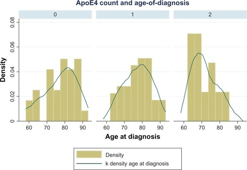
provides the basic demographics of the three genotype groups. Participants were predominantly nonsmokers and had mean MMSE scores between 22 and 24. There was a predominance of younger and female participants in the e4/e4 group. This nonnormal distribution of age of onset across the three genotype groups is consistent with published literature that e4/e4 individuals have a 5- to 10-year earlier onset of Alzheimer’s compared to the other genotype groups ().
Figure 1 Age of Alzheimer’s disease onset across apolipoprotein (ApoE) e4 status (0, 1, and 2 × e4 alleles).
One-way analysis of variance was applied to test for any difference in hippocampal volume (left then right cortex) across the three allele groups. No differences between the mean volume measurements across the individual genotype groups were demonstrated () (left hippocampal volume, P > 0.86; right hippocampal volume, P > 0.68). We also demonstrated that this lack of association between genotype and hippocampal volume remained when adjusted for age and sex using linear regression ().
Figure 2 Left and right hippocampal volumes across apolipoprotein E e4 load (0, 1, and 2 × e4 alleles).
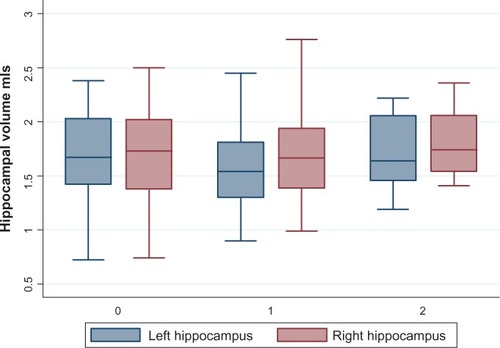
Table 2 Multiple linear regression of hippocampal volume
Discussion
The study demonstrated no association between ApoE e4 status and hippocampal volume in early AD patients living in community settings, so confirming that the presence of one or more e4 alleles does not estimate the degree of hippocampal volume loss in early AD.
There were no significant differences in the distribution of baseline cognitive scores between the groups defined by ApoE status. As was consistent with previous literature, the expected difference in the age of onset of the double-e4 group compared to the single e4 and non-e4 groups was demonstrated.
The strengths of this study are its unique community setting of early, well-defined AD patients where the clinical utility of ApoE genotype would be of most benefit. The large sample size and consecutive recruitment of these community dwellers increases the study’s ability to reflect the true community AD ApoE e4 distribution. Also, all clinical assessments and disease-severity staging were performed by a single, senior geriatrician applying standard AD grading (CDR) and diagnostic criteria (DSM IV-TR), with a single observer conducting all MRI volume measurements blinded to both ApoE genotype and clinical details.
This study’s results rely on the published literature’s consensus of the correlation between AD pathological load and medial temporal lobe atrophy measured by MRI. If this correlation was disproved in the future, further studies into the possible association of ApoE e4 allele and early AD pathology would be required.
Disclosure
The authors report no conflicts of interest in this work.
References
- MorrisJCThe Clinical Dementia Rating (CDR): current version and scoring rulesNeurology19934324122414
- KimJBasakJHoltzmanDThe role of apolipoprotein E in Alzheimer’s diseaseNeuron20096328730319679070
- BakerRGholkarAScheltensPApoE e4 allele, temporal lobe atrophy, and white matter lesions in late life dementiasArch Neurol19995696196510448801
- GeroldiCPihlajamäkiMLaaksoMAPOE-epsilon4 is associated with less frontal and more medial lobe atrophy in ADNeurology1999531825183210563634
- JackCRJrPetersenRCXuYCHippocampal atrophy and apolipoprotein E genotype are independently associated with Alzheimer’s diseaseAnn Neurol1998433033109506546
- TanakaSKawamataJShimohamaSInferior temporal lobe atrophy and ApoE genotypes in Alzheimer’s disease. X-ray computed tomography, magnetic resonance imaging and Xe-133 SPECT studiesDement Geriatr Cogn Disord1998990989524800
- JuottonenKLehtovirtaMHelisalmiSRiekkinenPJSrSoininenHMajor decrease in the volume of the entorhinal cortex in patients with Alzheimer’s disease carrying the apolioprotein E epsilon4 alleleJ Neurol Neurosurg Psychiatry1998653223279728943
- MuellerSGSchuffNRaptentsetsangSElmanJWeinerMWSelective effect of Apo e4 on CA3 and dentate in normal aging and Alzheimer’s disease using high resolution MRI at 4 TNeuroimage200842424818534867
- LehtovirtaMLaaksoMSoininenHVolumes of hippocampus, amygdala and frontal lobe in Alzheimer’s patients with different apolipoprotein E genotypesNeuroscience19956765677477910
- HashimotoMYasudaMTanimukaiSApolipoprotein E e4 and the pattern of regional brain atrophy in Alzheimer’s diseaseNeurology2001571461146611673590
- BassoMGelernterJYangJApolipoprotein E epsilon4 is associated with atrophy of the amygdala in Alzheimer’s DiseaseNeurobiol Aging2006271416142416182410
- SchuffNWoernerNBoretaLMRI of hippocampal volume loss in early Alzheimer’s Disease in relation to ApoE genotype and biomarkersBrain20091321067107719251758
- American Psychiatric AssociationDiagnostic and Statistical Manual of Mental Disorders4th edtext revisionArlington (VA)APA2000
- FolsteinMFolsteinSMcHughP“Mini-mental state.” A practical method for grading the cognitive state of patients for the clinicianJ Psychiatr Res1975121891981202204