
Abstract
Background
The deep-seated infections caused by the Candida genus are associated with a high mortality rate, and Candida albicans is the most frequent species associated with these diseases. The fungal wall is composed of macromolecules not synthesized by the host, and therefore is a source of ligands recognized by innate immune cells.
Methods
We performed a comparative study analyzing the cell wall composition and organization of Candida tropicalis, Candida guilliermondii, Candida krusei, and Candida auris, along with their ability to stimulate cytokine production and phagocytosis by human innate immune cells.
Results
We found that the wall of these species had the basic components already described in C. albicans, with most of the chitin and b1,3-glucan located underneath the mannan layer. However, the walls of C. krusei and C. auris were rich in chitin and the former had a lower content of mannans. C. guilliermondii contained changes in the mannan and the b1,3-glucan levels. These species were differentially phagocytosed by human macrophages and stimulated cytokine production in a dectin-1-dependent pathway. C. krusei showed the most significant changes in the tested parameters, whereas C. auris behaved like C. albicans.
Conclusion
Our results suggest that the cell wall and innate immune recognition of C. tropicalis, C. guilliermondii, C. krusei, and Candida auris is different from that reported for C. albicans.
Introduction
The species of the Candida genus are responsible for superficial and systemic can-didiasis, and the latter represents a significant burden to health care systems and patients because of the high rate of morbidity and mortality associated with these infections, especially in immunocompromised patients.Citation1,Citation2 Although Candida albicans is the most frequently isolated species from patients with diagnosed candidiasis, other Candida species are responsible for about 35%–65% of candidemia cases.Citation3,Citation4 Among them, Candida tropicalis, Candida parapsilosis, Candida guilliermondii, Candida glabrata, and Candida krusei top the epidemiological lists of causative agents of systemic candidiasis.Citation5,Citation6
C. tropicalis is considered as an important causative agent of invasive candidiasis and colonizes 60%–80% of immunocompromised patients.Citation6 This species has been associated with similar or higher mortality rates than those reported for C. albicans.Citation7–Citation9 C. krusei and C. guilliermondii are also part of the emergent group of Candida species related to systemic candidiasis, being the causative agents in 2%–5% of the reported cases.Citation6,Citation7 A significant difference between infections associated with C. guilliermondii and C. krusei is the mortality rate: in C. guilliermondii this is similar to that recorded for C. albicans (27%–49%),Citation1,Citation10 while in C. krusei the mortality rate is higher (20%–67%), likely related to their poor response to standard antifungal therapies.Citation6,Citation8,Citation9,Citation11 Candida auris is an emergent fungal species, firstly identified in 2009 as an etiological agent of candidiasis, and has a natural resistance to several of the antifungal drugs used to treat these infections.Citation12 Despite not being related to high numbers of cases of nosocomial candidiasis, it is distributed worldwide, and most of the clinical isolates have shown high minimal inhibitory concentrations for azoles, polyenes, and echinocandins.Citation12
The fungal immune sensing is a key step in the establishment of a protective anti-fungal immune response, and among the first events that trigger this interaction is the recognition of the fungal cell wall.Citation13,Citation14 The wall is an essential cell component that provides protection from extracellular insults, controls communication with the extracellular environment, and is the molecular scaffold that displays virulence factors in fungal pathogens.Citation15 As it is composed of polysaccharides which are absent in the human cells, the cell wall is the main source of pathogen-associated molecular patterns that the immune system recognizes through pattern recognition receptors (PRRs), mainly localized at the cell surface of immune cells.Citation13,Citation15
The C. albicans cell wall has been thoroughly studied, and nowadays, there is enough information to propose general models regarding its composition, organization, and relevance for viability, virulence, and immune sensing.Citation13,Citation15–Citation17 Despite this significant advance, little is known about the cell wall organization and composition of other species of the Candida genus, such as C. guilliermondii, C. tropicalis, C. krusei, and C. auris. A possible reason is that it is assumed that the C. albicans cell wall model should apply to all the members of this genus. In C. albicans, the cell wall is composed of two well-defined layers,Citation13,Citation16 visible under electronic microscopy inspection: an inner layer composed of chitin, β1,3- and β1,6-glucans, and an outer layer rich in proteins modified with N- or O-linked mannans, named mannoproteins.Citation18 These components are not synthesized by the host cells, which in turn are recognized by a variety of PRRs that will ultimately contribute to the establishment of a protective antifungal response by the host immunity.Citation13 The b1,3-glucans are sensed by a heterodimer composed of toll-like receptor (TLR) 2 and TLR6 and by dectin-1, the latter being a specific receptor for this polysaccharide;Citation13,Citation19 while TLR4 participates in the recognition of O-linked mannansCitation20 and mannose receptor,Citation20 dendritic cell-specific intercellular adhesion molecule-3-grabbing non-integrin,Citation21 dectin-2,Citation22 mincle,Citation23 and dectin-3Citation24 recognize the N-linked mannans. The chitin–receptor interaction can block the fungal sensing,Citation25 or promote an anti-inflammatory state in a pathway dependent on the mannose receptor, nucleotide-binding oligomerization domain-containing protein 2, and TLR9.Citation26
So far, little is known about the immune sensing of C. tropicalis, C. krusei, C. guilliermondii, and C. auris. It has been shown that neutrophils and murine phagocytic cells are capable of discriminating among Candida species, the former displaying reduced uptake against C. krusei,Citation27 and the latter an increased ability to kill C. guilliermondii and C. krusei.Citation28 In addition, C. tropicalis is more susceptible to damage by neutrophils than C. albicans.Citation29 Human monocytes differentially recognize Candida species, such as C. albicans, C. parapsilosis, C. tropicalis, and C. krusei, but not C. glabrata and C. guilliermondii, which are good stimuli for C3 and granulocyte-macrophage colony-stimulating factor.Citation30 On the contrary, there are studies indicating that different Candida species have similar abilities to interact with innate immune cells, triggering similar killing rates and phagocytosis as C. albicans.Citation31–Citation33 Although the C. auris-immune effector interplay is an area that is poorly explored, it has been demonstrated that neutrophils are not capable of forming extracellular traps when interacting with C. auris.Citation34
Here, in order to assess insights about the relevance of the cell wall in the immune sensing of C. tropicalis, C. krusei, C. guilliermondii, and C. auris by human peripheral blood mononuclear cells (PBMCs) and human monocyte-derived macrophages, we determined the basic composition of the cell wall of these fungal species and their ability to stimulate cytokine production and phagocytic uptake.
Materials and methods
Strains and culture media
C. albicans SC5314,Citation35 C. guilliermondii ATCC 6260,Citation36 C. krusei ATCC 6258, C. tropicalis ATCC MYA-3404,Citation37 and C. auris VPCI 479/P/13,Citation38 a genome-sequenced strain, were used in this study. Cell cultures were propagated at 30°C in Sabouraud broth (1% [w/v] mycological peptone, 4% [w/v] glucose). Unless otherwise indicated, aliquots containing 500 µL of overnight-grown cells were inoculated in 100 mL of fresh medium and further incubated at 30°C with constant shaking (200 rpm) until reaching the mid-log growth phase (typically 5–6 hours). For heat inactivation, yeast cells were incubated at 56°C for 1 hour.Citation39 In all cases, loss of cell viability was confirmed by incubating cells in Sabouraud medium for 72 hours at 30°C. To remove O-linked mannans, cells were β-eliminated overnight with 100 mM NaOH as described.Citation40 Following this protocol, >96% of the cells retained their viability, as tested by cell growth before and after treatment with the alkali.Citation41
Cell wall analysis
Cell homogenates were obtained in a Braun homogenizer, as previously described,Citation41 and walls were separated from cellular debris by centrifuging at 21,000× g for 10 minutes at 4°C. The cell walls were washed six times with deionized water; adsorbed intracellular components were removed with hot 2% (v/v) SDS, 0.3 M β-mercaptoethanol, and 1 M NaCl; freeze-dried; and hydrolyzed with hot 2 M trifluoroacetic acid for 3 hours, as described previously.Citation39 The content of N-acetylglucosamine, glucose, and mannose in the wall hydrolysates was measured by high-performance anion-exchange chromatography coupled to pulsed amperometric detection (HPAEC-PAD) in a Dionex system (Thermo Fisher Scientific, Waltham, MA, USA) using similar separation conditions to those described earlier.Citation41 Protein content was determined in alkali-hydrolyzed samples from the cell wall,Citation39 using the Bradford protein assay.
Alcian blue binding assays
Yeast cells grown at the exponential phase were pelleted, washed three times with deionized water, and the cell concentration adjusted at an OD600 of 0.2 in deionized water. Aliquots of 1 mL were pelleted, and cells were suspended in 1 mL of Alcian blue (30 µg·mL−1 in 0.02 M HCl; Sigma-Aldrich Co., St Louis, MO, USA) and assayed as described.Citation42
Cell wall porosity assay
The relative cell wall porosity to polycations was estimated as described previously.Citation43 Cells were grown until reaching the exponential phase, washed twice with PBS, and cell concentration was adjusted at 1×10⁸ cells mL−1. Aliquots containing 1 mL were centrifuged, the pellet was saved and resuspended in either 10 mM Tris-HCl, pH 7.4 (buffer A), buffer A plus 30 µg·mL−1 poly-L-lysine (Mw 30–70 kDa, Sigma-Aldrich Co.), or buffer A plus 30 µg·mL−1 diethylaminoethyl (DEAE)-dextran (Mw 500 kDa, Sigma-Aldrich Co.). Cells were incubated for 30 minutes at 30°C with constant shaking (200 rpm), centrifuged, and the supernatants were saved, centrifuged again, and used to measure the absorbance at 260 nm. The relative cell wall porosity to DEAE-dextran was quantified as reported elsewhere.Citation43
Analysis of the exposure of the cell wall polysaccharide at the fungal wall surface
Chitin was labeled by incubating cells with 1 mg·mL−1 fluorescein-5-isothiocyanate conjugated lectin from Triticum vulgaris (WGA-FITC; Sigma-Aldrich Co.) for 60 minutes at room temperature, as reported previously,Citation25 while b1,3-glucan was labeled with 5 µg·mL−1 IgG Fc-Dectin-1 chimeraCitation44 for 40 minutes at room temperature. The binding of the lectin to the fungal wall was revealed by incubating cells with 1 µg·mL−1 donkey anti-Fc IgG-FITC (Sigma-Aldrich Co.) for 40 minutes at room temperature.Citation45 Samples were examined by fluorescence microscopy using a Zeiss Axioscope-40 microscope and an Axiocam MRc camera. From the pictures acquired, the fluorescence associated to 300 cells was collected using the software Adobe Photoshop™ CS6, Adobe Systems Incorporated (San Jose, CA, USA) with the formula: ([total of green pixels – background green pixels] × 100)/total pixels.Citation46
Quantification of N- and O-linked mannans
Mannan trimming was performed as reported.Citation47 The N-linked mannans were removed from the cell wall by incubating yeast cells with 25 U endoglycosidase H (New England Biolabs, Ipswich, MA, USA) for 20 hours at 37°C; while removal of O-linked mannans was performed by β-elimination with 1N NaOH and gently shaking for 18 hours at room temperature. Mannan release and quantification was performed by HPAEC-PAD, as previously described.Citation48
Ethics statement
Healthy adult volunteers were enrolled in the study and venous blood samples were withdrawn after information about the study was disclosed and written informed consent was obtained. This procedure was conducted in accordance with the Declaration of Helsinki. The use of human cells was approved by the Ethics Committee of Universidad de Guanajuato (permission code 17082011).
Isolation of human PBMCs and cytokine stimulation
The venous blood was mixed with Histopaque-1077 (Sigma-Aldrich Co.) and density centrifugation was performed as described.Citation49 The PBMC–Candida interactions were carried out using 5 × 10⁵ PBMCs in 100 µL of RPMI 1640 Dutch modification (added with 2 mM glutamine, 0.1 mM pyruvate, and 0.05 mg·mL−1 gentamycin; all reagents from Sigma-Aldrich Co.) and 100 µL containing 1×10⁵ yeast cells freshly harvested or treated. The interactions were placed in round-bottom 96-well microplates and incubated for 24 hours at 37°C with 5% (v/v) CO2. Then, the plates were centrifuged for 10 minutes at 3,000× g at 4°C before the supernatants were saved and kept at –20°C until further use. The concentrations of tumor necrosis factor alpha (TNFα), IL-6, and IL-10 were quantified by ELISA using the kit ABTS ELISA Development from Peprotech, while the IL-1β levels were measured using a DuoSet ELISA Development kit (R&D systems). In all plates, mock interactions containing only PBMCs were included as controls and produced threshold levels that were subtracted to the quantifications of all the cytokines analyzed.
When indicated, human PBMCs were preincubated at 37°C for 60 minutes with 200 µg·mL−1 laminarin (Sigma-Aldrich Co.) before adding the fungal stimuli.
Phagocytosis assays
Human monocyte-derived macrophages were obtained by incubating the PBMCs with recombinant human granulocyte-macrophage colony-stimulating factor (Sigma-Aldrich Co.) as previously reported.Citation46 Yeast cells were washed twice with PBS, stained with 1 mg·mL−1 acridine orange (Sigma-Aldrich Co) as described,Citation50 washed again with PBS, and the cell concentration was adjusted at 3×10⁷ yeast cells mL−1. The macrophage–fungus interactions were performed in aliquots of 800 µL of DMEM medium (Sigma-Aldrich Co), in six-well plates with a macrophage-to-yeast ratio of 1:6. Plates were incubated for 2 hours at 37°C and 5% (v/v) CO2, and then macrophages were washed twice with cold PBS and suspended in 1.25 mg·mL−1 trypan blue as an external fluorescence quencher, as described.Citation37,Citation51 A MoFlo XDP (Beckman Coulter) fuorescence-activated cell sorting system was used to analyze samples by flow cytometry, collecting 50,000 events gated for macrophage cells. Signals were obtained using the FL1 (green fluorescence) and FL3 (red fluorescence) channels previously compensated with macrophage cells without any labeling. The phagocytosis of fungal cells was analyzed from counted events in the green channel (early stage of the phagocytic event) and red channel (cells within acidified phagolysosomes, ie, in the late stage of the phagocytic event).Citation37
Statistical analysis
Statistical analysis was performed using GraphPad Prism 6 software. Stimulation of cytokine production and phagocytosis by human cells were carried out in duplicate with samples from six healthy donors, while the other experiments were performed at least three times in duplicate. Data represent cumulative results of all experiments performed and are shown as the mean and the SD. The Mann–Whitney U test was used to analyze data with a significance level set at P<0.05.
Results
Cell wall composition and organization of the Candida species under study
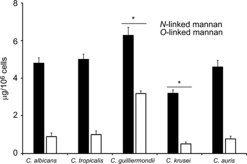
To quantify the basic polysaccharides and oligosaccharides of the cell wall of C. guilliermondii, C. krusei, C. tropicalis, and C. auris, the walls were extracted, depleted of cell components, acid-hydrolyzed, and analyzed by HPAEC-PAD. This methodology has been applied before in the analysis of other Candida species, including C. albicans, and allowed the quantification of chitin, glucan, and mannan, by assessment of the content of N-acetylglucosamine, glucose, and mannose, respectively.37,39,41,42,46–48,52–54 Although it has been extensively characterized previously, here we included the analysis of the C. albicans cell wall for comparison purposes. The cell walls of the species analyzed showed similar levels of chitin, glucan, and mannan (), with significant differences in some cases: C. krusei and C. auris showed higher content of chitin when compared to the other species, and they were also significantly different when compared between each other (). Mannan was significantly higher in C. guilliermon-dii cell wall, but lower in C. krusei wall, and the former also showed lower levels of glucan (). All strains tested showed similar levels of cell wall protein content, with exception of C. krusei that showed low levels of cell wall protein (). To confirm the changes in the cell wall mannan content in C. guilliermondii and C. krusei, we measured the content of O-linked and N-linked mannans decorating the cell wall glycoproteins, by trimming the oligosaccharides with b-elimination and endoglycosidase H digestion, respectively.Citation47 Results shown in indicated that upon β-elimination, the O-linked mannan content in the five species analyzed was lower when compared to the N-linked mannans, and C. guil-liermondii and C. krusei were the only two strains that showed changes in the mannan content: the former had increased levels of both mannans, whereas C. krusei showed low levels of both N-linked and O-linked mannans (). Interestingly, in C. albicans, C. tropicalis, C. krusei, and C. auris, O-linked mannans represented about 15% of the total content of cell wall mannan, but in C. guilliermondii these molecules were relatively more abundant than the N-linked mannans, representing around 33% of total mannan content.
Figure 1 The content of N-linked and O-linked mannan in the cell wall of Candida albicans, Candida tropicalis, Candida guilliermondii, Candida krusei, and Candida auris.
Table 1 Cell wall analysis of C. albicans, C. tropicalis, C. guilliermondii, C. krusei, and C. auris
Notes: Yeast cells were treated either with endoglycosidase H or b-eliminated to trim N-linked mannans or O-linked mannans, respectively. The released oligosaccharides were saved and used to measure the mannose content by HPAEC-PAD. Data are mean ± SD of three independent experiments performed in duplicates. *P<0.05, when compared with mannans from the other species analyzed.
Abbreviation: HPAEC-PAD, high-performance anion-exchange chromatography coupled to pulsed amperometric detection.
To provide additional evidence to these observations, we next assessed the content of cell wall phosphomannan and the cell wall porosity to polycations, as these parameters have been associated with the length of cell wall man-nans.Citation39,Citation43,Citation46,Citation47,Citation52–Citation56 The C. albicans and C. auris walls showed similar phosphomannan levels, while C. tropicalis and C. guilliermondii showed higher content of this oligosaccharide, and the C. krusei walls showed lower phosphomannan abundance (). The cell wall porosity was similar for C. tropicalis, C. guilliermondii, and C. krusei, but this parameter was significantly lower in the walls of C. albicans and C. auris ().
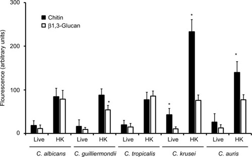
To analyze the organization of the structural polysaccharide within the cell wall, the b1,3-glucan was labeled with the IgG Fc-Dectin-1 chimera,Citation57 and then with FITC-conjugated IgG, whereas WGA-FITC was used for chitin labeling. The five species analyzed were barely stained with both lectins (); however, when cells were inactivated by heat, a treatment that artificially exposes chitin and b1,3-glucan on the surface of the cell wall,Citation58 the fluorescence associated with cells from the five species was significantly increased (). Therefore, chitin and b1,3-glucan are localized underneath other cell wall components that impair the proper lectin–polysaccharide interaction. It is noteworthy that the labeling of b1,3-glucan in heat-killed (HK) C. guilliermondii cells was significantly lower compared to cells from other species under the same treatment (), and HK C. krusei and C. auris cells were more labeled by WGA-FITC, suggesting an increased chitin content (). These results correlated with the cell wall composition provided in . Interestingly, cells from C. krusei showed increased chitin labeling in the live form, suggesting that more of this polysaccharide is naturally exposed on the surface of this organism than in the other species under analysis (). Overall, the data indicates that the cell wall of C. tropicalis, C. guilliermondii, C. krusei, and C. auris differs to which has been described in C. albicans.
Figure 2 Fluorescent labeling of chitin and b1,3-glucan in the cell wall of Candida albicans, Candida tropicalis, Candida guilliermondii, Candida krusei, and Candida auris.
Notes: Live or heat-killed (HK) yeast cells were incubated with either fluorescein isothiocyanate-wheat germ agglutinin conjugate (closed bars, labels chitin) or IgG Fc-Dectin-1 chimera (open bars, labels b1,3-glucan) as described in the “Materials and methods” section, inspected under fluorescence microscopy, and the fluorescence associated to 300 individual cells was recorded. *P<0.05, when compared with cells under the same treatment.
Stimulation of cytokine production by C. albicans, C. tropicalis, C. guilliermondii, C. krusei, and C. auris
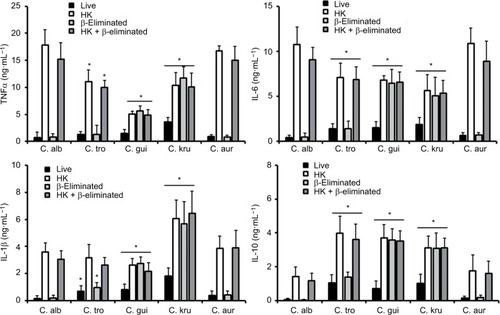
The differences in the cell wall described earlier are likely to affect the Candida–immune cell interaction when compared to that described for C. albicans. Thus, to get some insights about such interaction, yeast cells were coincubated with human PBMCs and the level of secreted cytokines was measured as a read-out of this interaction. C. albicans and C. auris cells barely stimulated the production of TNFα, IL-6, IL-1β, or IL-10 (); however, C. tropicalis, C. guilliermondii, and C. krusei stimulated higher levels of the four cytokines analyzed (). Among the tested strains, one that is capable of inducing a high cytokine production was C. krusei, and this strain significantly stimulated the secretion of higher levels of TNFα and IL-1b () in comparison with the remaining strains. To assess the role of components of the inner part of the cell wall and O-linked mannans, we compared the cytokine profile stimulated with either live or HK cells with or without b-elimination that trimmed the O-linked mannans from the cell wall.Citation40,Citation58 As previously reported for C. albicans,Citation41,Citation58 HK yeast cells stimulated the production of higher levels of TNFα, IL-6, IL-1β, and IL-10 than live cells (). The trimming of O-linked mannan did not affect significantly the cytokine profile stimulated either by live or HK C. albicans cells (). The C. auris cells showed similar ability to C. albicans to stimulate cytokine production, as the profiles of the four cytokines tested were similar for both species when live, HK, b-eliminated or HK + β-eliminated cells were used for the interaction with the human PBMCs (). For C. tropicalis, C. guilliermondii, and C. krusei, the HK cells stimulated higher cytokine levels than live cells (), but some differences were observed when their cytokine profiles were compared with those stimulated by C. albicans. These three species stimulated lower production of TNFα and IL-6 than C. albicans, including when cells were HK and b-eliminated (). Interestingly, upon β-elimination, live cells stimulated high levels of these two cytokines, comparable with those obtained with HK cells (). In the case of the production of IL-1β, the HK C. tropicalis cells stimulated similar levels of this cytokine to those produced upon interaction of the human cells with C. albicans, but live and b-eliminated cells stimulated higher levels of this cytokine when C. tropicalis cells were used during the coincubation period (). For C. guilliermondii, HK and HK + β-eliminated cells stimulated less IL-1β production than C. albicans, but higher levels of this cytokine were observed when β-eliminated cells were used in the interactions (). The HK, b-eliminated, and HK + β-eliminated cells from C. krusei stimulated similar IL-1β levels that were significantly higher than those produced by C. albicans cells (). A similar observation could be drawn for the stimulation of IL-10 production by cells of C. guilliermondii and C. krusei, whereas only HK and HK + b-eliminated C. tropicalis cells stimulated high production of this anti-inflammatory effector ().
Figure 3 Stimulation of cytokine production by Candida albicans, Candida tropicalis, Candida guilliermondii, Candida krusei, and Candida auris.
Notes: Human PBMCs were coincubated for 24 hours with live yeast cells, and then the supernatant was collected and used to quantify the cytokine levels. *P<0.05, when compared with the cytokine level stimulated by C. albicans cells.
Abbreviations: PBMCs, peripheral blood mononuclear cells; TNFα, tumor necrosis factor alpha.
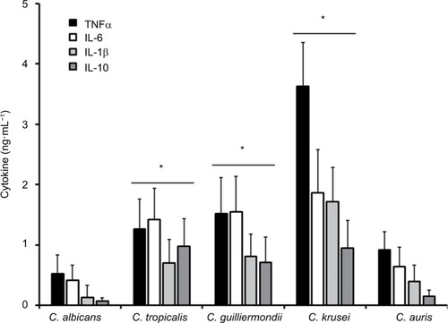
Figure 4 Stimulation of cytokine production by heat-killed and b-eliminated cells from Candida albicans, Candida tropicalis, Candida guilliermondii, Candida krusei, and Candida auris.
Notes: Yeast cells were heat-killed (HK), b-eliminated, or subjected to both treatments before being coincubated with human PBMCs for 24 hours. The supernatants of interactions were collected and used to quantify the cytokine levels. * P<0.05, when compared with the cytokine level stimulated by C. albicans cells under the same treatment. Abbreviations: PBMCs, peripheral blood mononuclear cells; TNFα, tumor necrosis factor alpha; C. alb, Candida albicans; C. tro, Candida tropicalis; C. gui, Candida guilliermondii; C. kru, Candida krusei; C. aur, Candida auris.
To explore whether the significant increment in the ability to stimulate cytokine production by HK cells was dependent on the recognition of b1,3-glucan by dectin-1, the human PBMCs were preincubated with laminarin, an antagonist of dectin-1,Citation41,Citation46,Citation47 and then used in the interaction with the yeast cells. Results given in indicated that preincubation of the human cells with laminarin did not affect the ability of live C. albicans, C. tropicalis, and C. auris to stimulate cytokine production, but a significant reduction in the cytokine levels were observed when HK cells were used for the interactions. Similar data were obtained when cells were subjected to β-elimination (). For the case of live and HK cells from C. guilliermondii and C. krusei, similar results were observed, but the cytokine production stimulated by both live β-eliminated and HK + β-eliminated cells was significantly affected by preincubation of human PBMCs with laminarin ().
Table 2 Effect of laminarin on the ability of C. albicans, C. tropicalis, C. guilliermondii, C. krusei, and C. auris to stimulate cytokine production
Human monocyte-derived macrophages differentially uptake Candida spp
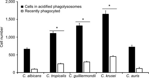
Next, to assess whether other cellular players of the innate immunity differentially interact with the analyzed Candida species and thus affecting the recognition and phagocytosis, we evaluated the ability of human monocyte-derived macrophages to uptake the yeast cells, using a flow cytometry-based protocol that has been used previously to characterize the phagocytic process of fungal cells.Citation37,Citation51,Citation59 Yeast cells were labeled with acridine orange, which emits a green fluorescence that turns reddish once they are in an acidified microenvironment of the mature phagolysosomes.Citation37,Citation59 Results given in shows that most of the fungal species were uptaken and internalized into acid phagolysosomes, but the number of fungal cells undergoing this process was significantly higher for C. tropicalis, C. guilliermondii, and C. krusei than for C. albicans and C. auris, which showed a similar behavior when interacting with these human cells. Similar results were observed for green cells associated to macrophages, ie, recently phagocyted, with low cell numbers associated to C. albicans and C. auris and relatively high cell numbers associated to C. tropicalis, C. guilliermondii, and C. krusei (). Collectively, these data indicate a differential ability of human monocyte-derived macrophages to uptake Candida spp. cells.
Figure 5 Phagocytosis of Candida albicans, Candida tropicalis, Candida guilliermondii, Candida krusei, and Candida auris by human monocyte-derived macrophages.
Notes: Acridine orange-labeled yeast cells were incubated with the human cells at an MOI ratio of 1:6 for 2.5 hours at 37°C under a CO2 atmosphere. Then, macrophages were gated by FACS system and 50,000 cells were counted/sample. Results represent macrophages interacting with at least one green fluorescent cell (recently phagocyted), and those associated with red fluorescence that were classified as macrophages with yeast cells within acidified phagolysosomes. The data represent the mean ± SD of three independent biological replicates performed in duplicate. * P<0.05, when compared with C. albicans cells.
Abbreviations: MOI, multiplicity of infection; FACS, fluorescence-activated cell sorter.
Discussion
C. albicans is a model organism popular to study fundamental aspects of a human fungal pathogen, and a vast amount of information about its biology is currently available. Because of their taxonomical classification, it is usual to find literature assuming that most of the members of the Candida genus should have biological traits like those described in C. albicans, with minimal differences in the phenotype or the molecular determinants related to virulence.Citation41 Nonetheless, the genomic evidence clearly suggests that these organisms have significant differences in the fitness and metabolic processes,Citation60 which are likely to affect the interaction with the host’s components, such as the immune system. The comparative analysis performed here underscored the fact that even though the species under study had a similar composition of the cell wall, they have differences that are likely to affect the interaction with components of the innate immunity such as PPRs. As a proof of the concept, we have the case of C. guilliermondii cells, which had lower levels of b1,3-glucan and as a consequence, they induced the lowest levels of cytokines when this polysaccharide was exposed on the cell surface. Since the protein content in the C. guilliermondii cell wall was similar to the one obtained from the other species under analysis, we hypothesized that the increment in both N- and O-linked mannans in the cell wall was related to the different variety of cell wall proteins that are likely to contain more sites of these posttranslational modifications. Supporting this notion, a different cell secretome has been predicted for C. guilliermondii, which may affect the kind of proteins associated with the wall.Citation61 The lower C. krusei cell wall protein content fits with the low mannan levels, which is likely to be the reason behind this observation. In agreement, the phosphomannan content in this species was also low. The high porosity of the cell wall has been associated with mannans containing short lateral chains;Citation41,Citation46,Citation47 therefore, it is likely this will be the case of mannans on the surface of C. tropicalis, C. guilliermondii, and C. krusei.
Results presented here clearly demonstrate that in an in vitro setting, the human PMBCs are differentially stimulated to produce cytokines by the analyzed Candida species. It was interesting to observe that C. albicans and C. auris had similar abilities to interact with these immune cells and with monocyte-derived macrophages, even though they are species relatively distant in the phylogeny.Citation62 Thus far, only the interaction of C. auris with neutrophils has been studied, and differences in the ability to evade the neutrophil attack have been documented.Citation34 Nonetheless, our results reported here suggest that if escaped from interaction with these immune cells, the next line of defense, the mononuclear cells and macrophages are likely to control this pathogen. Further experiments are required to confirm this hypothesis.
Live cells of C. tropicalis, C. guilliermondii, and C. krusei stimulated higher cytokine levels than C. albicans cells, which might be related to the increased wall porosity. In addition, these three species showed different wall phosphomannan levels that might also contribute to this observation. It was interesting to note that upon removal of O-linked mannans from live cells from these three species, the ability to stimulate cytokine production changed, which in some cases was comparable to the quantified value when HK cells were used for immune stimulation. These data suggest that O-linked mannans play a negative role during recognition of these cell walls by the human mononuclear cells, and once they are removed, relevant ligands for cytokine production are accessible for immune receptors. Here, we demonstrated that the pathway behind this observation requires dectin-1 engagement with β1,3-glucan. Similar observations have been reported for Candida parapsilosis sensu lato and sensu stricto,Citation41,Citation46 and for the interaction of C. albicans with macrophages.Citation63
The therapeutic strategies to control candidiasis are extremely limited when compared to the wide repertoire of drugs to treat bacterial infections, and this has negatively impacted the sensitivity of fungal cells to the currently available drugs. Moreover, some Candida species are naturally resistant to some antifungal drugs or have the ability to rapidly develop resistance, such as Candida glabrata and C. kruseii.Citation64,Citation65 Therefore, it is imperative to expand the portfolio of antifungal drugs to treat candidiasis. An alternative approach to control these infections has been explored using immunomodulators such as b1,3-glucan. Administration of this cell wall polysaccharide primes PBMCs and monocytes, which acquire prolonged enhanced functional state mediated by epigenetic mechanisms.Citation66 Although these could lead to a promising strategy to help in the control of this disease, our data here demonstrated that this approach might not be beneficial for the treatment of infections caused by C. guilliermondii, as this species does not induce a strong cytokine production via engagement of dectin-1 with β1,3-glucan.
Since the O-linked mannans of the cell wall of the species studied here showed a masking effect on the b1,3-glucan exposure at the cell surface and therefore precluded a proper activation of the cytokine production via dectin-1, it is tempting to speculate that inhibitors of the O-linked mannosylation pathway may have a positive effect on controlling infections caused by these fungal species.
The evaluation of the phagocytic process by human monocyte-derived macrophages pointed out again that C. tropicalis, C. guilliermondii, and C. krusei are species with a different ability to interact with these immune cells when compared to C. albicans. We have recently demonstrated that C. tropicalis is more phagocytosed than C. albicans by a phosphomannan-dependent mechanism,Citation37 and results reported here confirmed this previous observation. Since C. guilliermondii also showed higher phosphomannan content than C. albicans cells, it is tempting to offer a similar explanation for this observation. Although we do not currently have a proper explanation for data generated with C. krusei, a similar observation for this species and C. guilliermondii has been reported when interacting with murine macrophages.Citation28
Conclusion
In conclusion, our study shows that the current knowledge about C. albicans cell wall and its interaction with innate immune cells cannot be extrapolated to C. tropicalis, C. guilliermondii, C. krusei, and C. auris. These species have differences in the cell wall composition and with exception of C. auris, displayed different abilities to stimulate cytokine production by human PBMCs and to be phagocytosed by human monocyte-derived macrophages.
Acknowledgments
We thank Prof Gordon Brown from the University of Aberdeen for the donation of the IgG Fc-Dectin-1 chimera. We acknowledge the technical assistance of Maria F Rodriguez-Preciado (Universidad de Guanajuato) during the acquisition of data related to Candida krusei. This work was supported by Consejo Nacional de Ciencia y Tecnología (ref PDCPN2014-247109 and FC 2015-02-834), Universidad de Guanajuato (ref 1025/2016; CIIC 95/2018), and Red Temática Glicociencia en Salud (CONACYT-México).
Disclosure
The authors report no conflicts of interest in this work.
References
- PappasPGRexJHLeeJA prospective observational study of candidemia: epidemiology, therapy, and influences on mortality in hospitalized adult and pediatric patientsClin Infect Dis200337563464312942393
- BrownGDDenningDWGowNALevitzSMNeteaMGWhiteTCHidden killers: human fungal infectionsSci Transl Med20124165165rv113
- Lass-FlörlCThe changing face of epidemiology of invasive fungal disease in EuropeMycoses200952319720519391253
- TanTYTanALTeeNWSNgLSYCheeCWJThe increased role of non-albicans species in candidaemia: results from a 3-year surveillance studyMycoses201053651552119619263
- PfallerMABoykenLHollisRJMesserSATendolkarSDiekemaDJIn vitro activities of anidulafungin against more than 2,500 clinical isolates of Candida spp., including 315 isolates resistant to fluconazoleJ Clin Microbiol200543115425542716272464
- PfallerMAPappasPGWingardJRPfaller MichaelAPappas PeterGInvasive fungal pathogens: current epidemiological trendsClin Infect Dis200643S1S3S14
- KrcmeryVBarnesAJNon-albicans Candida spp. causing fun-gaemia: pathogenicity and antifungal resistanceJ Hosp Infect200250424326012014897
- TortoranoAKibblerCPemanJBernhardtHKlingsporLGrillotRCandidaemia in Europe: epidemiology and resistanceInt J Antimicrob Agents200627535936616647248
- WisplinghoffHBischoffTTallentSMSeifertHWenzelRPEdmondMBNosocomial bloodstream infections in US hospitals: analysis of 24,179 cases from a prospective nationwide surveillance studyClin Infect Dis200439330931715306996
- ChenSCAMarriottDPlayfordEGCandidaemia with uncommon Candida species: predisposing factors, outcome, antifungal susceptibility, and implications for managementClin Microbiol Infect200915766266919614718
- SaviniVCatavitelloCOnofrilloDWhat do we know about Candida guilliermondii? A voyage throughout past and current literature about this emerging yeastMycoses201154543444121039941
- BidaudALChowdharyADannaouiECandida auris: an emerging drug resistant yeast – A mini-reviewJ Mycol Med201828356857330030072
- NeteaMGBrownGDKullbergBJGowNARAn integrated model of the recognition of Candida albicans by the innate immune systemNat Rev Microbiol200861677818079743
- Martínez-ÁlvarezJAPérez-GarcíaLAFlores-CarreónAMora-MontesHMThe immune response against Candida spp. and Sporothrix schenckiiRev Iberoam Micol2014311626624252829
- Díaz-JiménezDFPérez-GarcíaLAMartínez-ÁlvarezJAMora-MontesHMRole of the fungal cell wall in pathogenesis and antifungal resistanceCurr Fungal Infect Rep201264275282
- GowNARvan de VeerdonkFLBrownAJPNeteaMGCandida albicans morphogenesis and host defence: discriminating invasion from colonizationNat Rev Microbiol2012102112122
- GowNARHubeBImportance of the Candida albicans cell wall during commensalism and infectionCurr Opin Microbiol201215440641222609181
- KlisFMGrootPDHellingwerfKMolecular organization of the cell wall of Candida albicansMed Mycol200139118
- BrownGDGordonSImmune recognition: a new receptor for β-glucansNature200141368513637
- NeteaMGGowNARMunroCAImmune sensing of Candida albicans requires cooperative recognition of mannans and glucans by lectin and Toll-like receptorsJ Clin Invest200611661642165016710478
- CambiANeteaMGMora-MontesHMDendritic Cell Interaction with Candida albicans Critically Depends on N-Linked MannanJ Biol Chem200828329205902059918482990
- SaijoSIkedaSYamabeKDectin-2 recognition of α-Mannans and induction of Th17 cell differentiation is essential for host defense against Candida albicansImmunity201032568169120493731
- WellsCASalvage-JonesJALiXThe macrophage-inducible C-type lectin, mincle, is an essential component of the innate immune response to Candida albicansJ Immunol2008180117404741318490740
- ZhuL-LZhaoX-QJiangCC-type lectin receptors dectin-3 and dectin-2 form a heterodimeric pattern-recognition receptor for host defense against fungal infectionImmunity201339232433423911656
- Mora-MontesHMNeteaMGFerwerdaGRecognition and blocking of innate immunity cells by Candida albicans chitinInfect Immun20117951961197021357722
- WagenerJMalireddiRKSLenardonMDFungal chitin dampens inflammation through IL-10 induction mediated by Nod2 and TLR9 activationPLoS Pathog2014104e100405024722226
- RichardsonMDDonaldsonFInteraction of Candida krusei with human neutrophils in vitroJ Med Microbiol19944163843887966213
- VecchiarelliABistoniFCenciEPeritoSCassoneAIn-vitro killing of Candida species by murine immunoeffectors and its relationship to the experimental pathogenicityMed Mycol1985235377387
- RoilidesEHolmesABlakeCPizzoPAWalshTJEffects of granulocyte colony-stimulating factor and interferon-gamma on antifungal activity of human polymorphonuclear neutrophils against pseudohyphae of different medically important Candida speciesJ Leukoc Biol19955746516567536791
- HøgåsenAKAbrahamsenTGGaustadPVarious Candida and Torulopsis species differ in their ability to induce the production of C3, factor B and granulocyte-macrophage colony-stimulating factor (GM-CSF) in human monocyte culturesJ Med Microbiol19954242912987707338
- LymanCAWalshTJPhagocytosis of medically important yeasts by polymorphonuclear leukocytesInfect Immun1994624148914938132358
- MaródiLForehandJRJohnstonRBMechanisms of host defense against Candida species. II. Biochemical basis for the killing of Candida by mononuclear phagocytesJ Immunol19911468279027941849938
- MaródiLKorchakHMJohnstonRBMechanisms of host defense against Candida species. I. Phagocytosis by monocytes and monocyte-derived macrophagesJ Immunol19911468278327891901885
- JohnsonCJDavisJMHuttenlocherAKernienJFNettJEEmerging fungal pathogen Candida auris evades neutrophil attackMBio201894e01403e0141830131360
- GillumAMTsayEYHKirschDRIsolation of the Candida albicans gene for orotidine-5′-phosphate decarboxylase by complementation of S. cerevisiae ura3 and E. coli pyrF mutationsMol Gen Genet198419811791826394964
- MilleriouxYClastreMSimkinAJDevelopment of a URA5 integrative cassette for gene disruption in the Candida guilliermondii ATCC 6260 strainJ Microbiol Methods201184235535821182877
- Hernández-ChávezMJFrancoBClavijo-GiraldoDMHernándezNVEstrada-MataEMora-MontesHMRole of protein phosphoman-nosylation in the Candida tropicalis-macrophage interactionFEMS Yeast Res2018185 01 08 2018
- SharmaCKumarNMeisJFPandeyRChowdharyADraft genome sequence of a fluconazole-resistant Candida auris strain from a Candidemia patient in IndiaGenome Announc201534e007221526184929
- Mora-MontesHMBatesSNeteaMGEndoplasmic reticulum alpha-glycosidases of Candida albicans are required for N glycosylation, cell wall integrity, and normal host-fungus interactionEukaryot Cell20076122184219317933909
- Díaz-JiménezDFMora-MontesHMHernández-CervantesALuna-AriasJPGowNARFlores-CarreónABiochemical characterization of recombinant Candida albicans mannosyltransferases Mnt1, Mnt2 and Mnt5 reveals new functions in O- and N-mannan biosynthesisBiochem Biophys Res Commun20124191778222326920
- Estrada-MataENavarro-AriasMJPerez-GarciaLAMembers of the Candida parapsilosis complex and Candida albicans are differentially recognized by human peripheral blood mononuclear cellsFront Microbiol20156152726793173
- HobsonRPMunroCABatesSLoss of cell wall mannosylphosphate in Candida albicans does not influence macrophage recognitionJ Biol Chem200427938396283963515271989
- de NobelJGKlisFMMunnikTPriemJvan den EndeHAn assay of relative cell wall porosity in Saccharomyces cerevisiae, Kluyveromyces lactis and Schizosaccharomyces pombeYeast1990664834902080665
- GrahamLMTsoniSVWillmentJASoluble Dectin-1 as a tool to detect beta-glucansJ Immunol Methods20063141–216416916844139
- MarakalalaMJVautierSPotrykusJDifferential adaptation of Candida albicans in vivo modulates immune recognition by dectin-1PLoS Pathog201394e100331523637604
- Pérez-GarcíaLACsonkaKFlores-CarreónARole of protein glycosylation in Candida parapsilosis cell wall integrity and host interactionFront Microbiol2016763330627014229
- Navarro-AriasMJDefosseTADementhonKDisruption of protein mannosylation affects Candida guilliermondii cell wall, immune sensing, and virulenceFront Microbiol195120167
- Mora-MontesHMMckenzieCBainJMInteractions between macrophages and cell wall oligosaccharides of Candida albicansMethods Mol Biol201284524726022328379
- EndresSGhorbaniRLonnemannGvan der MeerJWMDinarelloCAMeasurement of immunoreactive interleukin-1β from human mononuclear cells: optimization of recovery, intrasubject consistency, and comparison with interleukin-1α and tumor necrosis factorClin Immunol Immunopathol19884934244382461270
- AbramsWRDiamondLWKaneABA flow cytometric assay of neutrophil degranulationJ Histochem Cytochem19833167377446404983
- González-HernándezRJJinKHernández-ChávezMJPhosphomannosylation and the functional analysis of the extended Candida albicans MNN4-like gene familyFront. Microbiol20178215629163439
- Mora-MontesHMBatesSNeteaMGA multifunctional mannosyltransferase family in Candida albicans determines cell wall mannan structure and host-fungus interactionsJ Biol Chem201028516120871209520164191
- BatesSMaccallumDMBertramGCandida albicans Pmr1p, a secretory pathway P-type Ca2+/Mn2+-ATPase, is required for glycosylation and virulenceJ Biol Chem200528024234082341515843378
- WestLLowmanDWMora-MontesHMDifferential virulence of Candida glabrata glycosylation mutantsJ Biol Chem201328830220062201823720756
- BatesSHughesHBMunroCAOuter chain N-glycans are required for cell wall integrity and virulence of Candida albicansJ Biol Chem20062811909816263704
- Lopes-BezerraLMLozoya-PerezNELopez-RamirezLAFunctional characterization of Sporothrix schenckii glycosidases involved in the N-linked glycosylation pathwayMed Mycol2015531606825526779
- GrahamLMTsoniSVWillmentJASoluble Dectin-1 as a tool to detect beta-glucansJ Immunol Methods20063141–216416916844139
- GowNARNeteaMGMunroCAImmune recognition of Candida albicans beta-glucan by dectin-1J Infect Dis2007196101565157118008237
- Lozoya-PérezNECasas-FloresSMartínez-ÁlvarezJAGeneration of Sporothrix schenckii mutants expressing the green fluorescent protein suitable for the study of host-fungus interactionsFungal Biol2018122101023103030227928
- ButlerGRasmussenMDLinMFEvolution of pathogenicity and sexual reproduction in eight Candida genomesNature2009459724765766219465905
- SorgoAGHeilmannCJBrulSde KosterCGKlisFMBeyond the wall: Candida albicans secret(e)s to surviveFEMS Microbiol Lett20133381101723170918
- SharmaCKumarNPandeyRMeisJFChowdharyAWhole genome sequencing of emerging multidrug resistant Candida auris isolates in India demonstrates low genetic variationNew Microbes New Infect201613778227617098
- MckenzieCGJKoserULewisLEContribution of Candida albicans cell wall components to recognition by and escape from murine macrophagesInfect Immun20107841650165820123707
- GongJXiaoMWangHGenetic differentiation, diversity, and drug susceptibility of Candida kruseiFront Microbiol20189271730524386
- Vale-SilvaLASanglardDTipping the balance both ways: drug resistance and virulence in Candida glabrataFEMS Yeast Res2015154
- van der MeerJWMJoostenLABRiksenNNeteaMGTrained immunity: a smart way to enhance innate immune defenceMol Immunol2015681404426597205