Abstract
Colon cancer is one of the most common causes of cancer-related mortality. Adenocarcinoma with mucinous features accounts for 10–15% of colon carcinoma. Distal nodal metastatic colorectal cancer is uncommon, and metastasis of colorectal cancer to the left supraclavicular lymph node is extremely rare without signs of metastatic organ involvement. We present a case of a 54-year-old Caucasian male with colonic adenocarcinoma that presented initially as a left-sided neck mass that had progressively increased in size over 9 months. On physical exam, a left supraclavicular soft tissue mass 6 cm in diameter was appreciated, it was non-tender with no submandibular lymphadenopathy. Soft tissue mass was palpable on the anterior abdominal wall in the epigastric region. Open excisional tissue biopsy of the left supraclavicular mass revealed metastatic adenocarcinoma with mucinous features and colonoscopy revealed a 6 cm obstructing mass in the transverse colon with biopsy revealing primary adenocarcinoma of the mucinous type. Palliative care with comfort measures was agreed upon. Typically, the most common sites of colon cancer metastasis are regional lymph nodes, liver, lung, bone and brain, and ours demonstrated an extremely rare pattern of colon cancer metastasis. The presentation to metastasize to the left supraclavicular node without solid end organ involvement makes this case even more novel.
Introduction
Colon cancer is one of the most common causes of cancer-related mortality. A total of 700,000 new cases are diagnosed annually, accounting for 400,000 mortalities worldwide.Citation1 Adenocarcinoma with mucinous features accounts for 10–15% of colon carcinoma.Citation2 Mucinous carcinoma is defined according to WHO as a subtype of colorectal carcinoma (CRC) with mucin lakes comprising at least 50% of the tumor mass.Citation3 Distal nodal metastatic colon cancer is uncommon and metastasis of CRC to the left supraclavicular lymph node, also known as Virchow’s node, is extremely rare without signs and symptoms of metastatic organ involvement.Citation4,Citation5
Typically, the most common sites of colon cancer metastasis are regional lymph nodes, liver, lung, bone and brain, with ours demonstrating an extremely rare pattern of colon cancer metastasis.Citation4 The novelty of this case revealed metastatic involvement to the left supraclavicular node without end organ involvement, ie (liver, lungs) conjugated with the patients carcinomic history. CRCs develop metastatic disease in (60%) of cases and commonly spread to the liver.Citation1 To account, only 6% of the cancers originate in the transverse colon.Citation1 We present a case of a 54-year-old Caucasian male who presented to a community hospital with a left-sided neck mass (Virchow’s node) with biopsy findings of metastatic adenocarcinoma with mucinous features originating from a transverse colon mass.
Case report
A 54-year-old Caucasian male presented to our emergency department with severe epigastric pain with intractable nausea and vomiting over 48–72 hrs. The patient reported early satiety, a 30 lb. weight loss over 3 weeks and was unable to tolerate anything by mouth. Additionally, the patient had developed a left lateral neck mass which progressively increased in size over the past 9 months. Patient denied tenderness to palpation of the mass and dysphagia. His past medical history was significant for papillary carcinoma of the thyroid, GERD, chronic hepatitis C, polycystic kidney disease (PKD) and chronic kidney disease (CKD). His past surgical history was remarkable for thyroid malignancy treatment with radioactive iodine and total thyroidectomy in addition to a gunshot wound. Family and social history were not significant. On physical exam, a left supraclavicular soft tissue mass 6 cm in diameter was appreciated, it was non-tender with no submandibular lymphadenopathy. A soft tissue mass was also palpable on the anterior abdominal wall in the epigastric region.
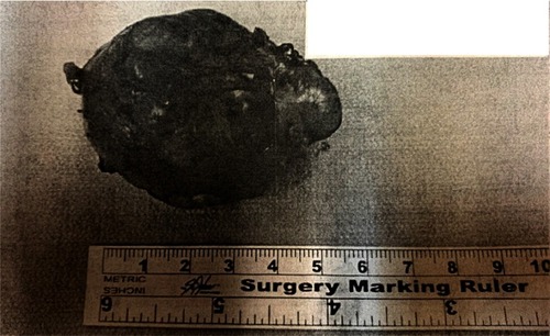
Given the patient's malignant history, significant weight loss in the past 3 weeks with increasing left mass size, nausea, vomiting and abdominal pain, the patient was admitted for further evaluation. Initial studies included ultrasound of the abdomen for workup of acute pancreatitis with findings unremarkable for the body of the pancreas and the head and tail not being visualized. CT of the abdomen and pelvis revealed pulmonary nodules in the posterior segment of the right lower lobe of the lung and bronchiectasis of the left lower lobe, PKD and bilateral hepatic cysts. Oncology was consulted, and a CT of the chest and neck were ordered in addition to a chest X-ray that was remarkable for two pulmonary nodules with an enlarged paratracheal node. Pulmonology was consulted, and the patient was recommended to follow-up in 6 months for surveillance of the pulmonary nodules. ENT and Surgery were consulted, and an excisional lymph node biopsy was pursued due to suspicion of possible recurrence of previous thyroid malignancy or metastases (). Histopathology revealed metastatic mucin-producing adenocarcinoma. Immunoperoxidase studies performed on paraffin sections revealed the neoplastic cells were positive for CK20 and CDX2. The cells were negative for CK7, TTF-1, p63, prostate-specific antigen (PSA), prostatic acid phosphatase (PACP), chromogranin, synaptophysin and S-100. Gastroenterology was consulted, and an esophagogastroduodenoscopy was performed including an antral biopsy of the gastric mucosa, which was unremarkable. Colonoscopy of the lower GI tract revealed a 6 cm obstructing mass in the transverse colon and a biopsy was taken revealing findings of invasive well to moderately differentiated adenocarcinoma with mucinous features, focally present in subserosal tissue with ten of eleven regional lymph nodes involved. This confirmed the primary cancer originated from the colonic mass and metastasized to the left lateral neck mass (Virchow’s node).
Surgical intervention was pursued with resection of the transverse colon with primary end-to-end anastomosis. This resolved the intractable acute abdomen. The patient was offered a trial of chemotherapy; however, given the patient’s diagnosis of metastatic colonic carcinoma, further treatment options were not pursued. Further discussion with the patient and his family led to a decision to proceed with comfort care measures.
Discussion
Approximately 1.2 million people develop CRC worldwide annually and it is the fourth most frequent cause of cancer-related mortality.Citation2 CRC is the second leading cause of death from gastrointestinal tract (GIT) cancer in the US and the third most common malignancy in both men and women.Citation1
CRC most commonly spreads to local lymph nodes (50–70%) and the liver (35–50%). Other sites of metastatic spread include the lungs (21%), peritoneum (15%), ovaries (13.1%), central nervous system (8.3%), bone (8.7%), kidney (6.6%), testes, penis, uterus and oral cavity. Very rare sites include the adrenal glands, hilar lymph nodes, skin and muscles among others.Citation5 To add, supraclavicular lymph node involvement is an unusual metastatic site for CRC and is more common for gastric carcinoma. To the best of our knowledge, this is the sixth reported case of CRC to metastasize to a distal node site ie, Virchow’s node without metastatic organ involvement.Citation5–Citation9
Virchow’s node is commonly referred to as a lymph node in the left supraclavicular fossa, typically the area above the left clavicle. Literature also classifies Virchow’s node as a deep cervical node.Citation10 This finding on physical exam is referred to as Troisier’s sign.Citation1 Majority of the gastrointestinal cavity drains to this node which lies near the junction of the thoracic duct and the left subclavian vein.Citation1,Citation11 Tumor spread from the thoracic duct usually leads to enlargement of this node. This gives important clues to a possible abdominal cavity malignancy, and other sites such as breast, esophagus and lymphomas which tends to be a sign of advanced disease as was the case here for our patient.Citation11
Statistics vary regarding the primary carcinomas that spread to the supraclavicular lymph node. CNS tumors (oligodendroglioma, glioblastoma multiforme, ependymoma), breast, lung, esophageal and genitourinary tract (testicular, cervical, uterine, ovarian, bladder, prostate) carcinomas range between (0.1–33%) of cases that metastasize to Virchow’s node.Citation12 When discussing CRC, literature states that 20% of the cases present initially as stage IV ie, distant node involvement.Citation13 Currently, the most common carcinoma that spreads primarily to Virchow’s node is gastric carcinomas. Gastric tumors involving mucosa, submucosa and T2/T3 involvement will spread in about (3–83%) of cases.Citation14 When focusing on the composition of the tumor, according to one study, 39% of the histopathological tumors biopsied from left supraclavicular lymph node are adenocarcinoma in form and originate from primaries of the breast, lung, prostates, stomach, pancreas and endometrium.Citation15 From current literature, it is evident metastatic spread can occur from a variety of primary sites with varying compositions. Distant spread to non-regional lymph nodes is quite low in CRCs, and spread without metastatic organ involvement is also rare with only a handful of cases reported.Citation5–Citation9
The relatively distant metastatic site lead to a stage IV diagnosis.Citation12 With a history of thyroidectomy due to papillary carcinoma and the spread of PKD, this is a unique presentation that begs the question why the metastasis from the primary colonic cancer did not involve the common metastatic sites before spreading to an uncommon distal site ie, supraclavicular lymph node. Currently, there is no literature stating the mechanism or process of metastatic spread of tumor cells to distant non-regional lymph nodes in patients with colorectal cancer. According to one theory, the process starts with spread of tumor cells into sequential lymphatic nodes, and studies have demonstrated that skip micrometastasis between regional lymph nodes stations can be seen in 18% of the cases.Citation16 Primary carcinoma metastatic spread without solid organ involvement is not typical as was presented in our case.Citation16 Early surveillance and workup for this patient could have improved his poor prognosis.
Conclusion
In summary, we report an uncommon pattern of metastasis in primary adenocarcinoma with mucinous features originating from a transverse colonic mass presenting as a left supraclavicular lymph node without solid end organ metastatic involvement. This case is significant due to the fact that liver and lung involvement were spared from the carcinoma. This case demonstrated an extremely rare pattern of involvement which has only been reported in five other cases to the best of our knowledge. In addition, there is no root cause or pathophysiologic etiology known for this type of “sparing” metastasis; however, in working up such a patient, it is important to explore a wide array of differentials by following each organ system individually.
Institutional approval
Institutional approval is not applicable nor required for publication of this manuscript.
Consent for publication
Written informed consent was obtained from the patient for publication of this case report.
Acknowledgment
We would like to acknowledge and thank Dr Muhammad Omer Jamil of Marshall Oncology at Edwards Comprehensive Cancer Center.
Disclosure
The authors report no conflicts of interest in this work.
References
- Achmad H, Hanifa R. Supraclavicular lymphnodes: unusual manifestation of metastase adenocarcinoma colon. Acta Med Indones. 2015;47(4):333–339.26932703
- Hugen N, Brown G, Glynne-Jones R, De Wilt JHW, Nagtegaal ID. Advances in the care of patients with mucinous colorectal cancer. Nat Rev Clin Oncol. 2016;13(6):361–369. doi:10.1038/nrclinonc.2015.14026323388
- Gonzalez R. Pathology outlines - mucinous carcinoma of colon. 2018 Available from: http://www.pathologyoutlines.com/topic/colontumorcolloid.html. Accessed 117, 2018.
- El-Halabi MM, Chaaban SA, Meouchy J, Page S, Salyers WJ. Colon cancer metastasis to mediastinal lymph nodes without liver or lung involvement: a case report. Oncol Lett. 2014;8(5):2221–2224. doi:10.3892/ol.2014.242625289100
- Reddy RR, Das P, Rukmangadha N, Manilal B, Kalawat TC. Colonic carcinoma presenting with axillary lymphadenopathy-a very rare clinical entity. IJSR. 2017;6(8):2015–2017. doi:10.12688/f1000research.12999.1
- Gubitosi A, Moccia G, Malinconico FA, et al. Unusual metastasis of left colon cancer: considerations on two cases. Acta Biomed l’Ateneo Parm. 2009;80(1):80–82.
- Chieco PA, Virgilio E, Mercantini P, Lorenzon L, Caterino S, Ziparo V. Solitary left axillary metastasis after curative surgery for right colon cancer. ANZ JSurg. 2011;81(11):845–846. doi:10.1111/j.1445-2197.2011.05877.x
- Perin T, Canzonieri V, Memeo L, Massarut S. Breast metastasis of primary colon cancer with micrometastasis in the axillary sentinel node: a metastasis that metastasized? Diagn Pathol. 2011;6(1):2–4. doi:10.1186/1746-1596-6-45
- Kawahara H, Yanaga K. Metastasis of colon cancer to axillary lymph nodes. Jikeikai Med J 2006; 53(4):167–70
- Healthline Medical Network. Cervical lymph nodes anatomy, diagram & function | body maps. Available from: https://www.healthline.com/human-body-maps/cervical-lymph-nodes#2. Accessed 117, 2018.
- Sundriyal D, Kumar N, Dubey SK, Walia M. Virchow’s node. Case Reports. 2013;2013(1):bcr2013200749–bcr2013200749. doi:10.1136/bcr-2013-200749
- Jr H, Bishop JA, StrojanP, Hartl DM. Cervical lymph node metastases from remote primary tumor sites.Head Neck 2016;38(Suppl1):1–24. doi:10.1002/hed.24344.
- Fujie Y, Ikeda M, Seshimo I, et al. Complete response of highly advanced colon cancer with multiple lymph node metastases to irinotecan combined with UFT: report of a case. Surg Today. 2006;36(12):1133–1138. doi:10.1007/s00595-006-3315-517123148
- Coburn NG. Lymph nodes and gastric cancer. J Surg Oncol. 2009;99(4):199–206. doi:10.1002/jso.2122419142901
- Ismi O, Vayisogl Y, Ozcan C, Gorur K, Unal M. Supraclavicular metastasis from infraclavicular organs: retrospective analysis of 18 patients. Int J Cancer Manag. 2017;10:4. doi:10.5812/ijcm.4720
- Aksel B, Dogan L, Karaman N, Demirci S. Cervical lymphadenopathy as the first presentation of sigmoid colon cancer. Middle East J Cancer. 2013;4(4):185–188. Available from: http://www.embase.com/search/results?subaction=viewrecord&from=export&id=L370219930%5Cnhttp://mejc.sums.ac.ir/index.php/mejc/article/Download/120/109%5Cnhttp://sfx.library.uu.nl/utrecht?sid=EMBASE&issn=20086709&id=doi:&atitle=Cervical+lymphadenopathy+as+t.

