
Abstract
Scabies is an infestation caused by the mite Sarcoptes scabiei var hominis. The clinical picture can vary and in children, scabies occurs in locations that are not usually found in adults. Complications can occur due to secondary impetiginization of scabies lesions. We presented a case of 8-year-old boy with painful red lumps, pus discharge, and alopecia on the back of the head since 2 weeks ago. Dermatological examination revealed multiple erythematous nodules with sinuses in the scalp of the parieto-occipital area and alopecia. Through microscopic examination we found the Sarcoptes scabiei mite; therefore, we conclude that the diagnosis in this patient was impetigenic scabies. Treatment with topical permethrin 5%, oral antihistamine and oral antibiotics resulted in good outcome.
Introduction
Scabies is one of the global health burdens that mainly affects both under-resourced and developed areas. The factors that contribute to the endemic of scabies in an area are poverty, densely populated areas, and difficulty in accessing health facilities.Citation1 It is estimated that the annual prevalence in the world is 300 million cases. In 2009, the World Health Organization declared scabies as a neglected skin disease. In developing countries, the prevalence of scabies is higher in children and adolescents than adults.Citation2 Karimkhani et al in 2015 found that Indonesia is the country with the highest burden of scabies.Citation3
Scabies is an infestation caused by the mite Sarcoptes scabiei var hominis.Citation4 Most transmissions occur between family members. Scabies in children can occur in locations that are not usually found in adults.Citation5 The clinical picture can vary from person to person, from asymptomatic to erythematous papules, nodules, and crusts.Citation4,Citation5 Sometimes scabies can be difficult to diagnose because not all symptoms can be found in patients and lesions are minimal.Citation2 In addition, complications can occur due to secondary impetiginization of scabies lesions. It is necessary to treat them using topical or oral antibiotics depending on their severity.Citation1 In the following, we present a case report of scabies with secondary infection which resembled kerion type tinea capitis.
Case Report
The 8-year-old boy patient presented with painful red lumps, pus discharge, and alopecia on the back of the head since 2 weeks ago. In the past two weeks the patient complained of pruritus and rashes on his head which were especially severe in the afternoon and evening. The patient was not given any treatment. However, 1 week ago, the patient’s mother noticed that the patient’s alopecia in the pruritic area and the rash had increased. The patient also began to complain of pain in the head area. Therefore, the patient decided to come for treatment at a dermatology and venereology specialist clinic. History of fever is denied. Similar complaints were also found in the patient’s twin, that consist of alopecia, red lumps, and pus discharge. But we did not found other family member with the same complaints. The patient has a history of playing with stray cats in front of his house and is said by the family to have poor hygiene. Informed consent was obtained from the patient and his family for his case and images to be published in this paper. This case details of this manuscript also has been approved to be published by the Department of Dermatology and Venereology, Faculty of Medicine, Universitas Sumatera Utara.
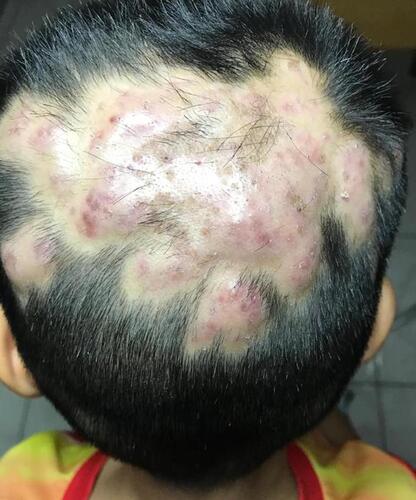
Dermatological examination revealed multiple erythematous nodules with sinuses in the scalp of the parieto-occipital area and alopecia. There is tenderness in the area of the lesion. () On palpation, there is no palpable enlargement of the lymph nodes. There is no lesion in the other parts of the patient’s body. Based on the history and physical examination, the differential diagnosis in the patient was kerion type tinea capitis and impetigenic scabies.
Figure 1 Dermatological examination showing multiple erythematous nodules with sinuses on the scalp of the parieto-occipital area and alopecia.
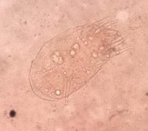
We examined skin scrapings in the lesion area using KOH 10% and examined it under light microscope, which did not show any fungal elements. The culture also showed a negative result. Through microscopic examination we found the Sarcoptes scabiei mite. The examination was done in Department of Microbiology, Faculty of Medicine, Universitas Sumatera Utara (). Based on the laboratory examination results, we conclude that the diagnosis in this patient was impetigenic scabies. The patient received treatment in the form of applying 5% permethrin cream once for 8 hours, cetirizine syrup 1 x 1 teaspoon and amoxicillin oral. In this case, the patient’s twin also given the same treatment as the patient. We also followed up both of them each week.
Figure 2 Sarcoptes scabiei on microscopic examination.
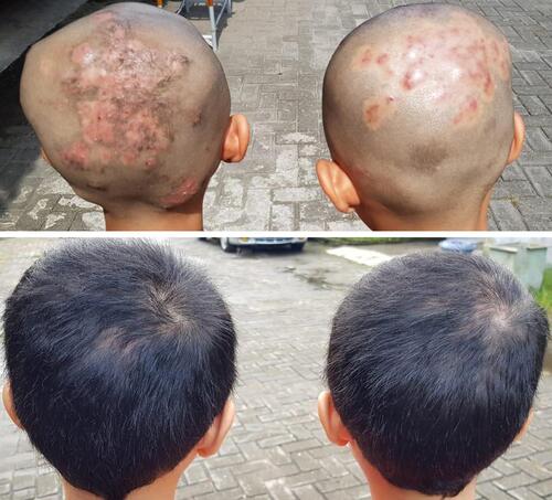
During the first until third control, the patient felt improvement in his pruritus and pain. By the third week, the patient’s hair was starting to grow. Dermatological examination revealed multiple erythematous macules. There is new hair growth in some areas that were previously alopecia. We followed up the patient for In the last visit, the patient’s hair has grown completely. () Similar result was also seen in patient’s twin. ()
Figure 3 Clinical improvement from first (A), second (B), third (C), and last visit (D).
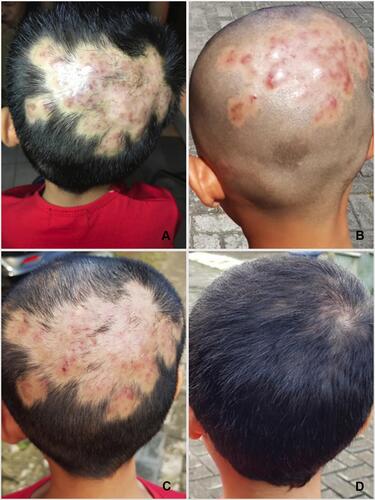
Figure 4 Comparison between second and last visit of the patient and his twin.
Discussion
The patient, an 8 years old boy, presented with painful, purulent red lumps and alopecia on his head since 2 weeks ago. The differential diagnosis in this patient were with kerion type tinea capitis and impetigenic scabies. Tinea capitis is a dermatophyte infection of the hair and scalp, usually caused by Trichophyton and Microsporum species. In the inflammation type, the most common causative pathogens are M. canis, M. gypseum, and T. verrucosum. Inflammatory reactions can range from follicular pustules to furunculosis or kerion.Citation6 The skin lesions in the patient provide a clinical appearance resembling kerion so that this differential diagnosis is considered.
Scabies is an infectious disease caused by the infestation of the parasite Sarcoptes scabiei var hominis.Citation7 Scabies in children is more often found in locations that are not usually found in adults, namely the scalp, face, palms and feet, and in the fold area. In addition, children are more at risk for developing scabies with bacterial superinfection.Citation5 The location of the lesion in a patient is an atypical location for scabies mite infestation, but the scalp is one of the locations for scabies in children. After examination of skin scrapings in the lesion area, scabies mites were found and no fungus components were found. This led to our elimination of a differential diagnosis of kerion-type tinea capitis and we concluded that scabies infestation was aggravated by secondary infection with furunculosis. Scabies transmission can occur due to direct and indirect contact.Citation7
Diagnosis of scabies is mainly based on clinical features.Citation1,Citation4 Classic manifestations in scabies consist of burrow, papule, and generalized pruritus.Citation8 Each individual can have different clinical manifestations. On physical examination, excoriation and eczematous dermatitis can be found between the fingers, sides of the fingers, volar wrist and sides of the palms, elbows, axillae, genital area. The head and neck area is usually not affected in healthy individuals, but this location can be involved in children, the elderly and the immunocompromised.Citation4 Symptoms are usually found in the form of persistent pruritus which is very disturbing and worsening, especially at night.Citation1 In some patients can be found fever due to secondary impetigo or cellulitis.Citation5
Complications that can occur are secondary impetiginization and poststreptococcal glomerulonephritis due to pyoderma such as impetigo, furuncles or cellulitis by Streptococcus pyogenes.Citation1,Citation4,Citation5 Scabies infestation is associated with an increased risk of impetigo. These bacteria can also be isolated from the skin tunnels and the scibala suggests that mites may contribute to the spread of bacteria. In addition, it is also known that mites can produce complement inhibitors that can support the growth of S. pyogenes.Citation1,Citation4 Patients have a secondary infection in the form of furunculosis. Therefore, patients get oral antibiotics so that secondary infections can be treated and prevent complications in patients.
Treatment of scabies through a combination of the use of scabies and handling contaminated goods.Citation4,Citation7 The two most commonly used treatments are topical permethrin and oral ivermectin which have good effectiveness and are generally well tolerated. Permethrin 5% cream is a first-line topical therapy and is safe for use in pregnant women and children.Citation1,Citation9 All householders and close patient contacts should be managed at the same time.Citation4 In addition, all clothes, pillowcases, towels and bed linen must be cleaned with hot water and dried over high heat.Citation4,Citation5 Our patient received 5% permethrin cream which was repeated weekly until the patient’s lesions improved. On the last control, it can be seen that there is an improvement in complaints and clinical picture of the patient. Until now, we have not found any similar case report with this atypical clinical manifestation in scabies. Even though we had difficulty in follow up these patients due to global pandemic, it showed that with simple clinical and laboratory examination, also with proper explanation to the family, proper treatment can be given to the patients.
Conclusion
Clinical manifestation of scabies can vary between each patient, which causes difficulty and dilemma in diagnosis. Secondary infection can also occur in this condition that can complicate the patient’s condition. Simple laboratory examination can help diagnosis; therefore making sure the appropriate treatment was given to the patients.
Disclosure
The authors report no conflicts of interest in this work.
References
- Chandler DJ, Fuller LC. A review of scabies: an infestation more than skin deep. Dermatology. 2019;235:79–90. doi:10.1159/000495290
- Anderson KL, Strowd LC. Epidemiology, diagnosis, and treatment of scabies in a dermatology office. J Am Board Fam Med. 2017;30(1):78–84. doi:10.3122/jabfm.2017.01.160190
- Karimkhani C, Colombara DV, Drucker AM, Norton SA, Hay R, Engelman D. The global burden of scabies: a cross-sectional analysis from the Global Burden of Disease Study 2015. Lancet Infect Dis. 2017;17:1247–1254. doi:10.1016/S1473-3099(17)30483-8
- Wheat CM, Burkhart CN, Burkhart CG, Cohen BA. Scabies, other mites, and pediculosis. In: Kang S, Amagai M, Bruckner AL, et al., editors. Fitzpatrick’s Dermatology in General Medicine. 9th ed. New York: McGraw Hill Companies; 2019:h.3274–8.
- Golant AK, Levitt JO. Scabies: a review of diagnosis and management based on mite biology. Pediatr Rev. 2012;33(1):e48–59. doi:10.1542/pir.33-1-e1
- Craddock LN, Schieke SM. Superficial fungal infection. In: Kang S, Amagai M, Bruckner AL, et al., editors. Fitzpatrick’s Dermatology in General Medicine. 9th ed. New York: McGraw Hill Companies; 2019:2941–2944.
- Salavastru CM, Chosidow O, Boffa MJ, Janier M, Tiplica GS. European guideline for the management of scabies. J Eur Acad Dermatol Venereol. 2017;31(8):1248–1253. doi:10.1111/jdv.14351
- Banerji A. Scabies. Paediatr Child Health. 2015;20(7):395–402. doi:10.1093/pch/20.7.395
- Kazeminejad A, Hajheydari Z, Dhahari MJ. Scabies treatment in children: a narrative review. J Pedicatr Rev. 2019;7(2):105–111.