Abstract
The case of a tender, isolated abdominal wall tumor within a Pfannenstiel incision due to a seeding deposit of endometrial tissue secondary to a previous obstetric operation (caesarean section) in a 39-year-old female without previously reported pelvic endometriosis is presented. The lesion clinically mimicked the appearance of an incarcerated incisional hernia at the outer corner of the healed Pfannenstiel incision. The preoperative differential diagnosis also included that of a locally forming post-operative tender granuloma and the remote possibility of an incisional endometrioma (although no link to menstruation could be made). Local malignancy was not taken as a serious possibility. Definitive diagnosis of the excised lesion was made at histology. The pre-operative diagnostic dilemma is presented, along with a short review of the literature.
Introduction
Endometriosis is defined as the presence of normal endometrial mucosa abnormally implanted in locations other than the uterine cavity. The ectopic implants are located in the minor pelvis, the ovaries, the fallopian tubes, and the uterosacral ligaments. More unusual implantation sites are the abdominal scars, scars of the perineum, the navel, the spleen, the kidney, the gallbladder, the pleura, and the nasal mucosa. This condition is not present before menarche, because the ectopic tissue possesses the same steroid and estrogen receptors as normal endometrium and is capable of responding to the normal cyclic hormonal milieu.Citation1,Citation2
Endometriosis is the more common cause of chronic pelvic pain in females. Its prevalence has been estimated to 1%–2% of reproductive age females and is more common (15%–25%) among women with infertility problems.Citation1
Case presentation
A 39-year-old female patient was referred to our Hospital’s Emergency Surgical Department with a palpable tender mass at the left corner of a Pfannenstiel incision. Medical history revealed that this mass presented four months ago (2 cm diameter at the left end of her abdominal incision), and since then it exhibited interrupted abdominal tenderness, but with no identifiable pattern of exacerbation. During the last 4 months she received analgesics for intermittent pain management. She had a typical Pfannenstiel skin incision having healed normally after having a child birth 4 years and 8 months before admission. Thus the patient was immediately admitted for further evaluation and treatment. The mass was immobile and clinically, aside from the possibility of an incarcerated incisional hernia, one could also entertain the diagnosis of a locally forming tender granuloma or that of a seeding deposit of ectopic endometrial tissue. There was no evidence to support the possibility of a soft tissue neoplastic growth. No other pathologic findings were recorded. The patient’s laboratory results (general blood count and serum biochemistry) were all within the normal range with no indication of an inflammatory response of significance. Plain chest and abdominal X-rays failed to provide conclusive evidence of any pathology. Thus the patient was programmed without further delay for an exploratory operation scheduled for the next available operation room.
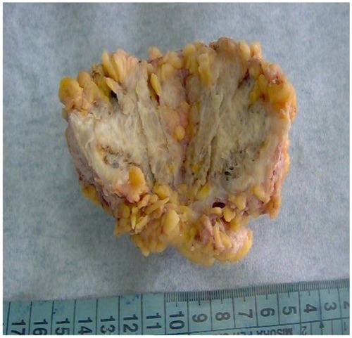
The abdominal skin was re-incised along the outer third of the previous incision and the subcutaneous layer was dissected free of the underlying abdominal musculature. Upon reaching the lower lateral border of the left rectus abdominus muscle, a hard mass was palpated. This mass infiltrated the external oblique, the internal oblique, and the transverse abdominus musculature and reached down to the preperitoneal fat layer. The mass was excised en-block with a small portion of surrounding abdominal wall musculature (). A small suction drain was placed preperitoneally and the abdominal wall defect was closed in one layer using a non-absorbable monofilament suture followed by the placement of a synthetic non-absorbable mesh. The excised specimen was sent for histological examination. The histology report concerned a specimen 3.5 × 2.2 cm in dimension containing fibrous, adipose, and skeletal muscular tissue. Within this tissue specimen, multiple sites of endometriosis were revealed. No findings of malignancy were reported.
Figure 1 Gross appearance of the surgical specimen.
Discussion
The finding of ectopic endometrial tissue within the abdominal wall seems to occur among 0.03% to 1% of women who have undergone prior gynecologic or obstetric surgery, and has been noticed particularly after cesarean section.Citation3–Citation6 Its prevalence as an isolated abdominal wall mass is reported in 4% of the endometrioma cases in another series.Citation7 Other studies report an incidence of 1.04% in patients previously operated on the uterus and 0.046% in those who underwent previous cesarean section.Citation8 Other authors have reported a much higher incidence of about 0.2% of the cesarean sections performed.Citation9
The literature seems to agree that the clinical appearance of a triad consisting of periodic abdominal scar pain (associated with menses), a history of cesarean section, and an underlying tumor inside a surgical scar is more often than not confirmed to be an endometrioma.Citation10 Moreover, the presentation is atypical and thus the preoperative diagnosis poses difficulties in differential diagnosis.Citation11,Citation12 What most authors agree about is the fact that definitive diagnosis can only be established by pathologic analysis of the specimen.
In our case, the patient did not link the unsteady nature of the periodic exacerbation of the pain followed by total pain remittance, with her menstruation. The clinical examination revealed a 2 cm hard, immobile, tender mass, which was definitely located within the abdominal wall musculature. Thus, an all too familiar irreducible incisional hernia with bouts of incarceration was viewed as the most likely diagnosis. The absence of specific intestinal symptoms allowed us to formulate an alternative diagnosis of the possible existence of a post-operative incisional granuloma formation, attributed to the possible presence of underlying suture material or the formation of an incisional endometrial tumor. Malignancy was unlikely, but a small soft tissue sarcoma could not be readily ruled out. During operative dissection no hernia was found and the operative diagnosis tilted towards the existence of a hard granulomatous lesion forming from the scar or fibrous tissue of the previous operation. The patient’s post-operative course was uneventful and she was discharged on the third post-operative day in excellent condition. The histology report unveiled the true nature of the malady. The patient was followed up 5 months post-operatively and was found well. No symptoms or clinical indications of relapse were evident.
This case comes to add to the well-known need to elicit an as accurate as possible history from the patient in which all pieces of information are pertinent, no matter how insignificant they may initially seem. The literature reports a limited number of cases similar to this one. It seems that it is a relatively rare finding, given the number of performed cesarean sections. Although endometriosis was suspected in most cases, in nearly all, the diagnosis was made histologically. Reliable pre-operative diagnosis has been reported with the use of ultrasonography in experienced hands, with the MRI Scan (T1/T2), which can detect blood within the endometrioma, and with the performance of a simple needle aspirate followed by cytological evaluation. The significance of having a reliable clinical diagnosis of an endometrioma lies in the fact that some patients may benefit from hormonal and anti-inflammatory therapy, thus reducing the need for operative intervention in a given number of patients.
Acknowledgments
All authors contributed equally to the writing and presentation of the case.
Disclosure
The authors report no conflicts of interest in this work.
References
- StreuliIde ZieglerDBorgheseBSantulliPBatteuxFChapronCNew treatment strategies and emerging drugs in endometriosisExpert Opin Emerg Drugs3232012 [Epub ahead of print.]
- RömerTTreatment of endometriosisMed Monatsschr Pharm20123524451 quiz 53–5422400429
- MerranSKarila-CohenPIncisional subcutaneous endometrioma of the abdominal wall: report of two casesJ Radiol2004854 Pt 140941015213651
- VélezSEPiccinniDJCaminosSSpitaleLSFerrariJCAbdominal wall endometriosis: case reportRev Fac Cien Med Univ Nac Cordoba2004611444715366236
- OzelLSagirogluJUnalAAbdominal wall endometriosis in the cesarean section surgical scar: a potential diagnostic pitfallJ Obstet Gynaecol Res32012383526530 Epub February 16, 201222381104
- DonatiMGandolfoLCavallaroGCiancioFBrancatoGEndometriosis of the abdominal wall (authors’ experience)Ann Ital Chir20047512934 discussion 3415283384
- BlancoRGParithivelVSShahAKGumbsMAScheinMGerstPHAbdominal wall endometriomasAm J Surg2003185659659812781893
- ZhaoXYLangJHLengJHClinical characteristics of abdominal wall endometrioma and its recurrence-related factorsZhonghua Fu Chan Ke Za Zhi20043929710015059586
- KhammashMROmariAKGasaimehGRBani-HaniKEAbdominal wall endometriosis. An overlooked diagnosisSaudi Med J200324552352512847630
- Esquivel-EstradaVBriones-GarduñoJCMondragón-BallesterosREndometriosis implant in cesarean section surgical scarCir Cir200472211311515175127
- CapassoLScianoDIarrobinoGExtrauterine endometriosis: three new casesG Chir2004251–2394215112760
- DivaniSVardouliAExarhosNLioupisAEndometriosis in the differential diagnosis of abdominal wall massesActa Cytol200347594494514526686