Abstract
The cases of two brothers training and competing as master cyclists and both preparing for a cycling tour are presented. The older brother aged 66 years went first to the primary care physician and presented with an asymptomatic depression in the exercise stress test of the ST segment in V5 and V6 during recovery after complete exhaustion. Coronary angiography revealed a multi vessel coronary artery disease and he underwent bypass surgery. One year later, he successfully completed his planned cycling tour of ~600 km in seven stages and covering ~12,000 m of total ascent. The younger brother aged 59 years went a few months later to the primary care physician and also performed asymptomatic exercise stress testing without changes in the ST segments. Unfortunately, 2 months later he suffered a cardiac arrest during his cycling tour and survived following immediate successful cardiopulmonary resuscitation on the road by his cycling colleagues. Immediate invasive coronary arteriography showed a complete stenosis of the trunk of arteria coronaria sinistra (left coronary artery), a 40%–50% stenosis of ramus circumflexus, and a 20% stenosis of arteria coronaria dextra (right coronary artery). The left coronary artery was dilated and he continued cycling 2 months later. In both brothers, familial hypercholesterolemia was the main cardiovascular risk factor for the multi vessel coronary artery disease. A negative exercise electrocardiogram in siblings with an increased risk for coronary artery disease seemed not to exclude an advanced multi vessel coronary artery disease. In master athletes with asymptomatic exercise electrocardiogram but a positive family history, further examinations should be performed in order to detect relevant stenosis in coronary arteries.
Introduction
The exercise electrocardiogram (ECG) is widely considered the best available test for screening asymptomatic adults without known cardiovascular disease prior to initiating a vigorous exercise program due to its prognostic value, widespread availability, and low cost.Citation1 Generally, the exercise ECG is used as a non-invasive method to evaluate coronary artery disease.Citation2 It has been reported that the exercise ECG has value in diagnosing coronary artery disease and predicting its severity.Citation3 The diagnostic accuracy of an exercise ECG varies depending on the age, the gender, and the clinical characteristics of the patient.Citation4 We report the case of two brothers without known cardiovascular disease training and preparing as master cyclists defined as athletes who exceeded the age requisite for success in elite competition; ie, typically 35–40 yearsCitation5,Citation6 for an event with a different outcome after exercise stress testing.
Case presentation
In June 2011, the 66 year old brother A went for a checkup prior to a planned cycling tour from Geneva (Switzerland) over the Alps to Nice (France) at the Mediterranean coast. He was healthy, had no medical problems, and trained frequently during his work life while he regularly competed in master races. During the year, he completed on average ~12,000 cycling kilometers mainly in road cycling. After his retirement, he intensified his training to prepare for the planned cycling tour and went for a routine check before starting the tour. The primary care physician performed an exercise ECG after a physical examination and some basic laboratory measurements. The physical examination of the lean master athlete with 1.76 m body height and 61 kg body mass (body mass index of 19.7 kg/m2) showed no abnormalities. The laboratory results are presented in and the ECG at rest is shown in . In the ECG at rest, high T waves in the precordial leads impress. In the exercise ECG (), he achieved 197 Watt at maximum load, equal to 146% of his target. Blood pressure was 120/60 mmHg at rest and achieved a maximum of 180/80 mmHg at maximum exertion. The ST segment decreased 0.9 mm in V5 and 1.0 mm in V6 during the recovery phase (4 minutes) without symptoms. The primary care physician decided to send him for invasive coronary angiography. This examination showed a 50%–70% stenosis of ramus interventricularis anterior (RIVA; left anterior descending, LAD), a 95% stenosis of arteria coronaria dextra (right coronary artery; RCA) and a 95% stenosis of ramus posterolateralis of the RCA. Due to these multiple stenoses, the cardiologist recommended cancelling the planned cycling tour and sent the patient to coronary artery bypass surgery. The heart surgeon performed a myocardial revascularization with arteria mammaria interna dextra on ramus posterolateralis dexter and ramus interventricularis posterior, and arteria mammaria interna sinistra on ramus diagonalis and ramus interventricularis anterior. The patient recovered well during a stationary rehabilitation and completed his planned cycling tour successfully in the summer of 2012.
Figure 1 ECG at rest from brother A.
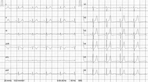
Figure 2 Exercise ECG from brother A.
Abbreviations: aVF, augmented unipolar limb lead in which the positive electrode is on the left leg; aVL, augmented unipolar limb lead in which the positive electrode is on the left arm; aVR, augmented unipolar limb lead in which the positive electrode is on the right arm; ECG, electrocardiogram.
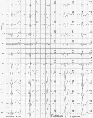
Table 1 Laboratory results of the two brothers at the time of the checkup
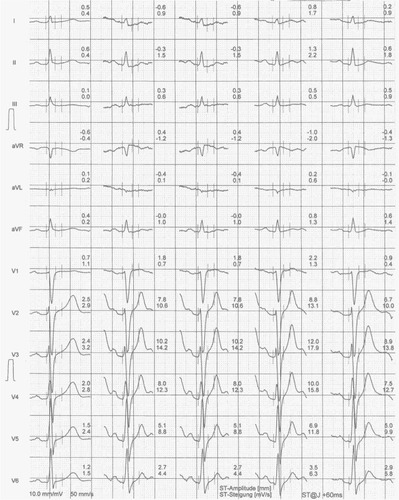
In March 2012, 59-year old brother B went to the same primary care physician also prior to a planned cycling tour in the Alps. He was also training frequently for mountain bike cycling and had neither cardiac symptoms nor any other medical problems. During the year, he completed on average ~9,000 cycling kilometers in both road and mountain bike cycling. The physical examination of the master athlete with 1.72 m body height and 69 kg body mass (body mass index of 23.3 kg/m2) showed no abnormalities. The laboratory results are presented in and the ECG at rest is shown in . In the exercise ECG (), he achieved 229 Watt, equal to 151% of his target. Blood pressure was 120/80 mmHg at rest and achieved a maximum of 220/90 mmHg at maximum exertion. The ST segment showed no changes at both maximum exertion and in the recovery phase. Blood pressure returned to 140/80 mmHg by the end of the test. Due to the high cholesterol and the increased thyroid stimulating hormone, the primary care physician decided to start with a lipid-lowering drug (40 mg of simvastatin) and 0.05 mg levothyroxine due to the latent hypothyroidism. The patient prepared for this planned cycling tour in the Alps starting May 17, 2012. The first day in a steep ascent in a mountain pass, he suffered an acute heart attack with cardiac arrest. His colleagues performed a successful cardiopulmonary resuscitation on the road and he was transferred by helicopter to the nearest hospital. The ECG by the rescue team showed ventricular fibrillation. Immediate invasive coronary arteriography showed a complete stenosis of arteria coronaria sinistra (LCA), a 40%–50% stenosis of ramus circumflexus, and a 20% stenosis of arteria coronaria dextra (RCA). The LCA was dilated. Due to the fall off the bike, he also suffered a fracture of his first vertebra of the lumbar spine (L1). The ejection fraction of the left ventricle was 55% upon discharge from hospital. He was sent to cardiac rehabilitation where he fully recovered. Two months after the cardiac arrest, he was again training and cycling.
Figure 3 ECG at rest from brother B.
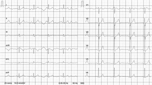
Figure 4 Exercise ECG from brother B.
Abbreviations: aVF, augmented unipolar limb lead in which the positive electrode is on the left leg; aVL, augmented unipolar limb lead in which the positive electrode is on the left arm; aVR, augmented unipolar limb lead in which the positive electrode is on the right arm; ECG, electrocardiogram.
In the family history of the two brothers, the father died at the age of 70 years due to a heart attack. The mother died at the age of 90 years with no history of coronary heart disease. In autumn 2012, the third brother of the two brothers described herein was sent for an exercise ECG in order to assess the risk of coronary artery disease. This brother also achieved more than the target in the exercise stress test and the exercise ECG revealed no ST segment depression. Due to hypercholesterolemia, he was treated since 2008 with 40 mg of atorvastatin. He agreed to invasive coronary arteriography due to his high risk of heart attack. This examination showed advanced atherosclerosis in all of his coronary arteries.
Discussion
In brother A, the ST segment depression appeared in the recovery phase of the exercise ECG, but not during exercise. Lanza et al investigated the diagnostic and prognostic value of an ST segment depression limited to the recovery phase of an exercise stress test and compared it to that of an ST segment depression during exercise.Citation7 They found that the diagnostic and prognostic power of the ST segment depression limited to the recovery phase of an exercise ECG was largely similar to that of the ST segment depression induced during effort. Also, Rashid et al reported that in patients undergoing exercise ECG, ST segment depression occurring only in the recovery phase of an exercise ECG had diagnostic value largely comparable with that of ST segment depression induced during the exercise phase of the exercise ECG.Citation8 The post-exercise ST segment depression without ST segment depression during exercise ECG may be associated with an abnormal response to imbalances in the cardiovascular sympathetic system in the post-exercise period rather than with myocardial ischemia.Citation9
Both brothers suffered from a multi vessel coronary artery disease. However, only in brother A did the exercise ECG reveal an ST segment depression in the recovery phase. Sun et al found that patients with multi vessel disease had a higher positive rate in exercise ECG compared to those with single vessel disease.Citation10 Wu et al compared the results of exercise ECG and coronary angiography in 461 patients.Citation11 They showed that positive results of exercise ECG with downslope or horizontal depression of the ST segment more than 0.1 mV during or after exercise may predict coronary artery disease, the sensitivity of which was 74.5% (322 cases) and the specificity as 84.2% (139 cases). With the use of ST depression more than 0.2 mV as the super-positive standard, the sensitivity and specificity of positive exercise ECG to predict triple vessel disease and left main coronary disease was 61.3% and 79.1%, respectively. The duration of the ST segment depression had a positive correlation with the number of diseased coronary arteries. The patients with multi vessel disease and left main coronary artery disease had a higher positive rate of exercise ECG testing compared to patients with single or double vessel disease. It was concluded that exercise ECG testing had some value in diagnosing coronary artery disease and predicting its severity. Some parameters in exercise ECG were also helpful for predicting the severity of coronary artery disease.
Pigozzi et al evaluated the prevalence and clinical significance of ST segment depression during an exercise ECG in 113 asymptomatic apparently healthy elderly male athletes.Citation12 A significant ST segment depression at peak exercise was detected in one athlete at the first evaluation. A further case was seen during the follow up period in a previously “negative” athlete. Both athletes were asymptomatic and single photon emission computed tomography (SPECT) and/or stress echocardiography (ECHO) were negative for myocardial ischemia. They concluded that athletes with exercise induced ST segment depression with no associated symptoms and/or complex ventricular arrhythmias and no adverse findings at second level cardiologic testing should be considered free from coronary disease and safe to continue athletic training.
For master athletes, Freeman et al recommend screening exercise ECG in asymptomatic men with diabetes and asymptomatic men over the age of 45 years with two or more cardiovascular risk factors prior to initiating a vigorous exercise program.Citation1 In the present case, it was pure luck that the coronary artery disease was discovered. The primary care physician performed the exercise stress test in the older brother only to show the wife that the master athlete would be able to complete the planned bike ride.
In these two brothers, the father died at the age of 70 years due to a myocardial infarction. Leander et al showed that family history of coronary heart disease is not only a strong risk factor for myocardial infarction in both sexes, but that its effect was synergistic with other cardiovascular risk factors, as well.Citation13 Siblings of patients with coronary artery disease would benefit from investigating and treating cardiovascular risk factors. Hengstenberg et al analyzed the risk profile as well as the current utilization of preventive strategies in asymptomatic siblings of patients with myocardial infarction.Citation14 Most asymptomatic individuals with positive fraternal family history had more than one modifiable risk factor. Rissanen investigated the occurrence of different clinical manifestations of coronary heart disease among the parents and siblings of 309 men with coronary heart disease, including 103 men with fatal and 100 with nonfatal myocardial infarction, and 106 men with angina pectoris and among the relatives of 106 reference men.Citation15 Coronary heart disease was equally common among relatives of all subjects. It was four times as common in case brothers and twice as common in case sisters as in the respective reference siblings. Cardiac deaths were most common in the siblings of men with fatal myocardial infarction, and uncomplicated angina in the men with angina pectoris. The familial resemblance in the clinical manifestations of coronary heart disease suggested familial influence in the mechanisms determining the clinical expression of the disease.
Most of the strong familial component in coronary heart disease of early onset thus appears to be mediated by familial hyperlipidemia and hypertension.Citation16 The risk of premature coronary heart disease in persons at highest risk could be largely eliminated if information about family history were used to identify such persons at an early stage and if they were treated properly for their correctable risk factors.Citation16 Both brothers showed hypercholesterolemia upon their first visit to the primary care physician. The high cholesterol values as a high risk for heart diseases can explain the result of both brothers, instead of the positive family history. The younger brother also had a subclinical hypothyroidism with increased thyroid stimulating hormone levels. High thyroid stimulating hormone levels are associated with a non-favorable lipid profileCitation17 and with an increased risk for all-cause and cardiovascular mortality in adults.Citation18 Most of the heart attacks, however, happen not with non-significant stenosis but with mild to moderated disease with plaque rupture, causing ST-elevation myocardial infarction.Citation19 Overall, the patient would have needed better risk factor management rather than a further cardiac evaluation as he was asymptomatic.
Apart from hypercholesterolemia, diabetes mellitus type II is also an important risk factor for coronary artery disease.Citation20,Citation21 Brother B had elevated blood glucose upon the check-up. However, the blood samples were not drawn in an absolute fasting state and subsequent determination of blood glucose showed normal values. Further, lipoprotein(a) is an established risk marker of coronary artery disease, which is independent from other risk factors.Citation22,Citation23 Unfortunately, this marker was not determined in these two brothers.
In brother A, impressive T waves were recorded in the precordial leads prior to the exercise ECG. A potential reason for high T waves in precordial leads could be hyperkalaemia. However, the electrolytes potassium (3.9 mmol/L), sodium (138 mmol/L), and creatinine (82 μmol/L) were in the normal range during the hospital stay for the coronary angiography.
A negative exercise ECG in siblings with an increased risk for coronary artery disease seemed not to exclude advanced multi vessel coronary artery disease. In master athletes with asymptomatic exercise ECG but a positive family history, further examinations should be performed in order to detect relevant stenosis in coronary arteries. Technological advances in recent decades have produced several methods of non-invasive cardiac imaging for the diagnosis of coronary artery disease, such as stress ECHO, myocardial perfusion radionuclide imaging, cardiac computed tomography, and cardiac magnetic resonance imaging. There is always a balance of cost and benefit for further examinations. The choice of an appropriate technique depends on the availability of the method, the patient’s clinical characteristics, and other factors, such as costs, risks, radiation, and potential contrast agent toxicity.Citation24,Citation25 Stress ECHO and myocardial perfusion scintigraphy are widely available and most extensively validated. Stress ECHO is a non-invasive technology that images the heart using ultrasound and is a useful imaging modality for the diagnosis of coronary artery disease in suspected patients.Citation26 Myocardial perfusion scintigraphy is a widely used nuclear, non-invasive image acquisition technique for investigating ischemic heart disease.Citation27
Disclosure
The authors report no conflicts of interest in this work.
References
- FreemanJFroelicherVAshleyEThe ageing athlete: screening prior to vigorous exertion in asymptomatic adults without known cardiovascular diseaseBr J Sports Med200943969670119734505
- SharmaKKohliPGulatiMAn update on exercise stress testingCurr Probl Cardiol201237517720222469057
- OvrehusKAJensenJKMickleyHFComparison of usefulness of exercise testing versus coronary computed tomographic angiography for evaluation of patients suspected of having coronary artery diseaseAm J Cardiol2010105677377920211318
- BanerjeeANewmanDRVan den BruelAHeneghanCDiagnostic accuracy of exercise stress testing for coronary artery disease: a systematic review and meta-analysis of prospective studiesInt J Clin Pract201266547749222512607
- RansdellLBVenerJHubertyJMaster athletes: an analysis of running, swimming and cycling performance by age and genderJ Exerc Sci Fit200976173
- ReaburnPDascombeBEndurance performance in masters athletesEur Rev Aging Phys Act200853142
- LanzaGAMustilliMSestitoAInfusinoFSguegliaGACreaFDiagnostic and prognostic value of ST segment depression limited to the recovery phase of exercise stress testHeart200490121417142115547017
- RashidMAMallickNHAlamSNNoemanAEhsanAHussainAUsefulness of ST segment depression limited to the recovery phase of exercise stress testJ Coll Physicians Surg Pak20091913619149971
- ShinmuraKSuganumaYHasegawaHClinical significance and mechanism of ST-segment changes during the post-exercise recovery period in male patients with coronary artery diseaseJ Cardiol1996275231239 Japanese8642510
- SunJLHanRGuoJHLiXYMaXLWangCYThe diagnostic value of treadmill exercise test parameters for coronary artery diseaseCell Biochem Biophys2013651697622872585
- WuLZhangJShaoGA comparative study on coronary angiography and exercise ECG testingZhonghua Nei Ke Za Zhi1996352107109 Chinese9275629
- PigozziFSpataroAAlabisoARole of exercise stress test in master athletesBr J Sports Med200539852753116046336
- LeanderKHallqvistJReuterwallCAhlbomAde FaireUFamily history of coronary heart disease, a strong risk factor for myocardial infarction interacting with other cardiovascular risk factors: results from the Stockholm Heart Epidemiology Program (SHEEP)Epidemiology200112221522111246583
- HengstenbergCHolmerSRMayerBSiblings of myocardial infarction patients are overlooked in primary prevention of cardiovascular diseaseEur Heart J2001221192693311428816
- RissanenAMFamilial occurrence of coronary heart disease according to clinical manifestationActa Med Scand198521843553634083078
- RissanenAMFamilial occurrence of coronary heart disease: effect of age at diagnosisAm J Cardiol19794416066453047
- DuntasLHBrentaGThe effect of thyroid disorders on lipid levels and metabolismMed Clin North Am201296226928122443975
- TsengFYLinWYLinCCSubclinical hypothyroidism is associated with increased risk for all-cause and cardiovascular mortality in adultsJ Am Coll Cardiol201260873073722726629
- AndersonJLAdamsCDAntmanEM2011 Writing Group MembersACCF/AHA Task Force Members2011 ACCF/AHA Focused Update Incorporated Into the ACC/AHA 2007 Guidelines for the Management of Patients With Unstable Angina/Non-ST-Elevation Myocardial Infarction: a report of the American College of Cardiology Foundation/American Heart Association Task Force on Practice GuidelinesCirculation201112318e426e57921444888
- Sheikh-AliMRahejaPBorja-HartNMedical management and strategies to prevent coronary artery disease in patients with type 2 diabetes mellitusPostgrad Med20131251173323391668
- MalikSBillimekJGreenfieldSSorkinDHNgo-MetzgerQKaplanSHPatient complexity and risk factor control among multimorbid patients with type 2 diabetes: results from the R2D2C2 studyMed Care201351218018523047130
- JaegerBRRichterYNagelDLongitudinal cohort study on the effectiveness of lipid apheresis treatment to reduce high lipoprotein(a) levels and prevent major adverse coronary eventsNat Clin Pract Cardiovasc Med20096322923919234501
- JaegerBRLabarrereCAGenetic variants in Lp(a) lipoprotein and coronary diseaseN Engl J Med20103621211461147 author reply 114720344827
- JaarsmaCLeinerTBekkersSCDiagnostic performance of noninvasive myocardial perfusion imaging using single-photon emission computed tomography, cardiac magnetic resonance, and positron emission tomography imaging for the detection of obstructive coronary artery disease: a meta-analysisJ Am Coll Cardiol201259191719172822554604
- DowsleyTAl-MallahMAnanthasubramaniamKDwivediGMcArdleBChowBJThe role of noninvasive imaging in coronary artery disease detection, prognosis, and clinical decision makingCan J Cardiol201329328529623357601
- Health Quality OntarioStress echocardiography for the diagnosis of coronary artery disease: an evidence-based analysisOnt Health Technol Assess Ser2010109161
- Health Quality OntarioSingle photon emission computed tomography for the diagnosis of coronary artery disease: an evidence-based analysisOnt Health Technol Assess Ser2010108164