
 ?Mathematical formulae have been encoded as MathML and are displayed in this HTML version using MathJax in order to improve their display. Uncheck the box to turn MathJax off. This feature requires Javascript. Click on a formula to zoom.
Abstract
The aim of this study was to compare the biological synthesis of gold nanoparticles (AuNPs) generated using the aqueous extracts of outer oriental melon peel (OMP) and peach. The synthesized OMP-AuNPs and peach extract (PE)-AuNPs were characterized by ultraviolet–visible spectroscopy, field emission scanning electron microscopy, energy dispersive X-ray analysis, X-ray powder diffraction, Fourier transform infrared spectroscopy, and thermogravimetric analysis. The surface plasmon resonance spectra were obtained at 545 nm and 540 nm for OMP-AuNPs and PE-AuNPs, respectively. The estimated absolute crystallite size of the synthesized AuNPs was calculated to be 78.11 nm for OMP-AuNPs and 39.90 nm for PE-AuNPs based on the Scherer equation of the X-ray powder diffraction peaks. Fourier transform infrared spectroscopy results revealed the involvement of bioactive compounds present in OMP and peach extracts in the synthesis and stabilization of synthesized AuNPs. Both the OMP-AuNPs and PE-AuNPs showed a strong antibacterial synergistic activity when combined with kanamycin (9.38–20.45 mm inhibition zones) and rifampicin (9.52–25.23 mm inhibition zones), and they also exerted a strong synergistic anticandidal activity (10.09–15.47 mm inhibition zones) when combined with amphotericin B against five pathogenic Candida species. Both the OMP-AuNPs and PE-AuNPs exhibited a strong antioxidant potential in terms of 1,1-diphenyl-2-picrylhydraxyl radical scavenging, nitric oxide scavenging, 2,2′-azino-bis(3-ethylbenzothiazoline-6-sulphonic acid) radical scavenging, and a reducing power, along with a strong proteasome inhibitory potential that could be useful in cancer drug delivery and cancer treatments. The PE-AuNPs showed comparatively higher activity than OMP-AuNPs, which could be attributed to the presence of rich bioactive compounds in the PE that acted as reducing and capping agents in the synthesis of PE-AuNPs. Overall, the results of the current investigation highlighted a novel green technology for the synthesis of AuNPs using food waste materials and their potential applications in the biomedical, pharmaceutical, and cosmetic industries.
Introduction
Nanotechnology is a field of applied science and technology that deals with materials in the nanoscale range with large surface areas.Citation1 These nanoscale particles, which are most commonly known as nanoparticles (NPs), have the ability to build micro-/macro-materials and products with atomic precision. NPs have recently drawn tremendous attention because of their potential applications in chemical, catalytic, electronic, optical, mechanical, magnetic, and medical fields.Citation2–Citation4 NPs are usually synthesized by chemical or physical approaches because of their inherent advantage in producing well-defined NPs with fairly controllable shapes and sizes.Citation5 However, these methods involve tedious treatments, such as microemulsion, laser photolysis, hydrothermal induction, and toxic chemical reduction, along with expensive physical techniques and equipment, and are potentially toxic to the environment.Citation6–Citation8 Thus, to avoid the adverse effects of NPs on the environment, the concept of green chemistry evolved. This method utilizes biological organisms for the fabrication of NPs.
In general, the synthesis of NPs using biological organisms such as microorganisms, plants or plant extracts, algae, and fungi is environmentally benign and cost-effective.Citation9 However, there are certain drawbacks in the synthesis of NPs using microorganisms because of difficulties in separation and purification procedures.Citation10 In contrast, plant biomass-mediated biosynthesis of NPs has recently attracted a great deal of attention because of their easy synthesis and extraction processes.Citation11 Naturally available biological, food, and agricultural waste materials have not been extensively investigated for the synthesis of different types of NPs.
Oriental melons and peaches are commonly consumed in East Asia. Basically, the juicy internal portions of both fruits are edible, while the outer peels are generally discarded and considered useless. The oriental melon (Cucumis melo L. var. makuwa) that belongs to the family Cucurbitaceae is primarily grown in South Korea, People’s Republic of China, and Japan.Citation12 The word melon is derived from the Latin name “melopepo” meaning melon. The fruit is ~10 cm long and typically weighs slightly more than one pound (0.45 kg).Citation12 This fruit has been utilized in Korean folk medicine for treatment of acute gastritis, fever, mental disorders, dysuria, jaundice, alcoholism, and hyperesthesia/paralysis.Citation13,Citation14 Peach (Prunus persica L. Batsch) that belongs to the family Rosaceae is a fruit of the deciduous tree native to Northwest China, between the Tarim basin and the northern slopes of the Kunlun Shan mountains.Citation15 This fruit is cultivated in many countries including the People’s Republic of China, Japan, South Korea, and Vietnam. Peaches are considered as important economic crops with potential bioactive compounds and medicinal benefits.Citation16,Citation17 The fruit is rich in a number of phenolic compounds, including chlorogenic acid, catechin, epicatechin, rutin, and cyanidin-3-glucoside,Citation18 and is utilized as a demulcent, antiscorbutic, and a stomachic agent.Citation19
NPs, especially gold, have recently attracted a great deal of attention due to their nontoxic nature and extensive applications in biomedical, chemistry, and electronics fields.Citation20,Citation21 Gold NPs (AuNPs) can be easily synthesized and exhibit intense surface plasmon resonance (SPR) with high chemical and thermal stability.Citation21 Investigation of the morphological behavior of gold nanostructures is important because of their wide usage in catalysis, optics, optical electronics, microelectronics, biodiagnostics, imaging, and biological and chemical sensing techniques.Citation21,Citation22 Biomedical applications of AuNPs such as drug and gene delivery, protein and pathogen detection, deoxyribonucleic acid labeling, fluorescent labeling, tissue engineering, photothermal ablation, and as contrast agents for magnetic resonance imaging and other imaging methods have become a highly active area of research during recent years.Citation23,Citation24
Many studies have investigated the biosynthesis of AuNPs using various plant extracts.Citation25–Citation28 However, only a few studies have investigated the synthesis of AuNPs using food waste materials.Citation29,Citation30 In the current study, the reducing ability of aqueous extracts of the outer peels of two common fruits, oriental melon and peach, were investigated to determine their usefulness for the synthesis of AuNPs. In addition, their antibacterial, anticandidal, antioxidant, and proteasome inhibitory potentials were evaluated.
Materials and methods
Preparation of aqueous extract of fruit waste materials
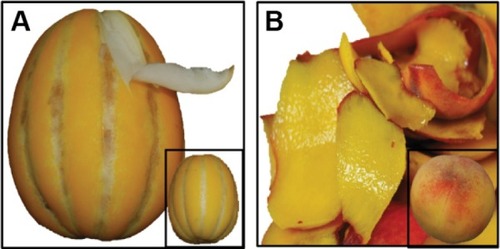
The fruits of oriental melon (Cucumis melo L. var. makuwa; ) and peach (Prunus persica L.; ) were purchased from a local market (Gyeongsan, South Korea). The fruits were washed thoroughly with double-distilled water and dried with tissue paper, and the outer nonedible portion of the fruits was peeled off using a peeler. The outer peels were subsequently cut into small pieces (~10 mm) with a knife, divided into 50 g aliquots, and immersed in 250 mL of deionized water in two separate 500 mL conical flasks. The mixtures were subsequently boiled for 15 minutes with continuous stirring using a magnetic stirrer, after which the aqueous oriental melon peel (OMP) extract and the aqueous peach extract (PE) were cooled to room temperature. Both the OMP and peach extracts were then filtered through Whatman No 1 filter paper, collected into separate sterilized bottles, and kept at 4°C until further use.
Figure 1 Fruit peels of oriental melon (Cucumis melo) (A) and peach (Prunus persica) (B) used for the synthesis of AuNPs.
Abbreviation: AuNPs, gold nanoparticles.
Biosynthesis of AuNPs using OMP and peach extracts
Auric chloride (AuCl3; Sigma-Aldrich Co., St Louis, MO, USA) was used as the precursor element for the synthesis of AuNPs. Prior to the synthesis, 1 mM AuCl3 solution was prepared in double-distilled water in an amber bottle. Approximately 100 mL of AuCl3 solution was then placed into two separate 500 mL conical flasks, after which 10 mL of OMP extract was added to one flask and 10 mL of PE to another flask dropwise using a separating funnel. Samples were then incubated at room temperature with continuous stirring for 24 hours.Citation31 The bioreduction and color change of AuCl3 to AuNPs was then monitored with respect to the incubation time. After 24 hours, the synthesized AuNP solution (OMP-AuNPs and PE-AuNPs) was centrifuged at 12,000 rpm for 30 minutes in a high-speed centrifuge machine (Supra 22K; Hanil Science Industrial Co., Ltd., Daejeon, South Korea). The supernatant was then discarded, and the pellets were washed twice with distilled water and dried to powder using a vacuum dryer (LVS 201T; ILMVAC GmbH, Ilmenau, Germany).
Characterization of the synthesized AuNPs
The synthesized OMP-AuNPs and PE-AuNPs were characterized by ultraviolet (UV)–visible spectroscopy, field emission scanning electron microscopy (FE-SEM), energy dispersive X-ray (EDX) analysis, X-ray powder diffraction (XRD), Fourier-transform infrared (FT-IR) spectroscopy, and thermogravimetric (TG) analysis.Citation31,Citation32
The bioreduction of Au ions to AuNPs in the reaction medium by the action of OMP and peach extracts was monitored by measuring the absorption spectra of the colloidal solution between 400 nm and 700 nm using a microplate reader (Infinite 200 PRO NanoQuant; Tecan, Mannedorf, Switzerland). The surface morphology of both OMP-AuNPs and PE-AuNPs was analyzed using an FE-SEM. Prior to the analysis, the OMP-AuNPs and PE-AuNPs were ground into fine powder using an agate mortar and pestle and then uniformly spread on the carbon tapes attached over the sample holder. The samples were subsequently sputter coated with platinum in an ion coater for 120 seconds, after which they were observed using a FE-SEM (S-4200; Hitachi Ltd., Tokyo, Japan). The elemental composition of the OMP-AuNPs and PE-AuNPs was determined using an EDX detector (energy dispersive spectroscopy; EDAX Inc., Mahwah, NJ, USA) attached to the SEM machine. The nature of the powdered OMP-AuNPs and PE-AuNPs was determined using an XRD machine (X’Pert MRD model; PANalytical, Almelo, the Netherlands). The NPs were dried at 60°C in a vacuum oven and ground into fine powder using an agate mortar and pestle. XRD analysis was performed at 30 kV and 40 mA with Cu Kα radians (l=1.5406 Å) at an angle of 2θ, while scanning from 20° to 90°.Citation31 FT-IR analysis of OMP, PE, powdered OMP-AuNPs, and powdered PE-AuNPs was conducted using an FT-IR spectrophotometer (Jasco 5300; Jasco, Easton, MD, USA) in the wavelength range of 400–4,000 cm−1. Prior to use, the powdered OMP-AuNPs and PE-AuNPs were blended with powdered potassium bromide (KBr) in a 1:100 ratio, then compressed into a 2 mm semitransparent disk to prepare a thin film of NPs. The OMP and peach extracts were used directly for FT-IR analysis. Both the NPs and OMP and peach extracts were analyzed for various modes of vibrations for the presence of different types of functional groups. The TG/derivative TG (DTG) analysis of the powdered OMP-AuNPs and PE-AuNPs was conducted using a TG analysis machine (SDT Q600; TA Instruments, New Castle, DE, USA). Prior to use, the powdered OMP-AuNPs and PE-AuNPs were placed in an alumina pan and heated from 20°C to 700°C at a ramping time of 10°C/min under nitrogen atmosphere.
Synergistic antibacterial activity of synthesized AuNPs
The synergistic antibacterial potential of both OMP-AuNPs and PE-AuNPs was determined against five different foodborne bacteria, namely Bacillus cereus American Type Culture Collection (ATCC) 13061, Listeria monocytogenes ATCC 19115, Staphylococcus aureus ATCC 49444, Escherichia coli ATCC 43890, and Salmonella typhimurium ATCC 43174, using the standard disk diffusion method.Citation33,Citation34 The pathogenic bacteria were obtained from the American Type Culture Collection (ATCC, Manassas, VA, USA) and maintained in nutrient broth (Difco; Becton, Dickinson and Company, Sparks Glencoe, MD, USA) medium at −80°C. Immediately before use, a colloidal solution of OMP-AuNPs and PE-AuNPs was prepared by dissolving 1,000 μg/mL in 5% dimethyl sulphoxide (DMSO), then sonicated at 30°C for 15 minutes. Filter paper disks containing 25 μg of the test compound per disk were used for the initial antibacterial activity test. OMP and peach extracts were taken as extract controls, and kanamycin and rifampicin at 5 μg/disk were taken as positive controls, while 5% DMSO was used as the negative control. To explore the synergistic effects of the AuNPs, 1,000 μg/mL of OMP-AuNPs or PE-AuNPs were mixed in a 1:1 ratio with 200 μg/mL of the two standard antibiotics separately and then sonicated for 15 minutes at room temperature. A total of 50 μL of the mixture of AuNP–antibiotics containing 25 μg AuNPs and 5 μg antibiotics were put on the filter paper disk, dried, and used for determination of the synergistic antibacterial effect. The diameter of zones of inhibition around each paper disk was measured after 24 hours of incubation at 37°C to determine the synergistic antibacterial effect of the mixture of AuNPs/antibiotics.
Synergistic anticandidal activity of synthesized AuNPs
The synergistic anticandidal activity of OMP-AuNPs or PE-AuNPs with a standard anticandidal agent, amphotericin B, was determined against five different pathogenic Candida species, namely Candida albicans KACC 30003 and KACC 30062, Candida glabrata KBNO6P00368, Candida geochares KACC 30061, and Candida saitoana KACC 41238, using the disk diffusion method.Citation35 All Candida strains were obtained from the Korean Agricultural Culture Collection (KACC, Suwon, South Korea), except for C. glabrata KBNO6P00368, which was obtained from Chonbuk National University Hospital (Cheongju, South Korea). Filter paper disks containing 50 μg/disk of AuNPs were initially tested for their anticandidal activity. To explore the synergistic anticandidal activity, the OMP-AuNPs or PE-AuNPs (2,000 μg/mL) were mixed at a 1:1 ratio with amphotericin B (200 μg/mL) separately and then sonicated for 15 minutes at room temperature. Filter paper disks were prepared by adding 50 μL of the AgNPs/amphotericin B mixture solution to the sterile paper disks, which finally contained 50 μg AuNPs and 5 μg amphotericin B/disk. Plates of potato dextrose agar (Difco) were spread uniformly with the Candida species grown in potato dextrose broth, after which the filter paper disks were placed over it and incubated at 28°C for 48 hours. OMP and peach extracts were taken as extract controls, amphotericin B at 5 μg/disk were taken as positive control, while 5% DMSO was used as the negative control. Finally, the diameters of the zones of inhibition around each paper disk were measured to determine the synergistic anticandidal activity of the mixture of AuNPs/amphotericin B.
Antioxidant activity of synthesized AuNPs
The in vitro antioxidant potential of the AuNPs was determined by four antioxidant assays, including 1,1-diphenyl-2-picrylhydraxyl (DPPH) free radical scavenging, nitric oxide scavenging, 2,2′-azino-bis(3-ethylbenzothiazoline-6-sulphonic acid) (ABTS) free radical scavenging, and a reducing power assay. The OMP-AuNPs, PE-AuNPs, and butylated hydroxyl toluene (BHT), which was taken as the standard reference compound, were used at 20–100 μg/mL for all assays. The DPPH free radical scavenging potential of both OMP-AuNPs and PE-AuNPs was determined as described by Braca et al.Citation36 The absorbance of the reaction mixtures was recorded at 517 nm using a microplate reader, and the results in terms of the percentage scavenging effect were calculated using the following equation:
The nitric oxide radical scavenging potential of both OMP-AuNPs and PE-AuNPs was determined by the standard method as described by Makhija et al.Citation37 The absorbance of the reaction mixtures was measured at 546 nm using a microplate reader, and the results in terms of the percentage scavenging effect were calculated according to EquationEquation 1(1) . The ABTS radical scavenging activities of OMP-AuNPs and PE-AuNPs were determined as described by Thaipong et al.Citation38 The absorbance of the reaction mixtures was recorded at 750 nm using a microplate reader, and the results in terms of the percentage scavenging effect were calculated according to EquationEquation 1
(1) . The reducing powers of the OMP-AuNPs and PE-AuNPs were determined using the procedure described by Patra and Baek.Citation39 The reducing power of AuNPs was determined in terms of the absorbance value at 700 nm.
Proteasome inhibitory activity of synthesized AuNPs
The proteasome inhibitory potentials of OMP-AuNPs and PE-AuNPs were assayed using the 20S Proteasome Assay Kit for drug discovery (ENZO Life Sciences, Farmingdale, NY, USA) according to the manufacturer’s protocols. Three different concentrations of both OMP-AuNPs and PE-AuNPs (1 μg/mL, 10 μg/mL, and 100 μg/mL) and epoxomicin, a standard reference compound (0.56 μg/mL, 2.78 μg/mL, and 5.56 μg/mL) were used for the assay. Suc-LLVY-AMC (37.5 mM) was used as the substrate to estimate the chymotrypsin-like proteasomal activity of the AuNPs in a 96-well microplate from 1 minute to 12 minutes of incubation. The fluorescence intensities were measured at an excitation wavelength of 350 nm and emission wavelength of 440 nm at a 30°C reaction temperature using the microplate reader.
Statistical analysis
All data were calculated as mean ± SD. Statistical analysis using one-way ANOVA followed by Duncan’s test at the 5% level of significance (P<0.05) was conducted using the Statistical Analysis Software (SAS, Version: SAS 9.4; SAS Institute Inc., Cary, NC, USA).
Results and discussion
Synthesis and characterization of AuNPs
There have been many reports on the use of whole fruits in various biological activities and medicinal potentials; however, only a few studies have investigated the importance of their peels. Fruit peels are rich in polyphenolic compounds, flavonoids, ascorbic acid, and a number of other biologically active components that have positive effects on health.Citation40–Citation43 In the recent past, there have been reports on the pharmacological effects of some fruit peel extracts against diseases such as cardiovascular problems, thyroid abnormalities, and diabetes mellitus.Citation29,Citation43,Citation44 Thus, we investigated the synthesis of AuNPs utilizing the fruit peels of two fruits common in East Asia, oriental melon and peach, and then compared their potential applications. AuNPs were synthesized by taking AuCl3 as the precursor element and both OMP and peach extracts as the reducing agents (). The bioreduction of Au+ to Au0 (AuNPs) first started vigorously after 1 hour of incubation in the PE-mediated synthesis compared to the OMP-mediated AuNP synthesis, during which time there was a gradual change in the color of the reaction mixture from colorless to purple (, inset). The solution was dark purple at 6 hours of incubation in both the cases; however, the reaction continued for up to 24 hours until the complete reduction of Au+ in the reaction mixture (, inset).
Figure 2 UV–visible spectroscopy and change in color of the reaction mixture at different time intervals of the synthesized OMP-AuNPs and PE-AuNPs.
Notes: (A) OMP-AuNPs; (B) PE-AuNPs.
Abbreviations: AuNPs, gold nanoparticles; OMP, oriental melon peel; PE, peach extract; UV, ultraviolet.
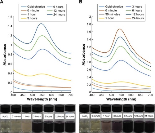
The dark purple color unveiled by the metallic colloidal AuNPs was due to the articulate excitation of all of the free electrons within the conduction band that led to the in-phase oscillation referred to as the SPR.Citation45 The bioactive compounds present in both fruit peels might have been responsible for the bioreduction of Au+ to AuNPs. The oriental melon is a rich source of vitamin A, vitamin C, 5-hydroxy-6,7,8-trimethoxy-2,3-dimethyl-4H-chromen-4-one, and anhalinine;Citation46,Citation47 therefore, it has potential for use as a reducing agent to synthesize OMP-AuNPs. Similarly, peach is rich in phenolic compounds, including chlorogenic acid, catechin, epicatechin, rutin, and cyanidin-3-glucoside;Citation18 therefore, it might be useful as a reducing agent to synthesize PE-AuNPs. Following synthesis, the OMP-AuNPs and PE-AuNPs are subjected to characterization of their structure, morphology, and chemical properties based on UV–visible spectroscopy, FE-SEM, EDX, XRD, FT-IR, and TG/DTG analysis.
UV–visible spectroscopy is a preliminary step in the confirmation of the synthesis and stability of AuNPs with dark purple coloration in aqueous reaction medium. The UV–visible spectra of both OMP-AuNPs and PE-AuNPs were recorded with an interval of 3 hours for 24 hours from 400 nm to 700 nm, and the results are presented in . The PE-AuNPs displayed sharp peaks with the SPR absorbance maxima at 540 nm (), whereas the OMP-AuNPs displayed a small flat peak with the SPR absorbance maxima at 545 nm (). Both sets of spectral data confirmed the synthesis of AuNPs, which were concordant with the results of earlier studies.Citation48 In general, AuNPs have been reported to exhibit a dark purple color in aqueous medium due to the intensity and size of the NPs owing to its SPR.Citation48,Citation49 The SPR absorbance is exceptionally sensitive to the nature, size, and shapes of the NPs formed, dielectric constant of the medium and temperature, and their interparticle distances.Citation50–Citation52
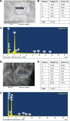
The surface morphology of the synthesized AuNPs was characterized by FE-SEM after 1 month of synthesis following completion of all reactions. Both the OMP-AuNPs and PE-AuNPs displayed slightly agglomerated distinct spherical structures within the nanoscale range that confirmed the synthesis of NPs (). Similar observations were reported in an earlier study.Citation28,Citation53 The elemental composition of the synthesized NP was confirmed by EDX analysis (). The results showed that OMP-AuNPs and PE-AuNPs contained 73.87% and 64.50% gold composition, respectively (). In addition, the OMP-AuNPs contained carbon (14.46%), oxygen (8.36%), chlorine (1.78%), and potassium (1.53%; ), while the PE-AuNPs contained carbon (23.48%) and oxygen (12.02%; ), possibly due to the presence of different types of secondary metabolites such as flavonoids and phenolic compounds in OMP and peach extracts. These secondary metabolites might have acted as the reducing and stabilizing agents during the synthesis of AuNPs.Citation53,Citation54
Figure 3 SEM image of synthesized OMP-AuNPs and PE-AuNPs.
Notes: (A) OMP-AuNPs; (B) PE-AuNPs.
Abbreviations: AuNPs, gold nanoparticles; OMP, oriental melon peel; PE, peach extract; SEM, scanning electron microscopy.
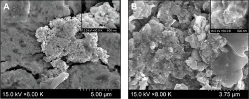
Figure 4 EDX analysis of synthesized OMP-AuNPs and PE-AuNPs.
Notes: OMP-AuNPs (A–C) and PE-AuNPs (D–F).
Abbreviations: AuNPs, gold nanoparticles; EDX, energy dispersive X-ray; OMP, oriental melon peel; PE, peach extract.
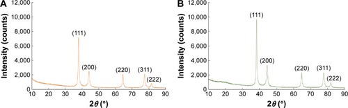
The synthesized AuNPs were further characterized for their structural information and crystallinity nature by XRD analysis (). Both OMP-AuNPs and PE-AuNPs displayed five distinct diffraction peaks in the 2θ range of 10°–90° that indexed the planes (111), (200), (220), (311), and (222) of the face-centered cubic (fcc) gold particles (). These peaks were located at 37.96°, 44.18°, 64.51°, 77.48°, and 81.74°, for OMP-AuNPs () and 38.35°, 44.52°, 64.82°, 77.80°, and 82.00° for PE-AuNPs (). The XRD analysis showed predominant peaks at (111) and (200), indicating the presence of crystallite particles displaying fcc lattice structures. The fcc structures of gold matched those in the database of the Joint Committee on Powder Diffraction Standards, USA (JCPDS No 00-004-0784), confirming that the synthesized AuNPs were composed of pure crystallite gold particles. The estimated absolute crystallite size of the synthesized AuNPs was calculated to be 78.11 nm for OMP-AuNPs and 39.90 nm for PE-AuNPs based on the Scherer equation.Citation55 The XRD patterns of both OMP-AuNPs and PE-AuNPs were consistent with the results of earlier studies of AuNPs.Citation56–Citation58
Figure 5 XRD spectra of synthesized OMP-AuNPs and PE-AuNPs.
Notes: (A) OMP-AuNPs; (B) PE-AuNPs.
Abbreviations: AuNPs, gold nanoparticles; OMP, oriental melon peel; PE, peach extract; XRD, X-ray powder diffraction.
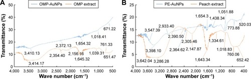
FT-IR analysis was conducted to identify possible functional groups responsible for the reduction of AuCl3 to AuNPs and their stabilization. FT-IR analysis of OMP, PE, OMP-AuNPs, and PE-AuNPs was performed in the range of 400–4,000 cm−1. FT-IR signals of OMP extract were observed at 3,414 cm−1, 2,350 cm−1, 2,156 cm−1, 1,645 cm−1, 1,039 cm−1, 761 cm−1, and 564 cm−1, while the FT-IR signals of OMP-AuNPs were observed at 3,410 cm−1, 2,372 cm−1, 1,654 cm−1, 1,018 cm−1, and 671 cm−1 (). Similarly, the FT-IR signals of PE were observed at 3,642 cm−1, 3,286 cm−1, 2,390 cm−1, 2,305 cm−1, 2,147 cm−1, 1,643 cm−1, 1,334 cm−1, 1,018 cm−1, 760 cm−1, and 652 cm−1, while the FT-IR signals of PE-AuNPs were observed at 3,547 cm−1, 2,933 cm−1, 2,364 cm−1, 1,654 cm−1, 1,438 cm−1, 1,051 cm−1, 773 cm−1, and 520 cm−1 (). In the case of the OMP extract, the strong peak band at 3,414 cm−1 corresponded to the O–H stretch vibration of alcohols and phenols that was shifted toward 3,410 cm−1 in the case of OMP-AuNPs.Citation59 Similarly, the strong peak band at 3,642 cm−1 in the peach extract corresponded to the O–H stretch vibration and free hydroxyl groups of the alcohols and phenols that was shifted toward 3,547 cm−1 in the case of the PE-AuNPs.Citation60,Citation61 The bands at 1,645 cm−1 and 1,643 cm−1 in OMP and peach extracts, respectively, were due to –C=C–stretching of carbonyl amide I vibrations that were shifted toward 1,654 cm−1 in both OMP-AuNPs and PE-AuNPs. The presence of this particular band in the AuNPs might have been due to the proteins/peptides or amino acid groups of OMP and peach extracts, which may have acted as the reducing and capping agent for the synthesis of NPs.Citation32,Citation62 The peak bands at 1,039 cm−1 and 1,018 cm−1 in both OMP and peach extracts corresponded to the C–N stretching of aliphatic amine groups and were shifted toward 1,018 cm−1 and 1,051 cm−1 in OMP-AuNPs and PE-AuNPs, respectively.Citation31 The peak bands at 651 cm−1 and 652 cm−1 in both OMP and peach extracts corresponded to the C–Br stretching of alkyl halide groups and were shifted toward 671 cm−1 and 520 cm−1 in OMP-AuNPs and PE-AuNPs, respectively.Citation63,Citation64 It is speculated that the components of OMP and peach extracts containing O–H, –C=C–, C–N, or C–Br functional groups might have been responsible for the reduction of Au+ to Au0.
Figure 6 FT-IR analysis of synthesized OMP-AuNPs and PE-AuNPs.
Notes: (A) OMP-AuNPs; (B) PE-AuNPs.
Abbreviations: AuNPs, gold nanoparticles; FT-IR, Fourier transform infrared spectroscopy; OMP, oriental melon peel; PE, peach extract.
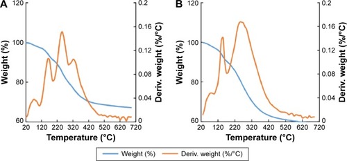
The TG/DTG analysis of both the OMP-AuNPs and PE-AuNPs at 20°C–700°C is presented in . The TG/DTG analysis of OMP-AuNPs exhibited a total weight loss of 32.75% in three phases, 20°C–210°C with 9.96% weight loss that is mainly due to the water molecules attached to the particles, then 210°C–440°C with 19.62% weight loss that corresponds to the loss of organic materials, and a third phase of weight loss of 3.17% from 440°C to 690°C due to residual compounds. Similarly, TG/DTG analysis of PE-AuNPs exhibited a total weight loss of 40.74% in three phases, 20°C–200°C, 200°C–420°C, and 420°C–690°C, during which there were losses of 12.32%, 24.88%, and 3.54%, respectively. The reduction in weight of the synthesized AuNPs confirmed the involvement of bioactive organic compounds from OMP and peach extracts in the reduction and stabilization of NPs.Citation31,Citation62
Figure 7 TG/DTG analysis of synthesized OMP-AuNPs and PE-AuNPs.
Notes: (A) OMP-AuNPs; (B) PE-AuNPs.
Abbreviations: AuNPs, gold nanoparticles; Deriv., derivative; DTG, derivative thermogravimetric; OMP, oriental melon peel; PE, peach extract; TG, thermogravimetric.
After characterization of both synthesized OMP-AuNPs and PE-AuNPs, comparative investigations of their biological potential in terms of their antibacterial, anticandidal, antioxidant, and proteasome inhibitory activities were conducted.
Antibacterial activity
Neither the OMP-AuNPs nor PE-AuNPs, nor the OMP and peach extracts (taken as extract control), exerted any antibacterial activity against the five tested foodborne pathogenic bacteria at 25 μg/disk (). Similarly, the positive controls, kanamycin and rifampicin, at 5 μg/disk and negative control, DMSO, did not exhibit any effects on the five tested foodborne pathogenic bacteria (). When the NPs and antibiotics were combined and the mixtures were tested for antibacterial effects, they exhibited positive synergistic responses against all the tested foodborne pathogenic bacteria (). Among the two mixtures of NPs and kanamycin, PE-AuNPs + kanamycin displayed higher synergistic activity against the four foodborne pathogenic bacteria than OMP-AuNPs + kanamycin. When the antibacterial activity of mixtures of NPs and rifampicin were compared, both exerted antibacterial activity against only three pathogenic bacteria, B. cereus ATCC 13061, E. coli ATCC 43890, and S. aureus ATCC 49444. Of the two mixtures of NPs/antibiotics, PE-AuNPs with kanamycin displayed stronger synergistic antibacterial activity than OMP-AuNPs. All the mixtures of NPs/antibiotics displayed strong antibacterial synergistic activity against S. aureus, with inhibition zones of 13.26–25.23 mm ().
Table 1 Comparative synergistic antibacterial activity of OMP-AuNPs (25 μg) and PE-AuNPs (25 μg) with standard antibiotics, kanamycin (5 μg) or rifampicin (5 μg), against foodborne pathogenic bacteria
The antibacterial activities of different types of NPs have been widely investigated using different pathogenic bacteria in recent years.Citation65–Citation67 Earlier reports have suggested an increase in the antibacterial activity of AuNPs in response to the addition of antibiotics,Citation68,Citation69 which was also evident in the present investigation. It has been reported that NPs could easily penetrate the bacterial cell membrane due to their small size, causing numerous holes in the bacterial cell wall and leakage of essential elements from the cell. When applied in combination with antibiotics, NPs could influence the bacterial nucleus and DNA, causing cell death.Citation66,Citation67 The antimicrobial potentials of different parts of Cucumis melo and Prunus persica have been reported.Citation19,Citation70–Citation75 The bioactive compounds present in peel extracts, which have been used for AuNPs synthesis and stabilization, might be responsible for the antibacterial activity of the AuNPs. The synergistic potential of AuNPs with antibiotics could be useful in minimizing the uncontrolled use of antibiotics, which would have resulted in the development of many antibiotic resistant strains.
Anticandidal activity
The synergistic anticandidal activities of both OMP-AuNPs and PE-AuNPs mixed with amphotericin B were evaluated against five different Candida species (). Initial testing of both OMP-AuNPs and PE-AuNPs at 50 μg/disk and amphotericin B at 5 μg/disk for their anticandidal activities revealed no inhibitory activity against any of the tested pathogenic Candida species (). However, these compounds were subsequently mixed together at the same concentrations, after which their synergistic anticandidal potentials were tested (). The extract control, positive control, and negative control did not display any inhibition activity against any of the pathogenic Candida species (). Both the OMP-AuNPs and PE-AuNPs combined with amphotericin B displayed a strong anticandidal activity against all the five tested Candida species. The AuNPs were more active against C. albicans KACC 30062 and C. geochares KACC 30061, as indicated by a 13.27–15.47 mm inhibition zone (). Among the two AuNPs, the PE-AuNPs + amphotericin B mixture exhibited a stronger positive activity than the OMP-AuNPs + amphotericin B. The comparative high anticandidal activity of PE-AuNPs could be correlated with their smaller particle size (average size =39.90 nm), which could have facilitated the easy penetration of the amphotericin B/PE-AuNPs into Candida cells, resulting in cellular lysis. The use of less amphotericin B and AuNPs using the mixtures could be beneficial to clinical applications of the mixture solution, avoiding the adverse effects caused by the use of high doses of drugs.Citation76
Table 2 Comparative synergistic anticandidal activities of OMP-AuNPs (50 μg) and PE-AuNPs (50 μg) mixed with amphotericin B (5 μg), a standard antifungal agent against pathogenic Candida species
Antioxidant activity
The potential application of both OMP-AuNPs and PE-AuNPs was evaluated in terms of its antioxidant activity by a number of in vitro antioxidant assays. The DPPH free radical scavenging potential of both OMP-AuNPs and PE-AuNPs are presented in . Among the two AuNPs, PE-AuNPs exhibited the stronger DPPH radical scavenging activity of 56.92% at 100 μg/mL, while OMP-AuNPs and BHT (the reference standard compound) showed activities of 26.21% and 34.82%, respectively (). The DPPH scavenging activity of AuNPs might be attributed to the presence of a number of secondary metabolites, mostly phenolic compounds, in the fruit peel extracts that were used for the biosynthesis of AuNPs. The potential effects of AuNPs might occur via its inhibitory effects on the peroxy radicals, which are responsible for lipid peroxidation.Citation77
Figure 8 Antioxidant potential of synthesized OMP-AuNPs and PE-AuNPs.
Notes: (A) DPPH free radical scavenging potential; (B) nitric oxide scavenging potential; (C) ABTS radical scavenging potential; and (D) reducing power potential. Values with different superscript letters are significantly different at P<0.05.
Abbreviations: ABTS, 2,2′-azino-bis(3-ethylbenzothiazoline-6-sulphonic acid); AuNPs, gold nanoparticles; BHT, butylated hydroxyl toluene; DPPH, 1,1-diphenyl-2-picrylhydraxyl; OMP, oriental melon peel; PE, peach extract.
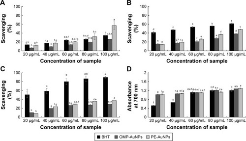
The nitric oxide scavenging potentials of AuNPs are presented in . Both OMP-AuNPs and PE-AuNPs displayed nitric oxide scavenging activity of 38.57% and 48.46%, respectively, at 100 μg/mL; however, the potentials were less than that of BHT, the standard reference compound, which showed 61.47% scavenging activity (). Nitric oxide is a very unstable compound responsible for various diseases, including cancer and inflammatory diseases;Citation78,Citation79 thus, the strong nitric oxide scavenging potentials of both PE-AuNPs and OMP-AuNPs could be beneficial for the biomedical and cosmetic industries.
PE-AuNPs and OMP-AuNPs also displayed moderate ABTS radical scavenging activities of 29.11% and 37.36%, respectively, while that of the standard reference compound BHT was 89.61% (). Moreover, they exhibited a strong reducing power of 1.28 and 1.31 absorbance at 700 nm, while the absorbance of BHT was 1.20 (). The strong reducing power of both OMP-AuNPs and PE-AuNPs might be attributed to the presence of phenolic compounds in the outer peel extracts. Among the AuNPs, PE-AuNPs exhibited the highest antioxidant potentials as indicated by all of the in vitro assays when compared to the OMP-AuNPs. The high antioxidant potentials might be attributed to the presence of a number of phenolic compounds in the outer peel of Prunus persica, which was used for the green synthesis of PE-AuNPs.Citation29
Proteasome inhibitory activity
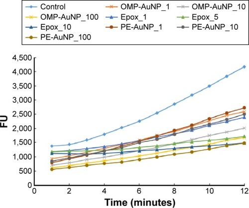
The proteasome inhibitory potential of both OMP-AuNPs and PE-AuNPs is presented in . The results showed that the values of the fluorescence unit decreased in all three PE-AuNPs, OMP-AuNPs and epoxomicin as the concentration increased, confirming their proteasome inhibitory potential (). PE-AuNPs displayed proteasome inhibitory activities of 33.77%, 36.04%, and 60.46% at 1 μg/mL, 10 μg/mL, and 100 μg/mL, respectively, while the OMP-AuNPs displayed activities of 31.74%, 47.67%, and 55.35% at 1 μg/mL, 10 μg/mL, and 100 μg/mL, respectively. Epoxomicin, the reference standard, displayed 30.28%, 40.06%, and 45.67% activity at concentrations of 0.56 μg/mL, 2.78 μg/mL, and 5.56 μg/mL, respectively. The results confirmed that PE-AuNPs displayed a stronger proteasome inhibitory activity than OMP-AuNPs and epoxomicin, which might have been due to the presence of a number of compounds, including chlorogenic acid, catechin, epicatechin, rutin, and cyanidin-3-glucoside in the outer peel of Prunus persica that was used for the biosynthesis of PE-AuNPs. It is well known that cancer cells have defective cell cycle checkpoints and DNA repair control mechanisms, while proteasomes are responsible for the degradation of many cytosolic proteins, including cyclins, misfolded, or denatured proteins, and transcription factors in cells.Citation80,Citation81 If these cancer cells are placed under stress conditions by the use of proteasome inhibitors such as AuNPs, they will then face tremendous problems and not be able to correct the cell cycle transition blockade. As a result, they may enter the apoptosis phase without affecting the normal cell.Citation82 Both PE-AuNPs and OMP-AuNPs with potent proteasome inhibitory potential could serve as potential candidates for targeted drug delivery and drug conjugation with efficient anticancer drugs in cancer treatment.
Figure 9 Proteasome inhibitory potential of both OMP-AuNPs and PE-AuNPs and standard reference compound, Epox.
Notes: 100 μg/mL (OMP-AuNP_100, PE-AuNP_100); 10 μg/mL (OMP-AuNP_10, PE-AuNP_10); 1 μg/mL (OMP-AuNP_1, PE-AuNP_1) and 5.56 μg/mL (Epox_10); 2.78 μg/mL (Epox_5) and 0.56 μg/mL (Epox_1).
Abbreviations: AuNPs, gold nanoparticles; FU, fluorescence units; Epox, epoxomicin; OMP, oriental melon peel; PE, peach extract.
Conclusion
In the current study, the biosynthesis of AuNPs was attempted using the outer peel extracts of Cucumis melo and Prunus persica fruits as the sole reducing agents. To the best of our knowledge, the use of naturally available fruit waste materials has not been investigated for such applications to date. The NPs were synthesized utilizing green technology, which is a nontoxic and environmental friendly procedure and is better than the conventional physical and chemical methods that give rise to highly toxic products. The synthesized NPs were subjected to a number of characterization techniques, including UV–visible spectroscopy, FE-SEM, EDX, XRD, FT-IR, and TG/DTG analysis, and confirmed to be AuNPs. The estimated absolute crystallite size of the synthesized AuNPs was calculated to be 78.11 nm for OMP-AuNPs and 39.90 nm for PE-AuNPs based on the Scherer equation. Both synthesized AuNPs were evaluated for antibacterial, anticandidal, antioxidant, and proteasome inhibitory potential. Among the synthesized NPs, the PE-AuNPs were found to be the most promising NPs, with strong antibacterial, anticandidal, antioxidant, and proteasome inhibitory activities. Bioactive compounds such as chlorogenic acid, catechin, epicatechin, rutin, and cyanidin-3-glucoside, which are the major constituents of peach fruits as evident from previous published literatures, might be responsible for the potential activity of PE-AuNPs. The significant biological properties of both synthesized ecofriendly NPs could make them promising candidates in biomedical, pharmaceutical, food, and cosmetic industries.
Disclosure
The authors report no conflicts of interest in this work.
Acknowledgments
This work was supported by grants from the Systems and Synthetic Agro-biotech Center through the Next-Generation Bio-Green 21 Program (PJ011117), Rural Development Administration, South Korea.
References
- NandaASaravananMBiosynthesis of silver nanoparticles from Staphylococcus aureus and its antimicrobial activity against MRSA and MRSENanomed Nanotechnol Biol Med200954452456
- DanielMCAstrucDGold nanoparticles: assembly, supramolecular chemistry, quantum-size-related properties, and applications toward biology, catalysis, and nanotechnologyChem Rev2004104129334614719978
- ZhanGHuangJLinLLinWEmmanuelKLiQSynthesis of gold nanoparticles by Cacumen Platycladi leaf extract and its simulated solution: toward the plant-mediated biosynthetic mechanismJ Nanopart Res2011131049574968
- KumarAKaurKSharmaSSynthesis, characterization and antibacterial potential of silver nanoparticles by Morus nigra leaf extractIndian J Pharma Biol Res2013141624
- XiaYXiongYLimBSkrabalakSEShape-controlled synthesis of metal nanocrystals: simple chemistry meets complex physics?Angew Chem Int Ed Engl20094816010319053095
- YuDBYamVWHydrothermal-induced assembly of colloidal silver spheres into various nanoparticles on the basis of HTAB-modified silver mirror reactionJ Phys Chem B2005109125497550316851589
- PalAShahSDeviSPreparation of silver, gold and silver-gold bimetallic nanoparticles in w/o microemulsion containing TritonX-100Colloid Surf A20073021–3483487
- LuXYavuzMSTuanHYKorgelBAXiaYUltrathin gold nanowires can be obtained by reducing polymeric strands of oleylamine-AuCl complexes formed via aurophilic interactionJ Am Chem Soc2008130288900890118540574
- MohanpuriaPRanaNKYadavSKBiosynthesis of nanoparticles: technological concepts and future applicationsJ Nanopart Res2008103507517
- GerickeMPinchesAMicrobial production of gold nanoparticlesGold Bull20063912228
- ShankarSSAhmadASastryMGeranium leaf assisted biosynthesis of silver nanoparticlesBiotechnol Prog20031961627163114656132
- LimTKCucumis melo (Makuwa Group)Edible Medicinal and Non-Medicinal Plants2LondonSpringer2012219
- ChenLKangYHIn vitro inhibitory effect of oriental melon (Cucumis melo L. var. makuwa Makino) seed on key enzyme linked to type 2 diabetes: assessment of anti-diabetic potential of functional foodJ Funct Foods201352981986
- ChenLKangYHSuhJKRoasting processed oriental melon (Cucumis melo L. var. makuwa Makino) seed influenced the triglyceride profile and the inhibitory potential against key enzymes relevant for hyperglycemiaFood Res Int201456236242
- FaustMTimonBOrigin and dissemination of peachJanickJHorticultural Reviews17Oxford, UKJohn Wiley & Sons, Inc1995
- YangZMaYChenLDifferential transcript abundance and genotypic variation of four putative allergen-encoding gene families in melting peachTree Genet Genomes20117903916
- ManzoorMAnwarFMahmoodZRashidUAshrafMVariation in minerals, phenolics and antioxidant activity of peel and pulp of different varieties of peach (Prunus persica L.) fruit from PakistanMolecules20121766491650622728349
- AndreottiCRavagliaDRagainiACostaGPhenolic compounds in peach (Prunus persica) cultivars at harvest and during fruit maturationAnnal Appl Biol20081531123
- AzizSRahmanHUBiological activities of Prunus persica L. batchJ Med Plants Res20137947951
- MagudapathyPGangopadhyayPPanigrahiBKNairKGMDharaSElectrical transport studies of Ag nanoclusters embedded in glass matrixPhysica B2001299142146
- JenningsTStrouseGPast, present, and future of gold nanoparticlesAdv Exp Med Biol2007620344718217333
- LeeKSEl-SayedMAGold and silver nanoparticles in sensing and imaging: sensitivity of plasmon response to size, shape, and metal compositionJ Phys Chem B200611039192201922517004772
- SalataOVApplications of nanoparticles in biology and medicineJ Nanobiotechnol2004213
- El-SayedIHHuangXEl-SayedMASelective laser photo-thermal therapy of epithelial carcinoma using anti-EGFR antibody conjugated gold nanoparticlesCancer Lett2006239112913516198049
- ShankarSSRaiAAnkamwarBSinghAAhmadASastryMBiological synthesis of triangular gold nanoprismsNat Mater20043748248815208703
- ChandranSPChaudharyMPasrichaRAhmadASastryMSynthesis of gold nanotriangles and silver nanoparticles using Aloe vera plant extractBiotechnol Progr2006222577583
- MubarakAliDThajuddinNJeganathanKGunasekaranMPlant extract mediated synthesis of silver and gold nanoparticles and its antibacterial activity against clinically isolated pathogensColloid Surface B2011852360365
- EliaPZachRHazanSKolushevaSPoratZZeiriYGreen synthesis of gold nanoparticles using plant extracts as reducing agentsInt J Nanomed2014940074021
- BankarAJoshiBKumaraARZinjardeSBanana peel extract mediated synthesis of gold nanoparticlesColloid Surface B20108014550
- PatraJKBaekKHNovel green synthesis of gold nanoparticles using Citrullus lanatus rind and investigation of proteasome inhibitory activity, antibacterial, and antioxidant potentialInt J Nanomedicine2015107253726426664116
- BasavegowdaNIdhayadhullaALeeYRPhyto-synthesis of gold nanoparticles using fruit extract of Hovenia dulcis and their biological activitiesInd Crop Prod201452745751
- PrakashPGnanaprakasamPEmmanuelRArokiyarajSSaravananMGreen synthesis of silver nanoparticles from leaf extract of Mimusops elengi, Linn. for enhanced antibacterial activity against multi drug resistant clinical isolatesColloid Surface B2013108255259
- DiaoWRHuQPFengSSLiWQXuJGChemical composition and antibacterial activity of the essential oil from green huajiao (Zanthoxylum schinifolium) against selected foodborne pathogensJ Agric Food Chem201361256044604923758080
- NaqviSZKiranUAliMICombined efficacy of biologically synthesized silver nanoparticles and different antibiotics against multidrug-resistant bacteriaInt J Nanomed2013831873195
- MurrayPRBaronEJPfallerMATenoverFCYolkeRHManual of Clinical Microbiology6th edWashington, DCASM Press1995
- BracaATommasiNDBariLDPizzaCPolitiMMorelliIAntioxidant principles from Bauhinia terapotensisJ Nat Prod200164789289511473417
- MakhijaIKAswatha-RamHNShreedharaCSVijay KumarSDevkarRIn vitro antioxidant studies of sitopaladi churna, a polyherbal ayurvedic formulationFree Rad Antioxidants201113741
- ThaipongKBoonprakobUCrosbyKCisneros-ZevallosLByrneDHComparison of ABTS, DPPH, FRAP, and ORAC assays for estimating antioxidant activity from guava fruit extractsJ Food Comp Anal200619669675
- PatraJKKimSHHwangHChoiJWBaekKHVolatile compounds and antioxidant capacity of the bio-oil obtained by pyrolysis of Japanese red pine (Pinus Densiflora Siebold and Zucc.)Molecules20152033986400625738540
- Higashi-OkaiKKamimotoKYoshiokaAOkaiYPotent suppressive activity of fresh and dried peels from Satsuma mandarin (Citrus unshiu Marcorv.) on hydroperoxide generation from oxidized linoleic acidPhytother Res200216878178412458489
- LeontowiczMGorinsteinSLeontowiczHApple and pear peel and pulp and their influence on plasma lipids and antioxidant potentials in rats fed cholesterol-containing dietsJ Agric Food Chem200351195780578512952433
- AnagnostopoulouMAKefalasPKokkalouEAssimopoulouANPapageorgiouVPAnalysis of antioxidant compounds in sweet orange peel by HPLC-diode array detection-electrospray ionization mass spectrometryBiomed Chromatogr200519213814815515108
- ParmarHSKarAProtective role of Mangifera indica, Cucumis melo and Citrullus vulgaris peel extracts in chemically induced hypothyroidismChem Biol Interact2009177325425819059228
- ParmarHSKarAProtective role of Citrus sinensis, Musa paradisiaca, and Punica granatum against diet-induced atherosclerosis and thyroid dysfunctions in ratsNutr Res200727710718
- MukherjeePSenapatiSMandalDExtracellular synthesis of gold nanoparticles by the fungus Fusarium oxysporumChem Bio Chem200235461463
- SasiKumarRPriyadharshiniSNandha KumarKPLNivedhaSIn vitro pharmacognostical studies and evaluation of bioactive constituents from the fruits of Cucumis melo L. (Muskmelon)Int J Pharma Phytochem Res20146936941
- MehraMPasrichaVGuptaRKEstimation of nutritional, phytochemical and antioxidant activity of seeds of musk melon (Cucumis melo) and water melon (Citrullus lanatus) and nutritional analysis of their respective oilsJ Pharma Phytochem2015398102
- MulvaneyPSurface plasmon spectroscopy of nanosized metal particlesLangmuir199612788800
- SosaMEEppingerSDRowlesCMIdentifying modular and integrative systems and their impact on design team interactionsJ Mech Des2003125240252
- LinkSEl-SayedMASize and temperature dependence of the plasmon absorption of colloidal gold nanoparticlesJ Phys Chem B199910342124217
- SunYXiaYIncreased sensitivity of surface plasmon resonance of gold nanoshells compared to that of gold solid colloids in response to environmental changesAnal Chem200274205297530512403584
- IslamNUJalilKShahidMMuhammadNRaufAPistacia integerrima gall extract mediated green synthesis of gold nanoparticles and their biological activitiesArabian J Chem2015110
- VarunSSellappaSRafiqKhanMVijayakumarSGreen synthesis of gold nanoparticles using Argemone mexicana L. leaf extract and its characterizationInt J Pharm Sci Rev Res2015324244
- Rimal IsaacRSSakthivelGMurthyCGreen synthesis of gold and silver nanoparticles using Averrhoa bilimbi fruit extractJ Nanotechnol201320136
- AftabtalabASadabadiHApplication of magnetite (Fe3O4) nanoparticles in hexavalent chromium adsorption from aquatic solutionsJ Pet Environ Biotechnol20156200
- KannanPJohnSASynthesis of mercaptothiadiazole functionalized gold nanoparticles and their self-assembly on Au substratesNanotechnol2008198085602
- LiZFriedrichATaubertAGold microcrystal synthesis via reduction of HAuCl4 by cellulose in the ionic liquid 1-butyl-3-methyl imidazolium chlorideJ Mater Chem200818910081014
- GhodakeGSDeshpandeNGLeeYPJinESPear fruit extract-assisted room-temperature biosynthesis of gold nanoplatesColloid Surface B2010752584589
- PrathnaTCChandrasekaranNRaichurAMMukherjeeABiomimetic synthesis of silver nanoparticles by Citrus limon (lemon) aqueous extract and theoretical prediction of particle sizeColloid Surface B2011821152159
- NarayananKBSakthivelNPhytosynthesis of gold nanoparticles using leaf extract of Coleus amboinicus LourMater Charact20106112321238
- BabuPJSharmaPKalitaMCBoraUGreen synthesis of biocompatible gold nanoparticles using Fagopyrum esculentum leaf extractFront Mater Sci20115379387
- SauTKRogachALNonspherical noble metal nanoparticles: colloid-chemical synthesis and morphology controlAdv Mater201022161781180420512953
- WilliamsDHFlemingISpectroscopic methods in organic chemistryNew York, NYThe McGraw-Hill1995
- CoatesJInterpretation of infrared spectra, a practical approachMeyersRAEncyclopedia of Analytical ChemistryChichesterJohn Wiley & Sons Ltd20001081510837
- YoonKYHoon ByeonJParkJHHwangJSusceptibility constants of Escherichia coli and Bacillus subtilis to silver and copper nanoparticlesSci Total Environ20073732–357257517173953
- RupareliaJPChatterjeeAKDuttaguptaSPMukherjiSStrain specificity in antimicrobial activity of silver and copper nanoparticlesActa Biomater20084370771618248860
- AzamAAhmedASOvesMKhanMSHabibSSMemicAAntimicrobial activity of metal oxide nanoparticles against Gram-positive and Gram-negative bacteria: a comparative studyInt J Nanomed2012760036009
- BuryginGLKhlebtsovBNShantrokhaANDykmanLABogatyrevVAKhlebtsovNGOn the enhanced antibacterial activity of antibiotics mixed with gold nanoparticlesNanoscale Res Lett20094879480120596384
- RavishankarRai VBaiAJNanoparticles and their potential application as antimicrobialsMendez-VilasAScience against microbial pathogens: communicating current research and technological advancesBadajoz, SpainFormatex Research Center2011197209
- RaturiRSinghHBahugunaPSatiSCBadoniPPAntibacterial and antioxidant activity of methanolic extract of bark of Prunus persicaJ Appl Nat Sci20113312314
- BaranwalAAroraSKumarRGEvaluation of the combinational antimicrobial effect of Prunus persia and Annona squamosa seeds methanolic extract on standard microbial strainsGlobal J Biosci Biotechnol20132571575
- ManzoorMNaseerSJabeenRManzoorMAntibacterial activity of fruits against Escherichia coliARPN J Agric Biol Sci20138258263
- SiddeegAAlsirEXuYJiangQXiaWChemical composition and antibacterial activity of the essential oil isolated from seinat (Cucumis melo Var. Tibish) SeedsInt J Technol Enhancements Emerging Engineer Res20142120124
- EdrahSAlafidFKumarAPreliminary Phytochemical screening and preliminary phytochemical screening and antibacterial activity of Pistacia atlantica and Prunus persica plants of Libyan originInt J Sci Res2015415521555
- ThakurHAAntimicrobial and antifungal activity of Cucumis melo L. (cucurbitaceae) and Pergularia daemia Frosk. (asclpiadaceae) an ethnomedicinal plantsInt J Bioassay2015436613665
- GajbhiyeMKesharwaniJIngleAGadeARaiMFungus-mediated synthesis of silver nanoparticles and their activity against pathogenic fungi in combination with fluconazoleNanomed Nanotechnol Biol Med200954382386
- ChengRGlynnSSantanaWFSwitzerCRidnourLWinkDANitric oxide and redox inflammation in cancerAdv Mol Toxicol20104157182
- SonejaADrewsMMalinskiTRole of nitric oxide, nitroxidative and oxidative stress in wound healingPharmacol Rep200557Suppl10811916415491
- RoyerMPradoMGarcia-PerezMEDioufPNStevanovicTStudy of nutraceutical, nutricosmetics and cosmeceutical potentials of polyphenolic bark extracts from Canadian forest speciesPharm Nutr20131158167
- KisselevAFJoining the army of proteasome inhibitorsChem Biol200815541942118482693
- TanakaTNakataniTKamitaniTInhibition of NEDD8-conjugation pathway by novel molecules: potential approaches to anticancer therapyMol Oncol20126326727522306028
- AlmondJBCohenGMThe proteasome: a novel target for cancer chemotherapyLeukemia200216443344311960320