
 ?Mathematical formulae have been encoded as MathML and are displayed in this HTML version using MathJax in order to improve their display. Uncheck the box to turn MathJax off. This feature requires Javascript. Click on a formula to zoom.
Abstract
Non-glucose biomarker-DNA oxidative damage biomarker 8-hydroxy-2′-deoxyguanosine (8-OHdG) has been successfully detected using a smartphone-enabled glucose meter. Through a series of immune reactions and enzymatic reactions on a solid lateral flow platform, 8-OHdG concentration has been converted to a relative amount of glucose, and therefore can be detected by conventional glucose meter directly. The device was able to detect 8-OHdG concentrations in phosphate buffer saline as low as 1.73 ng mL−1 with a dynamic range of 1–200 ng mL−1. Considering the inherent advantages of the personal glucose meter, the demonstration of this device, therefore, should provide new opportunities for the monitoring of a wide range of biomarkers and various target analytes in connection with different molecular recognition events.
Introduction
In recent years, much effort has been devoted toward developing point-of-care (POC) devices.Citation1–Citation9 Among them, paper-based POC devices are a special category due to the advantages of being simple, rapid, on-site, and cost-effective; they have been widely used in home health care and medical testing,Citation10–Citation12 even environmental monitoring.Citation13–Citation15 The lateral flow immunochromatographic assay, also called lateral flow test, is one of the simplest and most popular formats of paper-based POC devices and can be used to detect specific substances, including small molecules,Citation15–Citation18 large proteins,Citation19–Citation21 and even whole pathogens,Citation10 in a sample by using an immunological reaction. However, most of these tests are based on qualitative colorimetry, which is not sufficient in many cases. To overcome this limitation, a number of devices capable of quantitative analysis have been developed.
As we know, medical devices for POC are very costly to develop and manufacture and are difficult to commercialize. As a result, few have been commercialized successfully. The blood glucose meter (BGM) is one of the most successfully commercialized diagnostic devices on the market,Citation22,Citation23 and it is widely used by millions of diabetes patients to monitor their blood glucose levels every day because of its low cost, compact size, simple operation, and reliable quantitative results.Citation2 The successful launch of US Food and Drug Administration (FDA)-approved smartphone-enabled BGMs, which enable the wireless transmission of reminders, test results, and data analysis through smartphone apps, will attract an even larger number of users.Citation24 Generally, a glucose meter is capable of detecting glucose as the unique target. Recently, many groups have reported methods establishing a direct relationship between the concentrations of the targets in the samples and the glucose detected by a BGM, enabling a number of non-glucose targets to be detected quantitatively.Citation2,Citation25,Citation26 However, most of these methods included much liquid handling, such as adding and mixing solution, which should be avoided as much as possible to ensure ease of use. Conversely, a lateral flow device, as mentioned previously, is supposed to replace most of the manual solution addition and mixing involved in the above-mentioned methods.
Here, we describe a novel design that combines the traditional lateral flow strip with a commercialized smartphone-enabled BGM (iBGStar® Blood Glucose Monitoring System) for portable and quantitative detection of a non-glucose target. The concept is demonstrated by using an oxidative DNA damage biomarker, 8-hydroxy-2′-deoxyguanosine (8-OHdG). The basic design of the device is adapted from our previously reported colorimetric visual detection platformCitation17 based on a gold nanoparticles (AuNPs)-based competitive immunoassay. However, visual detection can provide only qualitative and semiquantitative results. Thus, to enable quantitative analysis, we establish a novel method that transforms the detection of the target to the detection of an enzyme invertase. The enzyme converts sucrose into glucose for BGM readout.
Materials and methods
Design and detection mechanism
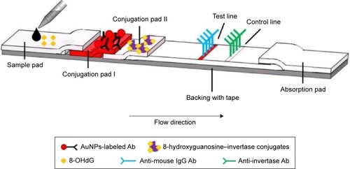
As shown in , the test strip is composed of five components, preloaded with different reagents and pasted onto a plastic backing plate. The sample pad pretreated with buffer (50 mM borate buffer, 1% bovine serum albumin [BSA], and 0.2% Tween 20) is where the strip contacts the liquid sample. Two conjugation pads, which are composed of glass fiber material, contain AuNPs-labeled mouse anti-8-hydroxyguanosine antibody (Ab; has equal binding affinity to 8-OHdG and 8-hydroxyguanosine) and 8-hydroxyguanosine–invertase conjugates, respectively. Next to the conjugation pad II is a flow membrane, which is made of nitrocellulose. There are two reaction zones on this membrane, which are called test line and control line. A goat anti-mouse immunoglobulin G (IgG) Ab is used as the test line capture reagent, whereas a goat anti-invertase Ab is used as the control line capture reagent. The sample moves along the strip due to capillary action and finally gets collected by the last part known as the absorption pad.
Figure 1 Design of a test strip for quantitative detection of 8-OHdG.
Abbreviations: Ab, antibody; AuNPs, gold nanoparticles; 8-OHdG, 8-hydroxy-2′-deoxyguanosine; IgG, immunoglobulin G.
To realize non-glucose target detection using a BGM, the relationship between target recognition and glucose generation must be established. In our design, as illustrated in , the detection is based on two steps: the first step is that of competitive-type immunoreactions in the lateral flow strip () and the second step contains an enzymatic reaction, which converts sucrose into detectable glucose for measurement by the smartphone-based BGMs ().
Figure 2 Mechanism for non-glucose target (8-OHdG) detection by a smartphone-based BGM.
Notes: (A) Sample loading, (B) immunoreactions and migration, (C) capture of antibody from the first conjugation pad on the test zone, (D) capturing the rest enzyme on the control zone, (E) cut the test and control zones and put them on a hydrophobic plate, (F) add the sucrose solution (20 µL) on top of each zone, (G) after a specific period of time, measure the concentration of glucose produced on each zone by iBGStar Blood Glucose Monitoring System.
Abbreviations: Ab, antibody; AuNPs, gold nanoparticles; BGM, blood glucose meter; 8-OHdG, 8-hydroxy-2′-deoxyguanosine; IgG, immunoglobulin.
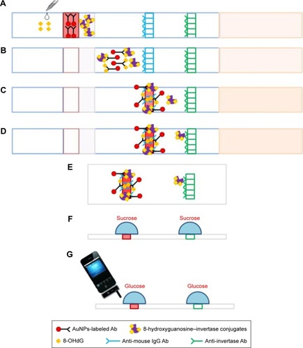
Upon target introduction (), the target moves along the strip due to capillary force and rehydrates conjugation pads I and II (). 8-OHdG in the sample solution and invertase-conjugated 8-hydroxyguanosine compete to bind to the Ab from the conjugation pad I. After that, the invertase that binds to AuNPs is captured by the test line (), whereas the free invertase is captured by the control line (). Ideally, the higher the concentration of 8-OHdG in the sample solution is the less invertase captured on the test line and the more invertase captured on the control line. Following the first step (~10 min), the test (3 mm) and control (3 mm) lines are cut by scissors and put on a hydrophobic plate (). Twenty microliters of 0.5 M sucrose solution is added on top of each line (). After a certain period of time, the invertase fixed on each line catalyzes the hydrolysis of sucrose to produce a specific amount of glucose for quantitative readout by the smartphone-based BGMs ().
Reagents
BSA, invertase from baker’s yeast (Saccharomyces cerevisiae), sucrose, gold chloride trihydrate, sodium borohydride (NaBH4), sodium periodate (NaIO4), ethylene glycol, potassium carbonate (K2CO3), boric acid, sodium chloride, and sodium phosphate were received from Sigma-Aldrich (St Louis, MO, USA). Trehalose, phosphate buffer saline (PBS, 1X, pH =7.4), and trisodium citrate dihydrate were purchased from VWR (West Chester, PA, USA). Tween 20 was obtained from Fisher Scientific (Fairlawn, NJ, USA). Plastic backing, nitrocellulose membrane, absorbing pad, and cellulose paper were acquired from Millipore (Billerica, MA, USA). 8-OHdG and 8-hydroxyguanosine were purchased from Cayman Chemical (Ann Arbor, MI, USA). Mouse monoclonal antibodies to 8-hydroxyguanosine, polyclonal goat anti-mouse IgG, and polyclonal goat anti-invertase IgG were purchased from Abcam (Cambridge, MA, USA). The iBGStar Blood Glucose Monitoring System and Blood Glucose Test Strips for iBGStar were obtained from Walgreens (Springfield, IL, USA).
Synthesis of AuNPs-labeled anti-8-hydroxyguanosine Ab conjugates for conjugation pad I
The AuNPs-labeled anti-8-hydroxyguanosine Ab conjugates were synthesized according to our previously published method.Citation17 The Supplementary materials also include a detail instruction for the synthesis. For the conjugation, the AuNPs solution (~20 nm) was concentrated fivefold and the pH was adjusted to 8.5–9.0 with 0.1 M K2CO3 in advance for labeling. The Ab with optimal concentration (45 µL, 0.54 mg mL−1) was added to the AuNPs solution (560 µL, 5X) and stirred gently at room temperature for 1 h. The conjugate was stabilized by adding 65 µL 10% BSA in 20 mM sodium borate for a final concentration of 1% and incubated for another 20 min. Then the mixture was centrifuged for 15 min at 7,000 rcf. The supernatant was discarded, and the pellet was resuspended in 600 µL 1% BSA/PBS. Following the same centrifugation step, the supernatant was removed and the soft sediment of conjugates was finally resuspended in 600 µL conjugation buffer, containing 20 mM sodium phosphate, 0.25% Tween 20, 10% trehalose, and 5% BSA. The conjugate was stored at 4°C until required for use. Characterization of AuNP and its conjugation is documented in the Supplementary materials.
Preparation of 8-hydroxyguanosine–invertase conjugates for conjugation pad II
8-hydroxyguanosine (0.8 mg) was dissolved in 800 µL 1X PBS. The solution (1 mg mL−1) was mixed with NaIO4 (200 µL, 50 mM) and the mixture was incubated for 1 h in the dark. The reaction was stopped by adding ethylene glycol (2.5 µL) for 5 min. Then the mixture (500 µL) was mixed with invertase (2.5 mL, 29 g L−1, pH =9.5, adjusted by K2CO3 [50 g L−1]) by adding it dropwise under constant stirring and incubated for another 1 h. After that, NaBH4 (2 mL, 12 g L−1) was added and the mixture was incubated in the dark at 4°C overnight (12–16 h). Finally, the conjugates were dialyzed against 1X PBS and stored at −20°C.
Assembly of the lateral flow immuno strip
The goat anti-mouse IgG Ab (1 mg mL−1) was used as the test line capture reagent, whereas the goat anti-invertase IgG Ab (3.3 mg mL−1) was used as the control line capture reagent. These capture reagents were dispensed by the Linomat 5 dispenser onto a nitrocellulose membrane as the test and control lines. The sample pad was pretreated with buffer (50 mM borate buffer, 1% BSA, and 0.2% Tween 20), then AuNPs-labeled anti-8-hydroxyguanosine Ab (20 µL) and 8-hydroxyguanosine–invertase conjugates (20 µL) were dispensed by pipette onto glass fiber pads, which were called the conjugate pad I and II, respectively. After drying these membranes, the sample pad, conjugate pads, nitrocellulose membrane, and absorbent pad were pasted onto a plastic backing plate, which was already cut into 5-mm wide strips using a strip cutter. Then the strips were stored in a self-sealing plastic bag until use.
Results and discussion
Check the properties of 8-hydroxyguanosine–invertase conjugates
One of the most important components in the design is 8-hydroxyguanosine–invertase conjugates. This is synthesized according to a well-established method.Citation27 The method requires several steps of chemical reactions that might affect the structure and function of the enzyme to a certain extent. Thus, two aspects of the functions of the conjugates were examined before being used for conjugation pad II: enzymatic activity (invertase part) for glucose production and antigenic activity (8-hydroxyguanosine part) for competitive immunoreactions.
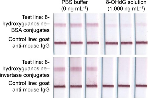
The enzymatic activity of the conjugates (invertase part) was tested by mixing 10 µL of conjugates solution with 50 µL of 0.5 M sucrose solution. The BGM readout of the modified invertase was 134±5 mg dL−1 (n=3) after 2 min, while that of the original invertase was 148±7 mg dL−1 (n=3), indicating that the modified invertase retains a high enzymatic activity after the chemical modifications. The antigenic activity of the conjugates was tested through our previously published method.Citation17 As illustrated in , competitive type strips are designed for testing small molecules with single antigenic determinants (ie, 8-OHdG), which cannot bind to two antibodies simultaneously. Both 8-OHdG in the sample solution and 8-hydroxyguanosine on the test line compete to bind to the Ab from the conjugation pad. As a result, the intensity of the test line is inversely proportional to the concentration in the sample. Our previous publication has verified that 8-hydroxyguanosine–BSA conjugates maintain considerable Ab capturing capacity. In this study, we compare 8-hydroxyguanosine–invertase conjugates with 8-hydroxyguanosine–BSA conjugates in terms of this capacity (see test line in ). These two conjugates show no difference at both low (0 ng mL−1) and high (1,000 ng mL−1) 8-OHdG sample loading, demonstrating the feasibility of the function of 8-hydroxyguanosine in the 8-hydroxyguanosine–invertase conjugates for the following application.
Figure 3 Mechanism of competitive lateral flow immunoassay for 8-OHdG testing.
Abbreviations: Ab, antibody; AuNPs, gold nanoparticles; BSA, bovine serum albumin; 8-OHdG, 8-hydroxy-2′-deoxyguanosine.
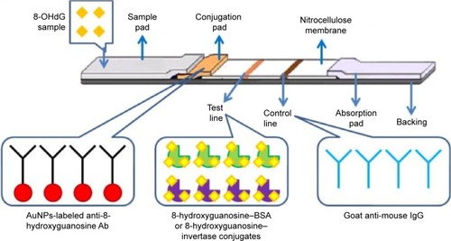
Figure 4 The photographs of test strips based on two samples (0 and 1,000 ng mL−1).
Notes: Top row: 8-hydroxyguanosine–BSA conjugates were used as the capture reagents on the test line; bottom row: 8-hydroxyguanosine–invertase conjugates were used as the capture reagents on the test line. Left three columns: samples with a concentration of 0 ng mL−1; right three columns: samples with a concentration of 1,000 ng mL−1.
Abbreviations: BSA, bovine serum albumin; 8-OHdG, 8-hydroxy-2′-deoxyguanosine; IgG, immunoglobulin G; PBS, phosphate buffer saline.
Optimization of the amount of 8-hydroxyguanosine–invertase conjugates used on conjugation pad II and of the time for enzymatic reaction
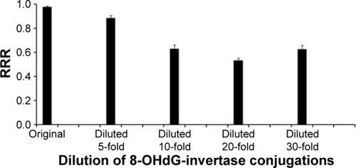
The amount of 8-hydroxyguanosine–invertase conjugates used on conjugation pad II played an important role in the performance of this detection system. We tested five concentrations of conjugates (original 14.5 mg mL−1, diluted 5-fold, diluted 10-fold, diluted 20-fold, and diluted 30-fold) on conjugation pad II. Two concentration levels, PBS buffer (0 ng mL−1) and high concentration (50 ng mL−1), were prepared and applied to these five types of strips. After 10 min, the test (3 mm) and control (3 mm) lines were cut by scissors and put on a hydrophobic plate. Twenty microliters of 0.5 M sucrose solution was added on top of each line. After a certain period of time, the invertase fixed on each line catalyzes the hydrolysis of sucrose to produce a specific amount of glucose for quantitative BGM readout. At the same time, the catalytic reaction time was also optimized. The readouts of the BGM at 20 and 40 min time points were collected respectively, from which we found that 20 min is not enough for some low concentrations (diluted 10-fold, 20-fold, and 30-fold). However, the relative readout ratio defined as
Figure 5 RRR for five concentrations of 8-hydroxyguanosine (8-OHdG) invertase conjugates used on the conjugation pad II.
Abbreviation: RRR, relative readout ratio.
Quantitative detection of 8-OHdG by a BGM
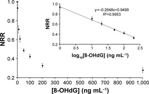
Under the optimized conditions, a series of 8-OHdG solutions with concentrations of 0, 1, 10, 50, 100, 200, and 1,000 ng mL−1 in 1X PBS buffer were prepared and applied to the strips. After 10 min, the test (3 mm) and control (3 mm) lines were cut by scissors and put on a hydrophobic plate. Twenty microliters of 0.5 M sucrose solution was put on top of each line, and a smartphone-enabled BGM was used for readout after 40 min. As shown in , the normalized readout ratio (NRR) defined as
Figure 6 Detection of 8-OHdG in buffer based on the smartphone-based blood glucose meter.
Notes: The concentration of 8-OHdG was varied from 0 to 1,000 ng mL−1. Insert: linear relationship between NRR and the logarithm of 8-OHdG concentration, three measurements for each point.
Abbreviations: 8-OHdG, 8-hydroxy-2′-deoxyguanosine; NRR, normalized readout ratio.
Conclusion
In conclusion, we have developed a simple approach for quantitative detection of 8-OHdG by using a low-cost strip and an easily accessible smartphone-based BGM. The cost for strips as demonstrated in this work is ~10 USD/pc, of which the majority comes from the purchase of 8-OHdG antibodies from commercial vendors. The device was able to detect 8-OHdG concentrations in PBS as low as 1.73 ng mL−1 with a dynamic range of 1–200 ng mL−1 in <1 h. The strip was easy to fabricate and convenient to operate, and the assay did not require any specialized skills or sophisticated instrument. In addition, because of the advantages of the BGM, the demonstration of this approach will provide new opportunities for the detection of other targets. Our current areas of focus are expanding the scope of the strip-BGM design to include other targets and even real biomedical samples testing, further simplifying the design to eliminate the need for target–invertase conjugates to be loaded on conjugation pad II enabling various types of target measurements and optimizing the experimental conditions to acquire a comparable sensitivity compared with other commercialized test kits. The results of these experiments will be reported in due course.
Acknowledgments
This research work is partially supported under grants NSF 1334417, NSFC 91543206, NSFC 21328501, and Shandong Taishan Scholar Project. We also thank Florida International University for Dissertation Year Fellowship (2014–2015) to X Zhu.
Disclosure
The authors report no conflicts of interest in this work.
Supplementary materials
Preparation of AuNPs
A well-established citrate reduction method was employed to generate AuNPs of various diameters. Different sizes of AuNPs produce different reflected color. AuNPs with a diameter of ~20 nm intrinsically give a better visibility on paper-based strip when accumulated on the test line, and therefore we used AuNPs with an average size ~20 nm in this work.
Briefly, the hydrogen tetrachloroaurate solution (50 mL, 0.01%) was added to a Erlenmeyer flask (250 mL), stirred, and brought to the boil on a hotplate. Trisodium citrate solution (1 mL, 1%) was added rapidly to the boiling solution under constant stirring. Gradually, the color changed from pale yellow to wine red. After the color change ceased, the solution was boiled for another 10 min and stirred without heating for another 10 min to complete the reduction of the gold chloride. The size of the particles was characterized by the Zetasizer after the solution reached room temperature.
Characterization of AuNPs pre- and post-conjugation
The wine red-colored solution contains AuNPs with a size of ~20 nm (Zetasizer, Figure S1A) and its corresponding maximum absorbance is exhibited at 520 nm as shown in Figure S1B (ultraviolet–visible spectrophotometer). It is well known that the strong absorbance at 520 nm is attributed to the surface plasmon resonance of AuNPs and the maximum absorption peak will be shifted or disappeared if the nanoparticles aggregate or get further modifications.
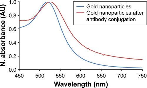
As a quality control step post-conjugation, the absorbance spectrum was again measured and compared with that of AuNPs that do not get antibody modifications. A ~3 nm red shift was observed as an indication for successful conjugations (Figure S2).
Figure S1 Characterization of synthesized AuNPs and antibody-AuNP conjugation. (A) Intensity distribution versus size of AuNPs solution (average size, ~20 nm) measure by Zetasizer; (B) ultraviolet–visible spectrum of ~20 nm AuNPs.
Abbreviations: AU, arbitrary unit; AuNPs, gold nanoparticles; Rel, relative.
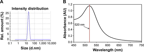
Figure S2 Antibody conjugation of gold nanoparticles produces a red shift in the ultraviolet–visible spectrometer.
Abbreviations: AU, arbitrary unit; N, normalized.
References
- MartinezAWPhillipsSTWhitesidesGMCarrilhoEDiagnostics for the developing world: microfluidic paper-based analytical devicesAnal Chem201082131020000334
- XiangYLuYUsing personal glucose meters and functional DNA sensors to quantify a variety of analytical targetsNat Chem20113969770321860458
- MartinezAWPhillipsSTCarrilhoEThomasSW3rdSindiHWhitesidesGMSimple telemedicine for developing regions: camera phones and paper-based microfluidic devices for real-time, off-site diagnosisAnal Chem200880103699370718407617
- WangSZhaoXKhimjiIIntegration of cell phone imaging with microchip ELISA to detect ovarian cancer HE4 biomarker in urine at the point-of-careLab Chip201111203411341821881677
- ShenLHagenJAPapautskyIPoint-of-care colorimetric detection with a smartphoneLab Chip201212214240424322996728
- WangSInciFChaunzwaTLPortable microfluidic chip for detection of Escherichia coli in produce and bloodInt J Nanomedicine201272591260022679370
- WangSEsfahaniMGurkanUAInciFKuritzkesDRDemirciUEfficient on-chip isolation of HIV subtypesLab Chip20121281508151522391989
- TennicoYHHutanuDKoesdjojoMTBartelCMRemchoVTOn-chip aptamer-based sandwich assay for thrombin detection employing magnetic beads and quantum dotsAnal Chem201082135591559720545301
- FoudehAMFatanat DidarTVeresTTabrizianMMicrofluidic designs and techniques using lab-on-a-chip devices for pathogen detection for point-of-care diagnosticsLab Chip201212183249326622859057
- LiCZVandenbergKPrabhulkarSPaper based point-of-care testing disc for multiplex whole cell bacteria analysisBiosens Bioelectron201126114342434821592765
- PattarawarapanMNangolaSCresseyTRTayapiwatanaCDevelopment of a one-step immunochromatographic strip test for the rapid detection of nevirapine (NVP), a commonly used antiretroviral drug for the treatment of HIV/AIDSTalanta200771146247019071328
- LiuGMaoXPhillipsJAXuHTanWZengLAptamer-nanoparticle strip biosensor for sensitive detection of cancer cellsAnal Chem20098124100131001819904989
- NieZNijhuisCAGongJElectrochemical sensing in paper-based microfluidic devicesLab Chip201010447748320126688
- ApiluxADungchaiWSiangprohWPraphairaksitNHenryCSChailapakulOLab-on-paper with dual electrochemical/colorimetric detection for simultaneous determination of gold and ironAnal Chem20108251727173220121066
- WangLMaWChenWAn aptamer-based chromatographic strip assay for sensitive toxin semi-quantitative detectionBiosens Bioelectron20112663059306221167704
- TangDSaucedaJCLinZMagnetic nanogold microspheres-based lateral-flow immunodipstick for rapid detection of aflatoxin B2 in foodBiosens Bioelectron200925251451819699076
- ZhuXHondroulisELiuWLiCZBiosensing approaches for rapid genotoxicity and cytotoxicity assays upon nanomaterial exposureSmall201399–101821183023417999
- ZhuXShahPStoffSLiuHLiCZA paper electrode integrated lateral flow immunosensor for quantitative analysis of oxidative stress induced DNA damageAnalyst2014139112850285724733353
- YangQGongXSongTQuantum dot-based immunochromatography test strip for rapid, quantitative and sensitive detection of alpha fetoproteinBiosens Bioelectron201130114515021963096
- LinYYWangJLiuGWuHWaiCMLinYA nanoparticle label/immunochromatographic electrochemical biosensor for rapid and sensitive detection of prostate-specific antigenBiosens Bioelectron200823111659166518406127
- WangWWuWYWangWZhuJJTree-shaped paper strip for semiquantitative colorimetric detection of protein with self-calibrationJ Chromatogr A20101217243896389920444459
- HellerAFeldmanBElectrochemical glucose sensors and their applications in diabetes managementChem Rev200810872482250518465900
- MontagnanaMCaputoMGiavarinaDLippiGOverview on self-monitoring of blood glucoseClin Chim Acta20094021–271319167374
- CarrollAEMarreroDGDownsSMThe HealthPia GlucoPack Diabetes phone: a usability studyDiabetes Technol Ther20079215816417425441
- XiangYLuYAn invasive DNA approach toward a general method for portable quantification of metal ions using a personal glucose meterChem Commun (Camb)201349658558723208450
- SuJXuJChenYXiangYYuanRChaiYPersonal glucose sensor for point-of-care early cancer diagnosisChem Commun (Camb)201248556909691122669465
- SenapathyPAliMAJacobMTMechanism of coupling periodate-oxidized nucleosides to proteinsFEBS Lett198519023373412995140