
Abstract
Cerium oxide nanoparticles (CeO2 NPs), used as a diesel fuel catalyst, can be emitted into the ambient air, resulting in exposure to humans by inhalation. Recent studies have reported the development of lung toxicity after pulmonary exposure to CeO2 NPs. However, little is known about the possible thrombotic effects of these NPs. The present study investigated the acute (24 hours) effect of intratracheal (IT) instillation of either CeO2 NPs (0.1 or 0.5 mg/kg) or saline (control) on pulmonary and systemic inflammation and oxidative stress and thrombosis in mice. CeO2 NPs induced a significant increase of neutrophils into the bronchoalveolar lavage (BAL) fluid with an elevation of tumor necrosis factor α (TNFα) and a decrease in the activity of the antioxidant catalase. Lung sections of mice exposed to CeO2 NPs showed a dose-dependent infiltration of inflammatory cells consisting of macrophages and neutrophils. Similarly, the plasma levels of C-reactive protein and TNFα were significantly increased, whereas the activities of catalase and total antioxidant were significantly decreased. Interestingly, CeO2 NPs significantly and dose dependently induced a shortening of the thrombotic occlusion time in pial arterioles and venules. Moreover, the plasma concentrations of fibrinogen and plasminogen activator inhibitor-1 were significantly elevated by CeO2 NPs. The direct addition of CeO2 NPs (1, 5, or 25 μg/mL) to mouse whole blood, collected from the inferior vena cava, in vitro neither caused significant platelet aggregation nor affected prothrombin time or partial thromboplastin time, suggesting that the thrombotic events observed in vivo may have resulted from systemic inflammation and/or oxidative stress induced by CeO2 NPs. This study concludes that acute pulmonary exposure to CeO2 NPs induces pulmonary and systemic inflammation and oxidative stress and promotes thrombosis in vivo.
Introduction
The use of nanotechnology has resulted in some novel and interesting medical and/or industrial uses, but also probable danger for human and environmental health.Citation1–Citation3 Accidental or involuntary contact during production or use of nanoparticles (NPs) may occur via various routes such as inhalation, ingestion, or skin penetration.Citation3,Citation4 Actually, the potential of NPs to pass across the alveolar–capillary barrier and to reach the circulating blood and thus have access to other organs is a reason for disquiet.Citation3,Citation5 It has been shown that NPs may penetrate the cells and be more biologically active than microsized particles because of their small size and large surface to volume ratio.Citation3,Citation5,Citation6
Cerium oxide (CeO2) is an important nanomaterial with a vast array of applications, including use in solar and fuel cells, gas sensors, and oxygen pumps and use as a fuel additive.Citation7,Citation8 CeO2 NPs have been widely used as a diesel fuel additive since 1999, because of their enhanced fuel-burning efficiency and decreased levels of greenhouse gases and particle numbers in vehicle exhaust.Citation7,Citation9–Citation11 However, it has been shown that, along with diesel exhaust particles (DEPs), CeO2 NPs are emitted in diesel exhaust, resulting in exposure to humans by inhalation.Citation8
Experimental studies in rodents have shown that acute (1 day) and subacute (28 days) intratracheal (IT) instillation of CeO2 NPs in rats induced lung inflammation, alveolar macrophage toxicity, air–blood barrier damage, and phospholipidosis.Citation12,Citation13 It has also been demonstrated that either acute inhalation or IT instillation of CeO2 NPs in mice caused pulmonary inflammation and small granulomas.Citation14,Citation15 More recently, it has been shown that CeO2 NPs induce pulmonary fibrosis and that the amorphous silica coating of CeO2 NPs reduced the inflammation, phospholipidosis, and fibrosis.Citation16,Citation17
It is well-established that particulate air pollution and some engineered NPs affect the cardiovascular system either through inflammatory mediators produced in the lungs and released into the circulation or via their ability to cross the alveolar–capillary barrier and reach the cardiovascular system.Citation5,Citation18 Since CeO2 NPs have been shown to cross the alveolar–capillary barrier to reach extrapulmonary sitesCitation19,Citation20 and cause lung inflammation,Citation14,Citation15,Citation20 the present study hypothesized that these NPs can plausibly interact with the circulating platelets and vasculature and induce thrombotic events. This has not been investigated before, and, consequently, the aim of this study was to assess the acute (24 hours) effect of CeO2 NPs on pulmonary and systemic inflammation and oxidative stress in mice and to evaluate their effects on coagulation in vivo and in vitro.
Material and methods
Particles
CeO2 NPs, 10%, w/w, in water with an average diameter of ~20 nm, were obtained from Sigma-Aldrich (St Louis, MO, USA). The CeO2 NP samples were diluted in sterile saline (0.9% NaCl). To minimize aggregation, particle suspensions were sonicated for 5 minutes in a Clifton ultrasonic bath (Clifton, NJ, USA) before dilution and IT administration. Particle suspensions were prepared immediately before use and were vigorously vortexed to obtain a well-mixed suspension prior to each instillation. The same particles from the same source were characterized and used recently by Ma et al.Citation12,Citation21
Animals and IT instillation
This project was reviewed and approved by the Institutional Review Board of the United Arab Emirates University, College of Medicine and Health Sciences, and the experiments were performed in accordance with the protocols approved by the Institutional Animal Care and Research Advisory Committee.
Both male and female BALB/c mice (Taconic Farms Inc., Germantown, NY, USA) weighing 23±2 g were housed in light- (12-hour light:12-hour dark cycle) and temperature-controlled (22°C ±1°C) rooms. The animals had free access to tap water and commercial laboratory chow.
Mice were anesthetized with sodium pentobarbital (60 mg/kg, i.p.) and placed supine with extended neck on an angled board. A Becton Dickinson (Sparks, MD, USA) 24-gauge cannula was inserted via the mouth into the trachea. Either CeO2 NP suspensions (0.1 or 0.5 mg/kg) or saline only were instilled IT (0.1 mL) via a sterile syringe, followed by an air bolus of 0.1 mL.
Blood collection and bronchoalveolar lavage (BAL) fluid analysis
Twenty-four hours after the IT administration of either CeO2 NPs or saline, the mice were anesthetized as described earlier, and blood was drawn from the inferior vena cava in ethylenediaminetetraacetic acid (4%). The collected blood was centrifuged at 4°C for 15 minutes at 900× g, and the plasma samples were stored at −80°C pending analysis.
Mice were then sacrificed with an overdose of sodium pentobarbital. The trachea was cannulated and the left bronchus was clamped. The right bronchi and right lungs were lavaged three times with 0.7 mL of sterile saline (NaCl 0.9%). The recovered fluid aliquots were pooled. No difference in the volume of collected fluid was observed between the different groups. BAL fluid was centrifuged (1,000× g ×10 minutes, 4°C). Cells were counted in a Thoma hemocytometer after resuspension of the pellets and staining with 1% gentian violet. The cell differentials (n=6–8 per group) were microscopically determined on cytocentrifuge preparations fixed in methanol and stained with Diff Quick (Dade, Brussels, Belgium). The supernatant was stored at -80°C until further analysis.
Histopathology
The unlavaged left lungs obtained from the animals above (n=6–8 per group) were fixed with 10% buffered formalin, excised, washed with ice-cold saline, blotted with filter paper, and weighed. Each left lung was sectioned, put in a cassette, and dehydrated in increasing concentrations of ethanol, cleared with xylene, and embedded in paraffin. Sections of 3 μm were prepared from paraffin blocks and stained with hematoxylin and eosin.Citation22,Citation23 The stained sections were evaluated by using light microscopy by a histopathologist participating in this project.
Determination of levels of tumor necrosis factor-α (TNFα), C-reactive protein (CRP), fibrinogen, and plasminogen activator inhibitor-1 (PAI-1) and activities of catalase and total antioxidant
In BAL fluid supernatant (n=6–8 per group), TNFα level was measured with a commercially available ELISA kit (Duo Set; R & D Systems, Minneapolis, MN, USA), and catalase activity was quantified by using a colorimetric assay kit (Cayman Chemical Company, Ann Arbor, MI, USA).
In plasma (n=6–8 per group), besides measuring TNFα level and catalase activity as described earlier, the levels of CRP (GenWay Biotech, Inc., San Diego, CA, USA), fibrinogen (Molecular Innovation, Southfield, MI, USA), and PAI-1 (Molecular Innovation) were measured by using ELISA kits. The total antioxidant activity was quantified with a colorimetric assay kit (Cayman Chemicals, Ann Arbor, MI, USA).
Experimental pial cerebral arteriole thrombosis model
In a separate experiment (n=6–8 per group), in vivo pial arteriolar and venular thrombogenesis was assessed 24 hours after IT instillation of either CeO2 NPs or saline according to a previously described technique.Citation24–Citation27
In vitro platelet aggregation in mouse whole blood
The platelet aggregation assay in whole blood (n=4–6 per group) was performed as described in previous studies.Citation28,Citation29 After anesthesia, blood from untreated mice was withdrawn from the inferior vena cava and placed in citrate (3.2%), and 0.1 mL aliquots were added to the well of a Merlin coagulometer (MC 1 VET; Merlin, Lemgo, Germany). The blood samples were incubated at 37°C with either saline (control) or CeO2 NPs (1, 5, or 25 μg/mL) for 3 minutes and then stirred for another 3 minutes. At the end of this period, 0.025 mL samples were removed and fixed on ice in 225 mL cellFix (Becton Dickinson). After fixation, single platelets were counted in a VET ABX Micros with a mouse card (ABX, Montpellier, France). The degree of platelet aggregation following CeO2 NP exposure was calculated as a fall in the number of single platelets counted and expressed as a % of control (saline-treated blood).Citation27
In vitro measurement of prothrombin time (PT) and activated partial thromboplastin time (aPTT)
After anesthesia, blood from untreated mice was withdrawn, as described earlier. The PT was measured on freshly collected, platelet-poor plasma with human, relipidated, recombinant thromboplastin (Recombiplastin; Instrumentation Laboratory, Orangeburg, NY, USA) in combination with an MC 1 VET (Merlin). Briefly, the plasma (n=4–6 per group) was incubated for 3 minutes at 37°C. After that, either CeO2 NPs (1, 5, or 25 μg/mL) or saline (control) was added for another 3 minutes, and then PT was measured as previously described.Citation25,Citation30,Citation31 The aPTT was measured with automated aPTT reagent (BioMerieux, Durham, NC, USA) by using an MC 1 VET (Merlin). Briefly, the plasma (n=4–6 per group) was incubated for 3 minutes at 37°C. After that, either CeO2 NPs (1, 5, or 25 μg/mL) or saline (control) was added for another 3 minutes, and then aPTT was measured as previously described.Citation25,Citation30,Citation31 Normal plasma was used as reference for both the PT and aPTT.
Statistics
All statistical analyses were performed with GraphPad Prism Software version 5 (San Diego, CA, USA). To determine whether parameters were normally distributed, the Kolmogorov–Smirnov statistic normality test was applied. Comparisons between groups were performed by one-way analysis of variance (ANOVA), followed by Newman–Keuls multiple range tests. Polymorphonuclear neutrophils (PMNs) in BAL fluid and TNFα in BAL fluid and plasma, which were not normally distributed, were analyzed with the Kruskal–Wallis test, followed by Dunn’s multiple comparison test. P-values <0.05 were considered as significant. All the data in the figures were expressed as mean ± standard error of the mean.
Results
Effect of CeO2 NPs on lung histology
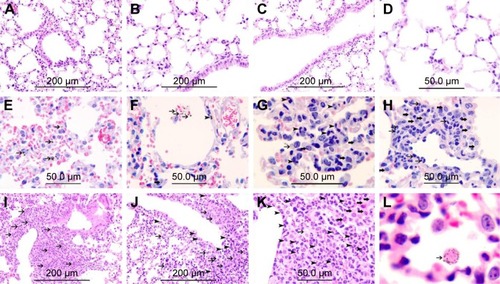
Histological sections from lungs of control mice had normal appearance (). In mice exposed to 0.1 mg/kg CeO2 NPs, particles were found inside the alveolar macrophages () as well as within alveolar air spaces (). Particles were also seen within the alveolar interstitial space (). There was focal damage of the alveolar wall and foci of expansion of the alveolar interstitial space due to neutrophil and macrophage infiltration of the interstitium (). In the group IT instilled with 0.5 mg/kg CeO2 NPs, particles were also found engulfed by alveolar macrophages (), and some particles were seen within macrophages within the interstitium (). There were foci of severe expansion of the alveolar interstitial space due to heavy neutrophil and macrophage infiltration of the interstitium ().
Figure 1 Representative histological lung sections obtained 24 hours after intratracheal instillation of either saline or 0.1 or 0.5 mg/kg cerium oxide nanoparticles (CeO2 NPs) in mice. (A–D) Are representative sections of control lungs. (A–D) Show normal lung tissue with unremarkable changes. (E–H) Are representative sections of lungs exposed to 0.1 mg/kg CeO2 NPs. (E) Shows the presence of CeO2 NPs within alveolar macrophages (thin arrows). (F) Shows the presence CeO2 NPs within alveolar space (thin arrow) and within the alveolar interstitial space (arrow head) associated with focal damage to the alveolar wall. (G) Shows the expansion of the alveolar interstitial space with neutrophil polymorphs (arrow head) and macrophages (thick arrow). CeO2 NPs are seen within alveolar macrophages (thin arrow). (H) Shows the expansion of the alveolar interstitial space with neutrophil polymorphs (thin arrow) and macrophages (thick arrow). (I–L) Are representative sections of lungs exposed to 0.5 mg/kg CeO2 NPs. (I) Shows severe expansion of the alveolar interstitial space with neutrophil polymorphs (thin arrow). (J) Shows severe expansion of the alveolar interstitial space with neutrophil polymorphs (arrow head) and macrophages (thin arrow). (K) Shows severe expansion of the alveolar interstitial space with neutrophil polymorphs (arrow head) and macrophages (thick arrow). CeO2 NPs are seen within a macrophage (thin arrow). (L) CeO2 NPs are seen within an alveolar macrophage (thin arrow). The scale bar on images A–C, I, and J is 200 μm. The scale bar on images D–H, and K is 50 μm.
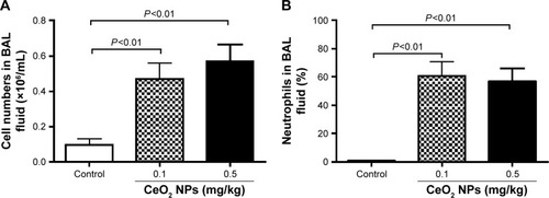
Effect of CeO2 NPs on cell composition and number in BAL fluid
represents the total cell numbers in the different groups, and shows the effects of CeO2 NPs on the influx of PMNs in BAL 24 hours after IT instillation. Compared with the saline-treated group (0.1±0.03×10⁶ cells/mL), the total number of cells in BAL fluid was significantly increased following the IT administration of 0.1 mg/kg (0.5±0.09×10⁶ cells/mL, P<0.01) and 0.5 mg/kg (0.6±0.09×10⁶ cells/mL, P<0.01) CeO2 NPs. In controls, PMNs made up 0.6%±0.4% of the total cells, the remaining cells being macrophages. The IT instillation of CeO2 NPs led to a PMN influx at 0.1 mg/kg (61%±10%, P<0.01) and at 0.5 mg/kg (57%±9%, P<0.01).
Figure 2 Total numbers of cells (A) and neutrophil numbers (B) in bronchoalveolar lavage (BAL) fluid 24 hours after intratracheal instillation of either saline or 0.1 or 0.5 mg/kg cerium oxide nanoparticles (CeO2 NPs) in mice. Data are mean ± standard error of the mean (n=6–8 in each group).
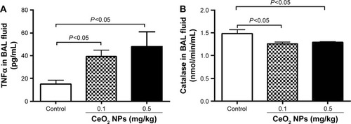
Effect of CeO2 NPs on TNFα concentration and catalase activity in BAL fluid
As shown in , compared with the control group, the IT instillation of CeO2 NPs induced significant increases in TNFα concentrations in BAL fluid at 0.1 and 0.5 mg/kg (P<0.05). On the contrary, IT instillation of CeO2 NPs caused a significant decrease of catalase activity in BAL fluid at 0.1 mg/kg (P<0.05) and 0.5 mg/kg (P<0.05) compared with the saline-treated group ().
Figure 3 Tumor necrosis factor-α concentration (TNFα) (A) and catalase activity (B) in bronchoalveolar lavage (BAL) fluid, 24 hours after intratracheal instillation of either saline or 0.1 or 0.5 mg/kg cerium oxide nanoparticles (CeO2 NPs) in mice. Data are mean ± standard error of the mean (n=6–8 in each group).
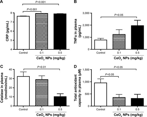
Effect in plasma of CeO2 NPs on concentrations of CRP and TNFα, catalase activity, and antioxidant capacity
Pulmonary exposure to CeO2 NPs induced a slight but a statistically significant increase in CRP concentration at 0.1 mg/kg (P<0.001) and 0.5 mg/kg (P<0.001) compared with the control group (). TNFα was dose dependently increased by CeO2 NPs, but the level of significance was only achieved at 0.5 mg/kg (P<0.05) (). On the other hand, compared with the control group, catalase activity was significantly decreased by the 0.5 mg/kg dose (P<0.01) (), whereas both doses of CeO2 NPs induced a significant decrease (P<0.05) of total antioxidant capacity ().
Figure 4 C-reactive protein (CRP) (A) and tumor necrosis factor-α (TNFα) (B) concentrations, and catalase (C) and total antioxidant (D) activities in plasma 24 hours after intratracheal instillation of either saline or 0.1 or 0.5 mg/kg cerium oxide nanoparticles (CeO2 NPs) in mice. Data are mean ± standard error of the mean (n=6–8 in each group).
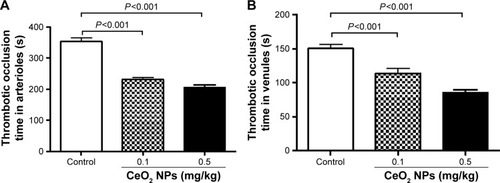
Effect of CeO2 NPs on pial arteriole and venule thrombosis
illustrates that the pulmonary exposure to CeO2 NPs induced a significant (P<0.001) and dose-dependent shortening of the thrombotic occlusion time in pial arterioles in photochemically injured vessels. Similarly, in pial venules, CeO2 NP administration caused a dose-dependent and significant (P<0.001) shortening of the thrombotic occlusion time compared with the saline-treated group.
Figure 5 Thrombotic occlusion time in pial arterioles (A) and venules (B) 24 hours after intratracheal instillation of either saline or 0.1 or 0.5 mg/kg cerium oxide nanoparticles (CeO2 NPs) in mice. Data are mean ± standard error of the mean (n=6–8 in each group).
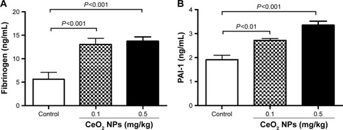
Effect of CeO2 NPs on fibrinogen and PAI-1 concentrations in plasma
shows that the IT instillation of CeO2 NPs induced a dose-dependent and significant increase of fibrinogen at doses of 0.1 mg/kg (P<0.001) and 0.5 mg/kg (P<0.001) compared with the control group. The concentration of PAI-1 was, in the same way, increased by the IT instillation of CeO2 NPs at 0.1 mg/kg (P<0.01) and 0.5 mg/kg (P<0.001) compared with the control group.
Figure 6 Fibrinogen (A) and plasminogen activator inhibitor-1 (PAI-1) (B) concentrations in plasma 24 hours after intratracheal instillation of either saline or 0.1 or 0.5 mg/kg cerium oxide nanoparticles (CeO2 NPs) in mice. Data are mean ± standard error of the mean (n=6–8 in each group).
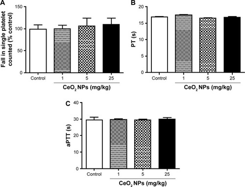
Effect of CeO2 NPs on platelet aggregation in vitro in whole blood, PT, and aPTT
shows that the direct addition of various concentrations of CeO2 NPs (1–25 μg/mL) to untreated whole blood in vitro did not induce any platelet aggregation. Also, compared with the control group, the incubation of untreated plasma with CeO2 NPs (1–25 μg/mL) neither altered the PT () nor the aPTT ().
Figure 7 In vitro platelet aggregation in whole blood (A), prothrombin time (PT) (B), and activated partial thromboplastin time (aPTT) (C) after incubation with either saline or cerium oxide nanoparticles (CeO2 NPs, 1, 5, or 25 μg/mL). Data are mean ± standard error of the mean (n=4–6 in each group).
Discussion
In this work, a histological analysis of the lung 24 hours after the IT instillation of CeO2 NPs revealed an expansion of the alveolar interstitial space due to infiltration of neutrophils and macrophages into the interstitium; particles were also seen within macrophages, and others were found within the alveolar interstitial space. BAL fluid analysis confirmed the influx of neutrophils and an increase of TNFα concentration and decrease in catalase activity. Likewise, in the plasma, the concentrations of CRP and TNFα were increased, whereas the activities of catalase and total antioxidant capacity were decreased. CeO2 NPs induced prothrombotic events in the pial arterioles and venules in vivo and increased fibrinogen and PAI-1 in plasma. However, the direct addition of CeO2 NPs to whole blood failed to induce platelet aggregation in vitro. Likewise, neither PT nor aPTT was affected by the direct addition of CeO2 NPs in vitro.
The present study used an IT instillation method as a mode of exposure to CeO2 NPs. The IT instillation is considered a valid, yet admittedly not perfect, mode of exposure to foreign compounds.Citation32,Citation33 The exact dose delivered to the lungs of each mouse can be established precisely, and this technique is simpler than inhalation, thus allowing the introduction of a range of doses to the lung in a short time.Citation32,Citation33 Moreover, IT instillation provides more accurate dosing, given that mice are nose breathers that filter most inhaled particles.Citation32,Citation33 The doses of particles used in the present study are comparable with those employed in previous animal models of IT administration of CeO2 NPs.Citation12,Citation34
In a previous study, it has been demonstrated that acute (24 hours) exposure of mice to DEPs causes impairment of cardiovascular homeostasis.Citation5,Citation29 This is in agreement with clinical and epidemiological studies that reported the development of myocardial complications within 24 hours of exposure to elevated levels of particulate air pollution,Citation35 and hence, the effect of CeO2 NPs 24-hour postexposure was assessed. Moreover, this time point is identical to the one used recently to assess the role of mast cells in vascular reactivity and ischemia reperfusion injury following IT administration of CeO2 NPs.Citation34
These data show that acute administration of CeO2 NPs caused a dose-dependent infiltration of neutrophils and macrophages, which caused an expansion of the alveolar interstitial space. Some CeO2 NPs were engulfed by macrophages, and others gained access to the interstitium. BAL fluid analysis confirmed the latter finding, showing a significant increase in neutrophils after the IT instillation of both doses of CeO2 NPs. Moreover, a significant and dose-dependent increase in the concentration of TNFα and a decrease in the activity of catalase were found, suggestive of a depletion of this antioxidant. These findings indicate the development of pulmonary inflammation and oxidative stress following acute exposure to CeO2 NPs. These results corroborate earlier findings which showed an induction of inflammation, cytotoxicity, and air–blood barrier damage, 24-hour post-IT instillation of CeO2 NPs in rats.Citation12 The findings of this study are also in agreement with previous studies that demonstrated an increase in neutrophil numbers in BAL fluid and the concentration of cytokine-induced neutrophil chemoattractant (CINC)-1, CINC-2, chemokine for neutrophil, and heme oxygenase-1, 3 days after the IT instillation of either 0.2 mg or 1 mg CeO2 NPs in rats, which persisted for up to 3 months postexposure.Citation36 Comparable findings were obtained, in the same study, following inhalation of CeO2 NPs (2 or 10 mg/m3) for 4 weeks (6 hours per day, 5 days per week).Citation36
Previous reports showed that pulmonary exposure to pollutant particles and some engineered NPs induce systemic and cardiovascular events.Citation22,Citation24,Citation37,Citation38 The latter effects were explained by the capacity of NPs to induce lung inflammation, which causes systemic inflammation and oxidative stress, and/or the aptitude of NPs to traverse the air–blood barrier to enter the blood and affect cardiovascular endpoints.Citation5,Citation18 It has been reported that IV administration of CeO2 NPs induce liver injury and oxidative stress.Citation39,Citation40 More recently, it has been shown that pulmonary, IV, and gastric exposures to CeO2 NPs cause endothelium-dependent and -independent arteriolar dysfunction.Citation20,Citation41 Nevertheless, the systemic effects of CeO2 NPs following pulmonary exposure are not fully understood, particularly their possible impact on thrombosis. The data of the present study show a significant increase in CRP caused by both doses of NPs. Moreover, a dose-dependent increase in TNFα concentrations and a decrease in catalase and total antioxidant activities following the acute exposure to CeO2 NPs were observed. It has been shown that exposure to particulate air pollution increases pulmonary inflammatory mediators that translocate to the circulation, contributing to systemic inflammation, with downstream effects such as cardiovascular dysfunction.Citation42 It has been shown that IT instillation of DEPs induces lung inflammation and oxidative stress responsible for systemic inflammation and oxidative stress at the 18–24-hour time point.Citation24,Citation37 It has been recently demonstrated that aortic RNA expression of endothelial nitric oxide synthase, tissue factor, and TNFα were significantly increased by 4-week inhalation exposure to whole CeO2 NPs added to diesel fuel (DECe) or gas-phase components of DECe.Citation19 However, the latter results are in conflict with the data from another study which implied that the addition of a Ce-based fuel-borne catalyst may decrease the atherosclerotic burden induced by exposure to diesel fuel in atherosclerosis-prone mice.Citation43 Also, it has been shown that CeO2 NPs improve microvascular reactivity in hypertensive rats.Citation44
This study evaluated the impact of CeO2 NPs on coagulation by measuring a set of relevant endpoints, ie, thrombosis in pial arterioles and venules in vivo and measurement of plasma concentrations of fibrinogen and PAI-1 and in vitro platelet aggregation, PT, and aPTT. As far as we are aware, the effect of CeO2 NPs on an animal model of thrombosis in vivo has not been reported so far. The present study used a well-established model of photochemical-induced thrombosis in pial arterioles and venules.Citation27,Citation37,Citation45 In this model, the damage to endothelial cells causes the platelets to adhere at the site of endothelial damage and then aggregate.Citation27,Citation37,Citation45 The data of the present study show that acute IT administration of CeO2 NPs induced a significant and dose-dependent shortening of the thrombotic occlusion time in pial arterioles and venules, indicating that CeO2 NPs possess prothrombotic effects. Along with thrombosis in vivo, a dose-dependent and significant increase in the concentrations of the coagulation factor, fibrinogen, and PAI-1 in the plasma following acute exposure to CeO2 NPs was found. PAI-1 is a potent endogenous inhibitor of fibrinolysis and has been reported to increase following exposure to DEPs, silica NPs, and carbon nanotubes.Citation46–Citation48 It has been shown that a 4-week inhalation exposure to DECe in rats induced an increase of tissue factor, but not in PAI-1 concentration.Citation19 The discrepancy between the latter study showing the absence of an increase of PAI-1 and this study could be related to the duration of exposure (24 hours versus 4 weeks), mode of exposure (IT instillation versus inhalation), or experimental animals used (mice versus rats). Additional studies are needed to clarify this point.
Recent studies have reported that pulmonary exposure to CeO2 NPs either by IT instillation or by inhalation resulted in systemic translocation and accumulation in various major organs such as the liver.Citation19,Citation49 This study wanted to verify whether the CeO2 NPs can directly induce platelet aggregation and affect PT and aPTT in vitro. The concentration of 1 μg/mL CeO2 NPs used here in vitro is similar to that employed recently to induce prothrombotic effects of DEPs in vitro.Citation29,Citation50 In addition to the latter concentration, two higher concentrations, that is, 5 and 25 μg/mL CeO2 NPs were used. These data show that CeO2 NPs (1–25 μg/mL) neither caused platelet aggregation nor affected either PT or aPTT. The latter findings suggest that the prothrombotic effects of CeO2 NPs observed in vivo may have resulted from systemic inflammation and oxidative stress rather than the direct contact of CeO2 NPs with platelets. These results also indicate that the in vitro effects of CeO2 NPs are different from those of DEPs reported previously, which showed that DEPs induced platelet aggregation in vitro and shortened PT and aPTT.Citation29,Citation51 These study data highlight the importance of conducting in vivo toxicity studies with NPs and suggest that the absence of toxicity of NPs in vitro does not necessarily mean that they are devoid of in vivo adverse effects.
This study has limitations. It did not thoroughly study the mechanisms underlying the systemic effects of CeO2 NPs and has investigated only the acute effects of these NPs. Further studies are warranted to assess the time effects (subacute and chronic) and the possible mechanism underlying the pulmonary and systemic effects of CeO2 NPs.
In conclusion, the data of this study provide novel evidence that pulmonary exposure to CeO2 NPs induces pulmonary and systemic inflammation and oxidative stress and promotes thrombosis in vivo.
Acknowledgments
This work was supported by funds of the College of Medicine and Health Sciences grant and United Arab Emirates University UPAR (31M189) and center-based interdisciplinary (31R052) grants. The authors wish to thank Professor Gerald Blunden (University of Portsmouth, Portsmouth, UK) for critically reading the manuscript.
Disclosure
The authors report no conflicts of interest in this work.
References
- RadomskaALeszczyszynJRadomskiMWThe nanopharmacology and nanotoxicology of nanomaterials: new opportunities and challengesAdv Clin Exp Med20162515116226935510
- ThorleyAJTetleyTDNew perspectives in nanomedicinePharmacol Ther201314017618523811125
- OberdorsterGSafety assessment for nanotechnology and nanomedicine: concepts of nanotoxicologyJ Intern Med20102678910520059646
- HoetPHBruske-HohlfeldISalataOVNanoparticles – known and unknown health risksJ Nanobiotechnol2004212
- NemmarAHolmeJARosasISchwarzePEAlfaro-MorenoERecent advances in particulate matter and nanoparticle toxicology: a review of the in vivo and in vitro studiesBiomed Res Int2013201327937123865044
- SotiriouGAPratsinisSEAntibacterial activity of nanosilver ions and particlesEnviron Sci Technol2010445649565420583805
- HEIEvaluation of human health risk from cerium added to diesel fuelHibbsJBJrHEI Communication 9Boston, MA, USAHealth Effects Institute2001
- CasseeFRvan BalenECSinghCExposure, health and ecological effects review of engineered nanoscale cerium and cerium oxide associated with its use as a fuel additiveCrit Rev Toxicol20114121322921244219
- ParkBDonaldsonKDuffinRHazard and risk assessment of a nanoparticulate cerium oxide-based diesel fuel additive – a case studyInhal Toxicol20082054756618444008
- WakefieldGWuXGardenerMParkBAndersonSEnvirox™ fuel-borne catalyst: developing and launching a nano-fuel additiveTech Anal Strat Manag2008127136
- SelvanAMAnandVUdayakumarMRBEffects of cerium oxide nanoparticle addition in diesel and diesel–biodiesel–ethanol blends on the performance and emission characteristics of a CI engineJ Eng Appl Sci20094716
- MaJYZhaoHMercerRRCerium oxide nanoparticle-induced pulmonary inflammation and alveolar macrophage functional change in ratsNanotoxicology2011531232520925443
- MaJMercerRRBargerMEffects of amorphous silica coating on cerium oxide nanoparticles induced pulmonary responsesToxicol Appl Pharmacol2015288637326210349
- ParkEJChoWSJeongJYiJChoiKKimYInduction of inflammatory responses in mice treated with cerium oxide nanoparticles by intratracheal instillationJ Health Sci201056387396
- SrinivasARaoPJSelvamGMurthyPBReddyPNAcute inhalation toxicity of cerium oxide nanoparticles in ratsToxicol Lett201120510511521624445
- MaJYMercerRRBargerMInduction of pulmonary fibrosis by cerium oxide nanoparticlesToxicol Appl Pharmacol201226225526422613087
- MaJMercerRRBargerMEffects of amorphous silica coating on cerium oxide nanoparticles induced pulmonary responsesToxicol Appl Pharmacol2015288637326210349
- MillsNLDonaldsonKHadokePWAdverse cardiovascular effects of air pollutionNat Clin Pract Cardiovasc Med20096364419029991
- SnowSJMcgeeJMillerDBInhaled diesel emissions generated with cerium oxide nanoparticle fuel additive induce adverse pulmonary and systemic effectsToxicol Sci201414240341725239632
- MinarchickVCStapletonPAPorterDWPulmonary cerium dioxide nanoparticle exposure differentially impairs coronary and mesenteric arteriolar reactivityCardiovasc Toxicol20131332333723645470
- MaJYYoungSHMercerRRInteractive effects of cerium oxide and diesel exhaust nanoparticles on inducing pulmonary fibrosisToxicol Appl Pharmacol201427813514724793434
- NemmarAMelghitKAl-SalamSAcute respiratory and systemic toxicity of pulmonary exposure to rutile Fe-doped TiO(2) nanorodsToxicology201127916717521073913
- NemmarAAl-SalamSYuvarajuPBeegamSAliBHEmodin mitigates diesel exhaust particles-induced increase in airway resistance, inflammation and oxidative stress in miceRespir Physiol Neurobiol2015215515726001677
- NemmarAAl SalamSDhanasekaranSSudhadeviMAliBHPulmonary exposure to diesel exhaust particles promotes cerebral microvessel thrombosis: protective effect of a cysteine prodrug l-2-oxothiazolidine-4-carboxylic acidToxicology2009263849219560508
- NemmarAZiaSSubramaniyanDFahimMAAliBHExacerbation of thrombotic events by diesel exhaust particle in mouse model of hypertensionToxicology2011285394521501650
- NemmarARazaHSubramaniyanDShort-term systemic effects of nose-only cigarette smoke exposure in mice: role of oxidative stressCell Physiol Biochem201331152423343613
- NemmarABeegamSYuvarajuPUltrasmall superparamagnetic iron oxide nanoparticles acutely promote thrombosis and cardiac oxidative stress and DNA damage in micePart Fibre Toxicol2016132227138375
- NemmarAMelghitKAliBHThe acute proinflammatory and prothrombotic effects of pulmonary exposure to rutile TiO2 nanorods in ratsExp Biol Med (Maywood)200823361061918375825
- NemmarAAlDRAlamiriJDiesel exhaust particles induce impairment of vascular and cardiac homeostasis in mice: ameliorative effect of emodinCell Physiol Biochem2015361517152626159184
- NemmarAAl SalamSZiaSDhanasekaranSShudadeviMAliBHTime-course effects of systemically administered diesel exhaust particles in ratsToxicol Lett2010194586520144906
- NemmarAYuvarajuPBeegamSJohnARazaHAliBHCardiovascular effects of nose-only water-pipe smoking exposure in miceAm J Physiol Heart Circ Physiol2013305H740H74623812392
- DriscollKECostaDLHatchGIntratracheal instillation as an exposure technique for the evaluation of respiratory tract toxicity: uses and limitationsToxicol Sci200055243510788556
- MorimotoYIzumiHYoshiuraYComparison of pulmonary inflammatory responses following intratracheal instillation and inhalation of nanoparticlesNanotoxicology20161060761826558952
- WingardCJWaltersDMCatheyBLMast cells contribute to altered vascular reactivity and ischemia-reperfusion injury following cerium oxide nanoparticle instillationNanotoxicology2011553154521043986
- BrookRDRajagopalanSPopeCAParticulate matter air pollution and cardiovascular disease. An update to the scientific statement from the American Heart AssociationCirculation20101212331237820458016
- MorimotoYIzumiHYoshiuraYPulmonary toxicity of well-dispersed cerium oxide nanoparticles following intratracheal instillation and inhalationJ Nanopart Res20151744226594128
- NemmarAAl-SalamSZiaSContrasting actions of diesel exhaust particles on the pulmonary and cardiovascular systems and the effects of thymoquinoneBr J Pharmacol20111641871188221501145
- BudingerGRSMckellJLUrichDParticulate matter-induced lung inflammation increases systemic levels of PAI-1 and activates coagulation through distinct mechanismsPlos One201164e1852521494547
- TsengMTLuXDuanXAlteration of hepatic structure and oxidative stress induced by intravenous nanoceriaToxicol Appl Pharmacol201226017318222373796
- YokelRAAuTCMacPhailRDistribution, elimination, and biopersistence to 90 days of a systemically introduced 30 nm ceria-engineered nanomaterial in ratsToxicol Sci201212725626822367688
- MinarchickVCStapletonPAFixNRLeonardSSSabolskyEMNurkiewiczTRIntravenous and gastric cerium dioxide nanoparticle exposure disrupts microvascular smooth muscle signalingToxicol Sci2015144778925481005
- KidoTTamagawaEBaiNParticulate matter induces IL-6 translocation from the lung to the systemic circulationAm J Respir Cell Mol Biol201144219720420378751
- CasseeFRCampbellABoereAJThe biological effects of subacute inhalation of diesel exhaust following addition of cerium oxide nanoparticles in atherosclerosis-prone miceEnviron Res201211511022507957
- MinarchickVCStapletonPASabolskyEMNurkiewiczTRCerium dioxide nanoparticle exposure improves microvascular dysfunction and reduces oxidative stress in spontaneously hypertensive ratsFront Physiol2015633926635625
- NemmarAYuvarajuPBeegamSAliBHShort-term nose-only water-pipe (shisha) smoking exposure accelerates coagulation and causes cardiac inflammation and oxidative stress in miceCell Physiol Biochem20153582984025634761
- NemmarASubramaniyanDAliBHProtective effect of curcumin on pulmonary and cardiovascular effects induced by repeated exposure to diesel exhaust particles in micePLoS One20127e3955422745783
- ErdelyAHuldermanTSalmenRCross-talk between lung and systemic circulation during carbon nanotube respiratory exposure. Potential biomarkersNano Lett20099364319049393
- NemmarAAlbarwaniSBeegamSAmorphous silica nanoparticles impair vascular homeostasis and induce systemic inflammationInt J Nanomedicine201492779278924936130
- HeXZhangHMaYLung deposition and extrapulmonary translocation of nano-ceria after intratracheal instillationNanotechnology20102128510320562477
- TaborCMShawCARobertsonSPlatelet activation independent of pulmonary inflammation contributes to diesel exhaust particulate-induced promotion of arterial thrombosisPart Fibre Toxicol201613626857113
- RadomskiAJuraszPAlonso-EscolanoDNanoparticle-induced platelet aggregation and vascular thrombosisBr J Pharmacol200514688289316158070