
Abstract
Carbon nanotubes (CNTs) are likely to transform the therapeutic and diagnostic fields in biomedicine during the coming years. However, the fragmented vision of their side effects and toxicity in humans has proscribed their use as nanomedicines. Most studies agree that biocompatibility depends on the state of aggregation/dispersion of CNTs under physiological conditions, but conclusions are confusing so far. This study designs an experimental setup to investigate the cytotoxic effect of individualized multiwalled CNTs compared to that of identical nanotubes assembled on submicrometric structures. Our results demonstrate how CNT cytotoxicity is directly dependent on the nanotube dispersion at a given dosage. When CNTs are gathered onto silica templates, they do not interfere with cell proliferation or survival becoming highly compatible. These results support the hypothesis that CNT cytotoxicity is due to the biomimetics of these nanomaterials with the intracellular nanofilaments. These findings provide major clues for the development of innocuous CNT-containing nanodevices and nanomedicines.
Introduction
In the past years, carbon nanotubes (CNTs) have emerged as promising materials in nanomedicine. Their unique structure-dependent properties have shown enormous potential in the design of groundbreaking tools in many different biological fields including biosensing, tissue engineering, intracellular probing, cancer therapy, nanodelivery, and bioimaging.Citation1–Citation5 Paradoxically, although no human pathology has been documented because of CNT exposure so far, their asbestos-like morphology and bio-persistence have relapsed their application in nanomedicine.
At the cellular level, there is ample information regarding CNT toxicity. These nanomaterials have been reported to produce a plethora of effects in many cell types including 1) genotoxic responses and DNA breakage,Citation6–Citation11 2) chromosomal malsegregation,Citation11–Citation14 3) oxidative stress,Citation15–Citation18 4) frustrated phagocytosis,Citation19–Citation23 5) biomechanical failure,Citation18,Citation24–Citation26 and mitochondrial damage.Citation27,Citation28 However, these results contrast with many other reports claiming CNTs as biocompatible in different biological systems.Citation29–Citation35 This confusing scenario is the result of many different issues.
As reviewed by Marchesan et al,Citation36 CNTs represent a highly heterogeneous type of nanomaterials that mainly differ in 1) their sizes – with diameters ranging from <1 nm up to 100 nm, 2) their lengths – that typically vary from a few hundred nanometers to several microns, 3) their purity – containing metal residues that may be present as traces or up to 30% in weight, and 4) their surface properties – that are strongly dependent on the surface functionalization, with all these factors being critical in the cellular response.Citation22,Citation23,Citation28,Citation31,Citation37 In addition to the heterogeneity of the startup material, many experimental toxicity assays have been performed following traditional in vitro approaches designed for soluble chemicals.Citation38 Unlike these, nanomaterials in general, and CNTs in particular, have a high surface-to-volume ratio, so they often aggregate and sediment. In addition, as the state of aggregation of CNTs in biological aqueous-based environments is critical in mitochondrial proapoptotic protein activation and reactive oxygen species production,Citation28 CNT dispersion adds extra experimental variables to toxicological studies.
To this effect, nanodispersed CNTs have been reported to trigger two major different phenotypes that include chromosomal breakage (clastogenic effects)Citation6–Citation10 and cytoskeletal incompetence,Citation12,Citation13,Citation18,Citation24–Citation26 and both can be attributed to the biomimetic properties of CNTs with the intracellular nanofilaments, namely DNA and microtubules. Multiwalled carbon nanotubes (MWCNTs) in particular share many properties with microtubules including 1) their tubular shape, 2) lengths, 3) resiliency, 4) and reactive surfaces,Citation39 which prompt their interaction in vitroCitation40 and in vivo.Citation14 The biosynthetic polymers assembled are moderately functional,Citation40 but highly stable compared to standard microtubules that typically undergo continuous polymerization/depolymerization cycles in the cell.Citation41 The cytoskeletal stability induced by CNT exposure leads to the disassembly of the microtubule nucleation center, known as the centrosome, triggering the disorganization of the typically radial microtubule array.Citation26 This effect produces serious biomechanical defects in ~72 h, frustrating cell migration and division and eventually killing the cell.Citation14,Citation18 This cytotoxic effect has been reported for different cell types including fibroblasts,Citation42 HeLa,Citation26 microglia,Citation43 melanoma,Citation44 or glioblastomaCitation26 cells, producing interesting in vivo antitumoral effects.Citation44
Here we investigate the intrinsic cytotoxicity of CNTs, disconnecting the biomimetic effect from other possible toxicity mechanisms attributed to their composition or physicochemical properties. To perform such studies, we have investigated the cellular response after exposure to well-dispersed CNTs versus controlled assemblies of identical nanotubes. The CNT-bearing structures, key issue in this study, have been obtained by means of the controlled deposition of CNTs onto spherical silica colloids.
Materials and methods
Chemicals and media
Rhodamine B isothiocyanate (70% labeling efficiency), (3-aminopropyl) triethoxysilane (97%), 4-(dimethylamino)pyridine (≥99%), dicyclohexylcarbodiimide solution (≥99%), triethylamine BioUltra ≥99.5%, poly(diallyldimethylammonium chloride) (MW <100,000 Da), poly-(sodium 4 styrenesulfonate) (MW =70,000 Da), nitric acid (HNO3, 65%), sodium hydroxide (NaOH, ≥97%), tetraethyl orthosilicate (TEOS, 98%), and EtOH were obtained from Sigma-Aldrich Química SL (Madrid, Spain). Multiwalled carbon nanotubes 9.5 nm in diameter and 1.5 μm in length, 95% C purity, syn thesized using Catalytic Chemical Vapor Deposition process, were purchased from Nanocyl as a powder. Ammonium hydroxide solution (NH4OH, 28%–30% NH3), cystamine hydrochloride 97%, N,N-dimethylformamide were obtained from Fluka. Sulfuric acid (H2SO4, 96%) was purchased from Panreac and sodium chloride (NaCl, ≥99%) from Merck.
Synthesis and characterization of the CNT-bearing structures
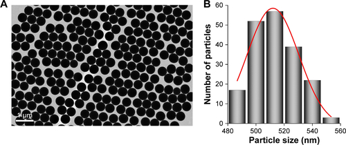
Monodisperse silica spheres (~500 nm) were prepared using a modified Stöber method (Figure S1). Typically, a tetraethyl orthosilicate (TEOS) solution (1.7 mL, 98%) was added to a solution containing ammonium hydroxide (1.97 mL, 28%–30% NH3), water (3.1 mL), and ethanol (18.2 mL), stirring at room temperature for 2 h. The excess of reagents was removed by three centrifugation/redispersion cycles with ethanol (9,000 rpm, 10 min). Particles were finally redispersed and stored in water. Silica particles were functionalized with (3-aminopropyl)triethoxysilane (APS) by means of the addition of 0.25 mL of APS in 5 mL of an ethanolic dispersion of SiO2 (8.7 mg/mL). After a 3-h stirring, the excess of reagents was removed by three repeated centrifugation/redispersion cycles with ethanol (7,000 rpm, 20 min). Then, the APS-functionalized SiO2 particles were diluted in 10 mL of EtOH and added to 10 mL of an ethanolic solution of rhodamine B isothiocyanate (RBITC) (0.32 mg/mL). After a 3-h stirring, the excess of dye was removed by three repeated centrifugation/redispersion cycles (7,000 rpm, 20 min), affording an aqueous solution of fluorescent-labeled silica particles (f-SiO2). CNT functionalization was performed using MWCNTs pretreated with acetone and ethanol to remove organic materials, frozen with N2 and lyophilized. Then, 100 mg of the purified MWCNTs was oxidized by sonication in 100 mL of a mixture of H2SO4/HNO3 (3:1) for 15 min with an ultrasonic probe (20 W) and 4 h in an ultrasonic bath. Then, the sample was washed with an NaOH aqueous solution by three centrifugation/redispersion cycles (13,000 rpm, 4 h). Upon stabilization of pH at 10, the sample was sonicated with the tip sonicator for 2 h. Then, the MWCNTs were washed with water by three centrifugation/redispersion cycles (13,000 rpm, 8 h). Finally, a stable dispersion (1.54 mg/mL) of oxidized MWCNTs (ox-MWCNTs) exhibiting a negative surface charge was obtained. Then, the ox-MWCNTs were assembled onto the labeled silica spheres. With this aim, the positively charged f-SiO2 nanoparticles (zeta potential =+40 mV) were functionalized by successive depositions of oppositely charged polyelectrolytes (poly-(sodium 4 styrenesulfonate)/poly(diallyldimethylammonium chloride)) giving rise to a positively charged surface. The deposition of the different polymers was performed following the same protocol. f-SiO2 spheres were added to a polymer solution (1 mg/mL, 0.5 M NaCl) under weak sonication for 1 h. The excess of polymer was removed by three centrifugation/redispersion cycles (20 min, 8,000 rpm). Then, 2 mL of an aqueous solution of NaCl (0.5 M) and 3 mL of an aqueous dispersion of ox-MWCNTs (1.54 mg/mL) were added to 100 mL of the positively charged f-SiO2 nanoparticles (0.87 mg/mL) and mixed under stirring for 15 h. Finally, the excess of MWCNTs was removed by three repeated centrifugation/redispersion cycles (7,000 rpm, 20 min), and the MWCNT-coated f-SiO2 spheres were dispersed in water (3.8 mg/mL with an f-SiO2:MWCNT wt. 22:1). The concentration of MWCNTs deposited onto the f-SiO2 nanoparticles was calculated by measuring the difference between the initial amount of nanotubes and that obtained in the supernatant of the centrifugation-washing step. The characterization of these composites was performed using transmission electron microscopy (TEM).
Cell culture, labeling, and microscopy imaging
HeLa cells (from the European Molecular Biology Laboratory Cell Bank) were cultured under standard conditions in Dulbecco’s Modified Eagle’s Medium containing 10% serum and containing antibiotics (from Gibco, Thermo Fisher Scientific, Waltham, MA, USA). Cells were incubated with 2–4 μg/mL of particles (unless otherwise indicated in the text) resuspended and functionalized by mild sonication in standard tissue culture medium containing serum. Cells were fixed in 4% paraformaldehyde. Microtubules were immunolabeled with the anti-α-tubulin (B512) (Sigma-Aldrich Química SL, Madrid, Spain) antibody that was recognized by a secondary goat anti-mouse immunoglobulin G (IgG) conjugated Alexa Fluor 488 (Molecular Probes, Thermo Fisher Scientific). Phalloidin-tetramethylrhodamine B isothiocyanate and Hoechst dye (Bisbenzimide) (both from Sigma-Aldrich Química SL) were used to stain actin and DNA, respectively. Confocal microscopy images were obtained with a Nikon A1R confocal microscope and were processed with the NIS-Elements Advanced Research software. High-resolution confocal imaging was performed using a Plan Apochromatic 100× oil numerical aperture 1.45 objective. All confocal cell images are pseudocolored. Live-cell phase-contrast imaging was performed on a live-cell Nikon-Ti live-cell station. TEM was also used to localize intracellular the CNT-bearing particles (CNPs) in pelleted HeLa cells fixed with 1% glutaraldehyde in 0.12 M phosphate buffer, washed in 0.12 M phosphate buffer, postfixed in 1% buffered osmium tetroxide, dehydrated, embedded in Araldite, and stained with lead citrate–uranyl acetate. Araldite sections (ca. 70 nm) were observed using a JEOL JEM 1011 microscope.
Particle quantification, live-cell assessment, and statistical analysis
Particle quantification was performed on random 20× magnification fluorescent images of the HeLa cell cultures. The Image-J software was used for random automatic quantification of extracellular versus intracellular particles. The total number of intracellular particles after overnight incubation was considered a 100%. The Student’s t-test was used to perform the statistical analysis and to evaluate significance. Cell death was identified and quantified using a standard Trypan blue exclusion assay. Cell proliferation blockage was performed using flow cytometry in a total of ca. 10,000 fixed and stained HeLa cells (per condition) in a Becton Dickinson FACS CantoII equipment. Data were analyzed using the FACS Diva software (Becton Dickinson, NJ, USA).
Microtubule depolymerization–repolymerization experiments
This assay was performed as previously describedCitation14 following classical protocols.Citation45 The microtubule tubulin polymer disassembles under low-temperature conditions (4°C, 30 min), repolymerizing in minutes under permissive temperatures (37°C). Microtubule depolymerization was performed with culture medium at 4°C, containing 2 μM nocodazole for 30 min. Microtubule regrowth was performed placing coverslips into fresh media at 37°C and cells were snap fixed in 1% glutaraldehyde at the indicated times.
Results
CNP design
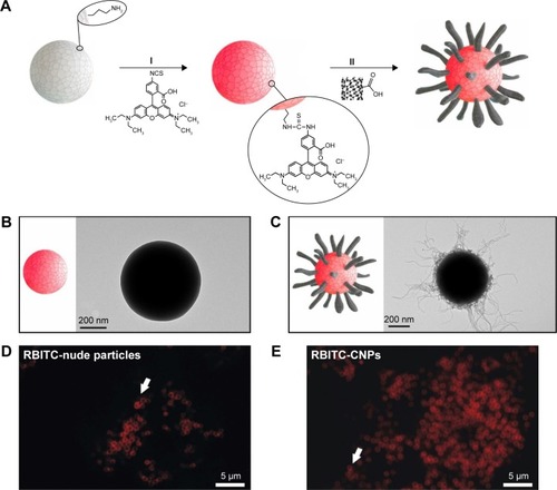
The synthesis of uniform CNPs was carried out following the approach depicted in . In a first step () SiO2 particles of ca. 500 nm were functionalized with RBITC, a fluorescence label that allows confocal microscopy intracellular particle tracking. The electrostatic deposition of negatively charged oxidized nanotubes onto fluorescence silica colloids (f-SiO2) was performed as previously described (, see “Materials and methods”).Citation46–Citation48 shows representative TEM images of the f-SiO2 particles used as uncoated controls, while shows the CNPs synthesized herein, as uniform and controlled assemblies of nanotubes. All these structures were finally coated with adsorbed serum proteins so they could be recognized by different cell-surface receptors on the cell membrane of HeLa cells, triggering receptor-mediated endocytosis as described for MWCNTs.Citation49,Citation50
Figure 1 Diagram of the fabrication process of the fluorescent CNPs.
Notes: (A) Sequential synthetic steps: (I) fluorescent labeling of the SiO2 particles (f-SiO2), and (II) deposition of CNTs onto the f-SiO2 spheres (CNP). TEM images of (B) f-SiO2 particles and (C) CNPs. Confocal microscopy image of the RBITC fluorescence on f-SiO2 particles (D) and (E) CNPs in buffered culture medium. Arrows point to individual particles in single Z-plane confocal images.
Abbreviations: CNP, CNT-bearing particle; CNTs, carbon nanotubes; f-SiO2, fluorescent-labeled silica particles; RBITC, rhodamine B isothiocyanate; TEM, transmission electron microscopy.
CNP engulfment by HeLa cells
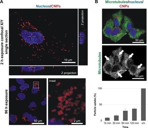
Particle engulfment was investigated using time-lapse video microscopy (Video S1). Endocytosis started a few minutes after the CNPs landed on the cell surface with ca. 50% of the particles being intracellular 2 h after the addition of the CNPs in the culture medium (). The intracellular distribution of the CNPs was determined using confocal microscopy imaging at different time points. CNP accumulation was significant in the vicinities of the nucleus, at the pericentrosomal region, 72–96 h after particle exposure. On the contrary to MWCNTs exposure, tubulin immunostaining revealed an intact microtubule cytoskeleton, irradiating from a well-organized centrosome (, arrows).
Figure 2 CNP cellular uptake and intracellular distribution.
Notes: (A) Confocal microscopy image of HeLa cells containing intracellular CNPs at 2 and 96 h. The white cross (also visible in the lateral projections) indicates the position of a cytoplasmic particle (red channel) 2 h after exposure. CNPs accumulate at the centrosomal region of the cells 96 h after initial contact (inset). (B) Confocal microscopy projection image of HeLa cells containing fluorescent CNPs displaying no abnormalities in the microtubule cytoskeleton. These tubulin nanofilaments irradiate from well-organized centrosomes (arrows). Cell nuclei (blue channel) of cells exposed to CNPs present a healthy appearance.
Abbreviations: CNP, CNT-bearing particle; CNTs, carbon nanotubes; o/n, overnight.
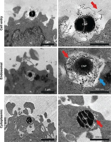
Further characterization of the CNP–cellular contact and the study of the intracellular fate of these particles was performed by TEM analysis. shows different steps in particle engulfment. First, the CNTs on the surface of the CNPs contact the HeLa surface, being eventually trapped inside endocytic membranes. The CNT-based coating surrounding the silica templates can be clearly visualized inside the endocytic vesicles (, arrows). HeLa cells exposed to CNPs during 72 h presented many particles localized inside the cytosol with their CNT coverture fully integrated in the surrounding cytoplasm, and no visible surrounding membranes. All these experiments support the idea that the entry of CNPs inside HeLa cells is similar to that described for dispersed CNTs.Citation49,Citation50
Figure 3 TEM imaging of resin section of HeLa cells exposed to CNPs.
Notes: CNP initial contact is CNT-mediated. The endocytic membranes are observed in some particles, presumably after endocytosis. Some particles also appear devoid of membranes, inside the cytoplasm. Red and blue arrows point at the CNTs attached on the surface of the CNPs and endosomal membrane, respectively. The nucleus (N) is also labeled.
Abbreviations: CNP, CNT-bearing particle; CNT, carbon nanotube; TEM, transmission electron microscopy.
CNTs attached to templates are not cytotoxic
Well-dispersed CNTs, and more particularly MWCNTs, trigger significant microtubule cytoskeleton disrupting effects that lead to antiproliferative effects and cell death in different cell types. Here we compare the effects of CNTs delivered to the cells in two different formats. On one hand, we exposed HeLa cells to fully dispersed one-dimensional (1D) functionalized CNTs as in previous studies.Citation14 On the other hand, cells were exposed to functionalized CNPs containing comparable amounts of CNTs attached to their surfaces. Cells were grown in the presence of these nanomaterials for 72 h before morphological and cell viability evaluation.
As previously reported,Citation14,Citation26,Citation51 cells exposed to dispersed CNTs displayed a disorganized microtubule cytoskeleton with no detectable centrosomes and a reorganized actin cortex (). Cell death quantification using the Trypan blue exclusion test revealed that exposure to dispersed CNTs triggered a reduction of ca. 6.5% cell viability during the first 48–72 h, thus coinciding with published observations.Citation14,Citation52
Figure 4 CNPs are highly biocompatible.
Notes: (A) Confocal microscopy images of the intracellular nanofilaments in interphase cells treated with CNPs (top) or CNTs (bottom) for 72 h. CNP-treated cells display a well-organized radial cytoskeleton with visible centrosomes (green channel, empty arrows). On the contrary, cells exposed to CNTs show a typically disorganized microtubule cytoskeleton displaying no visible centrosomes and a reactive actin cortex (red channel). (B) CNP-treated cells display a survival rate comparable to controls. (C) Control and HeLa cell cultures exposed to CNPs for 72 h. CNP-treated cells display well-assembled spindles (3, 4) compared to untreated control cells (1, 2). Dying cells were not detected.
Abbreviations: CNPs, CNT-bearing particles; CNTs, carbon nanotubes.
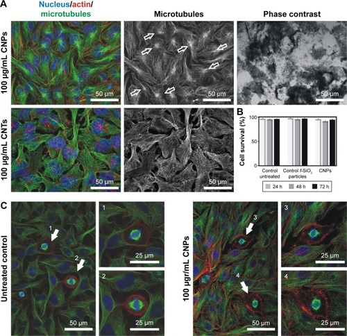
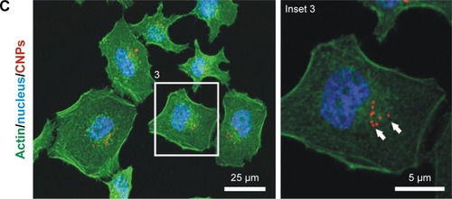
On the contrary, HeLa cells exposed to CNPs containing identical amounts of CNTs attached to their surfaces displayed a well-organized cytoskeleton, with a visible centrosome and nonreactive actin microfilaments on the cellular cortex despite the large number of CNPs per cell (>100 particles/cell) (, arrows). Interestingly, cells exposed to CNPs displayed identical survival rates to cells exposed to f-SiO2 particles or untreated controls ().
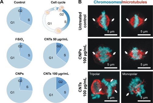
To investigate if intracellular CNPs interfered in cell division, we quantified cells at the different proliferation cycle stages using flow cytometry in a total population of ~10,000 cells per condition. As expected, cells treated with dispersed CNTs displayed a severe dosage-dependent proliferative blockage in G2 () just as previously reported. Conversely, cells exposed to CNPs displayed no detectable cell-cycle defects, dividing normally. Confocal microscopy morphological assessment of the proliferating cells in these cultures revealed normal bipolar spindle assemblies in cells exposed to CNPs that contrasted to the large collection of spindle abnormalities observed in cells exposed to dispersed CNTs. These aberrations included apolar, tripolar, or multipolar spindles, accompanied by many apoptotic figures (). In summary, we observed no obvious cytotoxic or proliferative effects in cells treated with CNTs attached to silica templates. These results stress the importance of the dispersion of CNTs in cytotoxicity.
Figure 5 (A) CNPs do not interfere with cell proliferation.
Notes: Quantification of cells at the different stages of the cell proliferation cycle (G1, S, G2) using flow cytometry. CNPs display a similar cell distribution to controls (untreated and f-SiO2-treated cells). Conversely, CNT-treated cells show an obvious dose-dependent cell proliferation blockage in G2. (B) Confocal microscopy projection images of mitotic spindles in untreated controls and cells treated with 100 μg/mL attached to CNPs or dispersed (bottom). Aberrations in the organization of the spindle microtubules (red channel) are observed only in CNT-treated cells. White arrows show the centrosomal location.
Abbreviations: CNPs, CNT-bearing particles; CNT, carbon nanotube; f-SiO2, fluorescent-labeled silica particles.
CNTs gathered on CNPs do not nucleate tubulin or actin nanofilaments
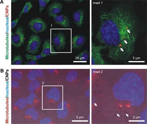
Tubulin and, to a lesser extent, actin have shown biomimetic properties with MWCNTs. To explore if these features are maintained when CNTs are gathered onto CNPs, we investigated tubulin and actin polymer nucleation on these templates following previous protocolsCitation14 that reproduced classical microtubule depolymerization–repolymerization experimentsCitation45 on live cells containing 5–15 particles/cell (“Materials and methods”). This procedure allowed to investigate if grouped CNTs could nucleate tubulin polymers ectopically, behaving as “artificial” centrosomes.
shows high-resolution confocal microscopy images of cells where microtubule ectopic nucleation sites coinciding with the CNPs were not detected after depolymerization–repolymerization. Actin recruitment on the CNPs was not detected either. In conclusion, these polymerization patterns suggest that neither tubulin nor actin interacted with CNTs gathered on CNPs, and validate the hypothesis of the biomimetic interaction of 1D CNTs with the intracellular cytoskeletal filaments.
Figure 6 Depolymerization/repolymerization experiment performed in live HeLa cells containing CNPs.
Notes: (A) Confocal microscopy image of HeLa cells containing CNPs (red channel, arrows). Repolymerizing microtubules (green channel) do not nucleate at the particles. (B) Fluorescence + phase contrast microscopy on HeLa cells treated with unlabeled CNPs (arrows). The CNT coating of the particle allows particle identification in the phase contrast image. No tubulin nucleation (red channel) is observed in the surroundings of the CNPs. Asterisks indicate the location of the centrosomes. (C) Actin nucleation (green channel) is not observed in the surroundings of the CNPs (arrows).
Abbreviations: CNPs, CNT-bearing particles; CNT, carbon nanotube.
Conclusion
These experiments demonstrate how the presence of intracellular CNTs is not sufficient to trigger cytotoxicity, and how this is determined by the dispersion of the CNTs. Our experiments support the idea that individualized CNTs can freely interact with the cellular filaments, triggering lethal biomimetic cytotoxic or genotoxic effects, while identical CNTs assembled onto templates cannot interfere with the function of intracellular nanofilaments, and thus, result innocuous to the same cells at comparable dosages. The simplest explanation is that dispersed nanotubes behave as nanomaterials, while CNPs behave like submicrometric materials. This fact has critical implications in biology because nanomaterials, and in particular CNTs, display highly reactive surfaces, as well as morphological resemblance to the cytoskeletal elements that favor their biomimetic interaction triggering a biomechanical impedance that leads to cell death. When CNTs are attached to templates, these nanofilaments do not interact with the cellular cytoskeleton behaving more like particulate materials, and thus becoming more biocompatible. These urge the revision of many of the nanotoxicological tests, often designed for standard chemicals now applied to nanomaterials. Here we demonstrate how the classical Paracelsus dogma “the dose makes the poison” is not applicable for CNTs.
Understanding the actual significance of the toxicity of CNTs provides new cues in the development of radically new delivery systems opening many new expectations and uses of CNTs in nanomedicine.
Acknowledgments
The authors are grateful to Drs Sumaira Ashraf and Iñigo Casafont for their technical assistance with microscopy analysis, and the Nikon A1R Laser Microscopy Unit of the IDIVAL Institute for the microscopy. This work was supported by the Spanish MINECO Project (references PI13/01074, PI16/00496 and CTM2014–58481-R, IDIVAL INNVAL15/15), Xunta de Galicia (Centro Singular de Investigación de Galicia-Accreditation 2016–2019 and EM2014/035), European Regional Development Fund – ERDF and Fundación Tatiana Pérez de Guzmán El Bueno.
Disclosure
The authors report no conflicts of interest in this work.
Supplementary material
Figure S1 (A) Transmission electron microscopy image of the synthesized silica nanoparticles, and (B) histogram of particle size distribution (mean size: 512±18 nm).
References
- MehraNKMishraVJainNKA Review of ligand tethered surface engineered carbon nanotubesBiomaterials20143541267128324210872
- De VolderMFTawfickSHBaughmanRHHartAJCarbon nanotubes: present and future commercial applicationsScience2013339611953553923372006
- Hernández-RiveraMZaibaqNGWilsonLJToward carbon nanotube-based imaging agents for the clinicBiomaterials201610122924027294540
- SajidMIJamshaidUJamshaidTZafarNFessiHElaissariACarbon nanotubes from synthesis to in vivo biomedical applicationsInt J Pharm20165011–227829926827920
- Correa-DuarteMAWagnerNRojas-ChapanaJFabrication and biocompatibility of carbon nanotube-based 3D networks as scaffolds for cell seeding and growthNano Lett200441122332236
- Di GiorgioMLDi BucchianicoSRagnelliAMAimolaPSantucciSPomaAEffects of single and multi walled carbon nanotubes on macrophages: cyto and genotoxicity and electron microscopyMutat Res20117221203121382506
- GhoshMChakrabortyABandyopadhyayMMukherjeeAMulti-walled carbon nanotubes (MWCNT): induction of DNA damage in plant and mammalian cellsJ Hazard Mater201119732733621999988
- GuoY-YZhangJZhengY-FYangJZhuX-QCytotoxic and genotoxic effects of multi-wall carbon nanotubes on human umbilical vein endothelial cells in vitroMutat Res2011721218419121296185
- CveticaninJJoksicGLeskovacAPetrovicSSobotAVNeskovicOUsing carbon nanotubes to induce micronuclei and double strand breaks of the DNA in human cellsNanotechnology201021115102
- SiegristKJReynoldsSHKashonMLGenotoxicity of multi-walled carbon nanotubes at occupationally relevant dosesPart Fiber Toxicol2014116
- MullerJDecordierIHoetPHClastogenic and aneugenic effects of multi-wall carbon nanotubes in epithelial cellsCarcinogenesis200829242743318174261
- SargentLMHubbsAFYoungSHSingle-walled carbon nanotube-induced mitotic disruptionMutat Res20127451–2283722178868
- GonzalezLDecordierIKirsch-VoldersMInduction of chromosome malsegregation by nanomaterialsBiochem Soc Trans20103861691169721118149
- Rodríguez-FernándezLValienteRGonzálezJVillegasJCFanarragaMLMultiwalled carbon nanotubes display microtubule biomimetic properties in vivo, enhancing microtubule assembly and stabilizationACS Nano2012686614662522769231
- SrivastavaRKPantABKashyapMPMulti-walled carbon nanotubes induce oxidative stress and apoptosis in human lung cancer cell line-A549Nanotoxicology20115219520720804439
- ReddyARReddyYNKrishnaDRHimabinduVMulti wall carbon nanotubes induce oxidative stress and cytotoxicity in human embryonic kidney (HEK293) cellsToxicology20102721–3111620371264
- MiglioreLSaracinoDBonelliACarbon nanotubes induce oxidative DNA damage in RAW 264.7 CellsEnviron Mol Mutagen201051429430320091701
- DongCEldawudRSargentLMCarbon nanotube uptake changes the biomechanical properties of human lung epithelial cells in a time-dependent mannerJ Mater Chem B Mater Biol Med201533983399226146559
- LacerdaLRussierJPastorinGTranslocation mechanisms of chemically functionalised carbon nanotubes across plasma membranesBiomaterials201233113334334322289266
- MaruyamaKHaniuHSaitoNEndocytosis of multiwalled carbon nanotubes in bronchial epithelial and mesothelial cellsBiomed Res Int2015201579318626090445
- PolandCADuffinRKinlochICarbon nanotubes introduced into the abdominal cavity of mice show asbestos-like pathogenicity in a pilot studyNat Nanotechnol20083742342818654567
- Ali-BoucettaHNunesASainzRAsbestos-like pathogenicity of long carbon nanotubes alleviated by chemical functionalizationAngew Chem Int Ed Engl20135282274227823319294
- NagaiHOkazakiYChewSHDiameter and rigidity of multiwalled carbon nanotubes are critical factors in mesothelial injury and carcinogenesisProc Natl Acad Sci USA201110849E1330E133822084097
- HoltBDShamsHHorstTAAltered cell mechanics from the inside: dispersed single wall carbon nanotubes integrate with and restructure actinJ Funct Biomater20123239841724955540
- WangJSunPBaoYLiuJAnLCytotoxicity of single-walled carbon nanotubes on PC12 cellsToxicol In Vitro201125124225021094249
- García-HeviaLValienteRFernández-LunaJLInhibition of cancer cell migration by multiwalled carbon nanotubesAdv Healthc Mater20154111640164426097131
- MaXZhangLHWangLRSingle-walled carbon nanotubes alter cytochrome c electron transfer and modulate mitochondrial functionACS Nano2012612104861049623171082
- WangLRXueXHuXMStructure-dependent mitochondrial dysfunction and hypoxia induced with single-walled carbon nanotubesSmall201410142859286924677813
- GaribaldiSBrunelliCBavastrelloVGhigliottiGNicoliniCCarbon nanotube biocompatibility with cardiac muscle cellsNanotechnology2006172391397
- MooneyEDockeryPGreiserUMurphyMBarronVCarbon nanotubes and mesenchymal stem cells: biocompatibility, proliferation and differentiationNano Lett2008882137214318624387
- VittorioORaffaVCuschieriAInfluence of purity and surface oxidation on cytotoxicity of multiwalled carbon nanotubes with human neuroblastoma cellsNanomedicine20095442443119341817
- JuLZhangGZhangXProteomic analysis of cellular response induced by multi-walled carbon nanotubes exposure in A549 cellsPLoS One201491e8497424454774
- YehiaHNDraperRKMikoryakCSingle-walled carbon nanotube interactions with HeLa cellsJ Nanobiotechnology20075817956629
- LoboAOAntunesEFMachadoAHAPacheco-SoaresCTrava-AiroldiVJCoratEJCell viability and adhesion on as grown multi-wall carbon nanotube filmsMater Sci Eng C2008282264269
- PacurariMQianYFuWCell permeability, migration, and reactive oxygen species induced by multi-walled carbon nanotubes in human microvascular endothelial cellsJ Toxicol Environ Health A201275211212822129238
- MarchesanSKostarelosKBiancoAPratoMThe winding road for carbon nanotubes in nanomedicineMater Today20151811219
- WeisskerUHampelSLeonhardtABüchnerBCarbon nanotubes filled with ferromagnetic materialsMaterials20103843874427
- Authors not listedThe dose makes the poisonNat Nanotechnol2011632921654642
- PampaloniFFlorinELMicrotubule architecture: inspiration for novel carbon nanotube-based biomimetic materialsTrends Biotechnol200826630231018433902
- DinuCZBaleSSZhuGDordickJSTubulin encapsulation of carbon nanotubes into functional hybrid assembliesSmall20095331031519148890
- JordanMAWilsonLMicrotubules and actin filaments: dynamic targets for cancer chemotherapyCurr Opin Cell Biol19981011231309484604
- ZhangYWangBMengXSunGGaoCInfluences of acid-treated multiwalled carbon nanotubes on fibroblasts: proliferation, adhesion, migration, and wound healingAnn Biomed Eng201139141442620824344
- VillegasJCÁlvarez-MontesLRodríguez-FernándezLGonzálezJValienteRFanarragaMLMultiwalled carbon nanotubes hinder microglia function interfering with cell migration and phagocytosisAdv Healthc Mater20143342443223950018
- García-HeviaLVillegasJCFernándezFMultiwalled carbon nanotubes inhibit tumor progression in a mouse modelAdv Healthc Mater2016591080108726866927
- ShelanskiMLGaskinFCantorCRMicrotubule assembly in the absence of added nucleotidesProc Natl Acad Sci U S A19737037657684514990
- Correa-DuarteMAKosiorekAKandulskiWGiersigMLiz-MarzánLMLayer-by-layer assembly of multiwall carbon nanotubes on spherical colloidsChem Mater2005171232683272
- Correa-DuarteMAKosiorekAKandulskiWGiersigMSalgueiriño-MaceiraVNanoengineered polymeric thin films by sintering CNT-coated polystyrene spheresSmall20062222022417193024
- Sanles-SobridoMSalgueiriño-MaceiraVCorrea-DuarteMALiz-MarzánLMMagnificent sea-anemone-like magnetic silica capsules reinforced with carbon nanotubesSmall20084558358618446796
- HaniuHSaitoNMatsudaYCulture medium type affects endocytosis of multi-walled carbon nanotubes in BEAS-2B cells and subsequent biological responseToxicol In Vitro20132761679168523648666
- YaronPNHoltBDShortPALöscheMIslamMFDahlKNSingle wall carbon nanotubes enter cells by endocytosis and not membrane penetrationJ Nanobiotechnology201194521961562
- HoltBDShortPARapeADWangYLIslamMFDahlKNCarbon nanotubes reorganize actin structures in cells and ex vivoACS Nano2010484872487820669976
- García-HeviaLValienteRGonzálezJFernández-LunaJLVillegasJCFanarragaMLAnti-cancer cytotoxic effects of multi-walled carbon nanotubesCurr Pharm Des201521151920192925732667