 ?Mathematical formulae have been encoded as MathML and are displayed in this HTML version using MathJax in order to improve their display. Uncheck the box to turn MathJax off. This feature requires Javascript. Click on a formula to zoom.
Abstract
Colloidal suspensions of iron oxide magnetic nanoparticles are known to dissipate energy when exposed to an oscillating magnetic field. Such energy dissipation can be employed to locally raise temperature inside a tumor between 41°C and 45°C (hyperthermia) to promote cell death, a treatment known as magnetic fluid hyperthermia (MFH). This work seeks to quantify differences between MFH and hot-water hyperthermia (HWH) in terms of reduction in cell viability using two cancer cell culture models, Caco-2 (human epithelial colorectal adenocarcinoma) and MCF-7 (human breast cancer). Magnetite nanoparticles were synthesized via the co-precipitation method and functionalized with adsorbed carboxymethyl dextran. Cytotoxicity studies indicated that in the absence of an oscillating magnetic field, cell viability was not affected at concentrations of up to 0.6 mg iron oxide/mL. MFH resulted in a significant decrease in cell viability when exposed to a magnetic field for 120 minutes and allowed to rest for 48 hours, compared with similar field applications, but with shorter resting time. The results presented here suggest that MFH most likely induces apoptosis in both cell types. When compared with HWH, MFH produced a significant reduction in cell viability, and these effects appear to be cell-type related.
Introduction
The physical, chemical, thermal, and mechanical properties of magnetic nanoparticles make them suitable for biomedical applications including cell labeling, targeting and separation, drug delivery, magnetic resonance imaging, and hyperthermia.Citation1–Citation6 For these applications, particles must have combined properties of high magnetic saturation, biocompatibility, and suitable functionalization at the surface.Citation7 One promising biomedical approach using magnetic nanoparticles is magnetic fluid hyperthermia (MFH), where cancer tissue placed in contact with magnetic nanoparticles is exposed to an alternating magnetic field.
During this process, heat is generated due to magnetic hysteresis loss.Citation8 The increase in temperature will depend on the magnetic properties of the material, the strength of the magnetic field, the frequency of oscillation, and the cooling capacity of the blood flow in the tumor site.Citation7 Nanoplatforms such as magnetite-dextran nanoparticles, magnetic cationic liposomes, and aminosilane iron oxide nanoparticles have been studied in vitro, in vivo, and in human trials, with success.Citation9–Citation16 In humans, treatment was tolerated by patients with minor or no side effects, whereas in vivo analysis demonstrated successful tumor remission.
Although significant work has been performed in the area of MFH in vitro, little has been done to compare the effects of MFH with conventional hyperthermia treatments such as hot water. Jordan et al were the first to study in vitro MFH caused by a suspension of magnetic nanoparticles, and compared its effectiveness to hyperthermia using a hot water bath.Citation17,Citation18 Results indicated differences regarding the effects on the viability of WiDr (human colonic adenocarcinoma) and BT-20 (human mammary adenocarcinoma) cells when treated by MFH and hot water. Silane-coated nanoparticles were used with the BT-20 cells, whereas dextran-coated nanoparticles where used with the WiDr cells. Results with the WiDr cell line indicated that there was no difference in reduction in cell viability for the two treatments, whereas the BT-20 cells did demonstrate a significant difference in reduction in viability due to MFH when compared with hot-water hyperthermia (HWH). Such differences are difficult to explain when dealing with different cell lines and different types of nanoparticle functionalization. Silane-magnetite nanoparticles showed higher internalization in BT-20 when compared with dextran ferrite particles on WiDr.Citation18 Higher levels of magnetic nanoparticle internalization could promote additional damage and injury to the cells when submitted to a magnetic field, generating a further effect on cell viability. Thus, it is unclear whether the observed differences are due to the use of two different types of particles or due to the differences between cell lines.
Because of the substantial interest in hyperthermia using traditional techniques in the clinical literature, and the potential of MFH to deliver heat locally, potentially avoiding side effects associated with regional and systemic forms of hyperthermia treatment, there is still interest in direct comparisons between hyperthermia and MFH.Citation19–Citation20 This is further motivated by debate in the literature as to whether hyperthermia at the nanoscale poses any advantages over traditional forms of hyperthermia.Citation21 This work seeks to quantify the differences between MFH and conventional HWH treatment using a temperature range of 41°C–46°C, which is commonly considered to be the range of hyperthermia. For this purpose, two cell models, Caco-2 (human epithelial colorectal adenocarcinoma cells) and MCF-7 (human breast cancer cells) and one nanoparticle platform composed of co-precipitated iron-oxide based magnetic nanoparticles functionalized with carboxymethyl dextran, henceforth referred to as IO-CMDX, were employed. A series of MFH and hyperthermia exposure sequences were designed and performed in both cell lines to establish a coherent comparison basis. These scheduled treatments took into consideration field exposure time and resting times after treatment. Such parameters were varied to establish differences. Potential cell death mechanisms, such as apoptosis or necrosis, were also investigated.
Materials and methods
Materials for nanoparticle synthesis
Iron (II) chloride tetrahydrate, iron (III) chloride hexahydrate, ammonium hydroxide (29% v/v), tetramethylammonium hydroxide (25% w/v), and carboxymethyl dextran sodium salt were obtained from Sigma Aldrich (St. Louis, MO). Nitric acid (HNO3) was obtained from Fisher Scientific (Pittsburg, PA).
Synthesis of IO-CMDX
Iron oxide nanoparticles were synthesized through co-precipitation of aqueous suspensions of ferric and ferrous chloride ions.Citation22 They were then functionalized with carboxymethyl dextran to improve particle dispersion in cell culture medium. To obtain nanoparticles, an aqueous solution of ferric chloride (0.36 M) was vigorously stirred with a solution of ferrous chloride (0.18 M), in the presence of a nitrogen stream, to avoid oxidation of the iron ions prior to precipitation. Ammonium hydroxide was added to the iron solution. The product of this reaction quickly changed color from brown to black, which is indicative of magnetite formation. This solution was heated to 80°C for approximately 1 hour under continuous stirring at pH 8.0 to promote reduction and precipitation. The solution was left to cool overnight. The iron oxide solution was centrifuged for 5 minutes at 1800 rpm, and the supernatant was discarded. The precipitated particles were peptized with 0.5 M HNO3. The peptized iron oxide was centrifuged for 15 minutes at 3500 rpm and then resuspended in water. A 0.013 M solution of carboxymethyl dextran at pH 11.0 was added. The final solution was maintained at a pH range of 3.0–5.0 using 1M HCl. The solution was mixed at 200 rpm for a period of 48 hours. The magnetic colloidal suspension was washed with an equivalent volume of ethanol at 3500 rpm for 10 minutes to precipitate the nanoparticles and remove the unattached carboxymethyl dextran. The precipitated particles were dialyzed to a conductivity of 10 μS/cm using de-ionized water and a dialysis membrane with a molecular weight cutoff of 20 kDa. Finally, the nanoparticles were dried in a vacuum oven.
Nanoparticle suspension
Nanoparticles were autoclaved for 60 minutes at a temperature of 121°C and 18 psi. Before experiments with cells, particles were suspended in supplemented Dulbecco’s Modified Eagle’s Medium (DMEM) (Sigma, St. Louis, MO) with phenol red and with 10% of fetal bovine serum (FBS) (Invitrogen, Carlsbad, CA). The final particle concentration was 10 mg particles/mL (0.6 mg iron oxide/mL). For cytocompatibility analysis, four dilutions of 0.3, 0.6, 0.9, and 1.5 mg iron oxide/mL suspensions in DMEM/FBS were prepared. For MFH, a concentration of 0.6 iron oxide/mL was used.
Nanoparticle characterization
The weight fraction of iron oxide core in the IO-CMDX nanoparticles was determined through thermogravimetric analysis using a TA Instruments TA-2950 Thermogravimetric Analyzer (TA Instruments, Newcastle, DE) with a heating rate of 10°C/min under air flow. The mass fraction of iron oxide was calculated from the remnant mass at a temperature of 600°C, divided by the initial sample mass. The hydrodynamic diameter of the resulting IO-CMDX nanoparticles was determined by dynamic light scattering (DLS) using a Brookhaven Instruments BI-90Plus Particle Size Analyzer (90° scattering angle) (Brookhaven Instrument Corp, Holtsville, NY). Measurements were performed with particles suspended after autoclaving using 0.1% w/w suspensions in de-ionized water. The capacity of the nanoparticles to dissipate energy as heat upon application of an oscillating magnetic field was parameterized through the so-called specific absorption rate (SAR).Citation23 Measurements were made by placing 1 mL of IO-CMDX suspension at 0.6 iron oxide/mL in a thermally insulated nonmetallic/nonmagnetic container. The sample was allowed to reach thermal equilibrium with the surroundings, and then a magnetic field of 20 kA/m and 238 kHz was applied using a four-turn induction coil connected to an RDO Induction HFI 3-135/400 (RDO Enterprises Inc, Washington, NJ). Temperature was monitored as a function of time using a ThermoVision™ A20 thermal imaging camera (FLIR systems, Boston, MA). The SAR value of the nanoparticles was calculated according to:
where Ĉ is the specific heat capacity, assumed equal to that of water, msample and miron oxide are the total sample mass and iron oxide mass, respectively, and dT/dtt→0 is the initial rate of increase in temperature upon application of the magnetic field.
Caco-2 cell culture
Caco-2 cells were purchased from the American Type Culture Collection (Rockville, MD). Cells were cultured on 75 cm2 cell culture flasks (canted neck) (Costar Corning, Lowell, MA) at a concentration of 250,000 cells/flask, using DMEM containing 10% FBS, 1% nonessential amino acids (Invitrogen), 100 units/mL of penicillin (Sigma), and 100 μg/mL of streptomycin (Sigma). Cells were maintained in a controlled atmosphere at 37°C, 95% relative humidity, and 5% CO2. The culture medium was changed every other day for approximately 5–6 days until cells reached approximately 80%–90% confluency. Cells were detached from the culturing flask by trypsinization, resuspended in culture media, and counted.
MCF-7 cell culture
MCF-7 cells were purchased from American Type Culture Collection (Rockville, MD). Cells were cultured as described above, but with a concentration of 1 × 10⁶ cells/flask.
Cytocompatibility analysis
Cells were seeded at a concentration of 10,000 cells/cm2 for Caco-2 and 40,000 cells/cm2 for MCF-7 in 96-well assay plates of 0.71 cm2/well (Costar Corning) and grown for 1 week in supplemented DMEM with phenol red, with particle concentrations of 1.0, 2.0, 3.0, and 5.0 mg/mL at 37°C and 5% CO2. Pure DMEM was used as a negative control and 1.5% hypochlorite solution was used as a positive control for cell viability. At the end of the week, nanoparticle suspensions were removed and the 96-well assay plate was rinsed with Hank’s balanced salt solution (Sigma). For cell viability analysis, the cells were incubated at 37°C and 5% CO2 with CellTiter-BlueTM (Promega, Madison, WI). Cell viability was analyzed with a spectrofluorometer (SpectraMax Gemini EM, Molecular Devices, Sunnyvale, CA) with an excitation wavelength of 560 nm and an emission wavelength of 590 nm required for the aforementioned assay. Cell viability results were interpreted as the viability ratio as a function of nanoparticle concentration. The viability ratio was calculated by normalizing all relative fluorescent unit values with the relative fluorescent unit value of the negative control (cells with DMEM).
Hyperthermia using a hot water bath
Cells were cultured as described. They were detached from the culturing flask by trypsinization, resuspended in culture media, and counted. A quantity of 3 × 10⁶ cells was seeded on a 20 mL test tube and suspended to a final volume of 10 mL with DMEM + 10% FBS. The test tube was then placed in a hot water bath at constant temperature (41°C or 45°C) for a period of 2 hours. Samples were gently shaken at short time intervals. After hyperthermia treatment, the cells’ viability was quantified and the presence of apoptotic markers was studied as described below.
MFH
Cells were cultured as described above. Cells were detached from the culturing flask by trypsinization, resuspended in culture media, and counted. A concentration of 3 × 10⁶ cells was placed in a 20 mL test tube and suspended to a final volume of 15 mL with DMEM + 10% FBS (negative control) or with a concentration of 0.6 mg/mL of IO-CMDX nanoparticle suspension. The test tube was then inserted into a four-turn induction coil connected to an RDO induction HFI 3-135/400 (RDO Enterprises Inc, Washington, NJ) and bubbled with air to maintain cells suspended during magnetic field application.
The magnetic field was applied for a period of 1 hour or 2 hours with a magnetic field amplitude of 20 kA/m and frequency of 238 kHz. Temperature was monitored using a ThermoVision™ A20 thermal imaging camera (FLIR Systems, Boston, MA) and maintained between 42°C and 45°C. Samples took an average of 20 minutes to reach the desired temperature, after which exposure time started. After the magnetic field treatment the cell’s viability and apoptosis were analyzed as described below.
Cell viability and apoptosis
The effect of hyperthermia on cell culture was analyzed by performing cell viability and testing for the presence of apoptotic markers. These assays were performed immediately, 24 hours, or 48 hours after hyperthermia application. The assays at 24 and 48 hours were performed to allow cell death to occur. Cell viability was measured by cell-counting using trypan blue. Cells were counted before and after hyperthermia application. A 1:1 dilution of cell sample and trypan blue dye was used for cell counting using a hematocytometer. Stained cells were considered dead. Viability results were interpreted as the ratio of viable cells after treatment and viable cells before treatment. Final results were normalized with the negative control. The presence of apoptotic markers was analyzed using the ApoPercentage assay (Biocolor, County Antrium, UK). This assay stains cells when the inner plasma membrane leaflet has been exposed, a classic marker of apoptosis. Cells were put in contact with the assay with an ApoPercentage concentration of 5% for a period of 30 minutes. The apoptotic and nonapoptotic cells were counted using a hematocytometer. The results were interpreted as the apoptosis ratio over the viability ratio.
Data analysis
Unless otherwise stated, the sample size was n = 3. T-test (two- tailed distribution, two-sample with unequal variances) analysis was employed to determine any significance in observed data. A P-value of <0.05 was considered statistically significant.
Results
Particle characterization
The hydrodynamic diameter of the IO-CMDX was found to be 72.2 ± 4.5 nm according to DLS. Thermogravimmetric analysis indicated that it consisted of 27% by weight inorganic core and 73% by weight organic material, on the basis of the remnant mass after heating to 600°C. The SAR of the particles was 245.26 W/g at a magnetic field amplitude of 20 kA/m and frequency of 238 kHz. Particle samples were also tested for stability in culture media and after autoclaving. Results indicated that particle samples used for hyperthermia experiments consisting of a particle concentration of 0.6 mg of iron oxide/mL were stable for the experimental time frame, and their size in suspension was not affected by the autoclaving process. Note that the particles used in this study are similar to those reported in our recent work, in which additional chemical, physical, and magnetic characterization is provided.Citation24
Cytocompatibility
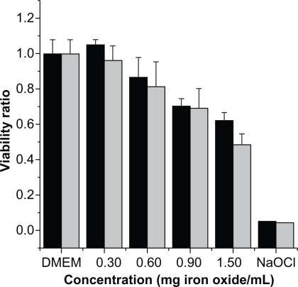
Cytocompatibilty analyses were first performed to rule out the effect of nanoparticle toxicity during MFH. Experiments were performed during cell seeding for a period of 1 week using two different cell culture models (Caco-2 and MCF-7). Cell viability analysis of IO-CMDX on both cell culture lines started to demonstrate cytotoxic effects at concentrations above 0.9 mg iron oxide/mL (see ).
Figure 1 Viability analysis of iron oxide nanoparticles coated with carboxymethyl dextran as a function of concentration.
Notes: ![]()
Abbreviation: DMEM - cells maintained in the incubator.
MFH and HWH
Two hyperthermia protocols were applied, HWH and MFH, with the purpose of comparing traditional hyperthermia with that resulting from energy dissipation from magnetic nanoparticles. Experiments were performed to measure the immediate and long-term impact of the hyperthermia treatments in cell viability using two cell lines, Caco-2 and MCF-7.
HWH experiments were performed at two temperatures, 41°C and 45°C, with an exposure period of 1 or 2 hours. These two temperatures were selected to examine possible differences between the minimum and maximum temperature of the established hyperthermia range. Cell viability was analyzed immediately after application and 24 and 48 hours after hyperthermia treatment. Apoptosis analysis was also performed on all treatments to investigate whether hyperthermia promoted apoptosis on our cell models. Apoptosis results are presented as the ratio between the apoptosis ratio and the viability ratio. Magnetic fluid hyperthermia was performed by varying the time of magnetic field application, with a magnetic field amplitude of 20 kA/m and frequency of 238 kHz. Temperature was continuously monitored during magnetic field application with an infrared camera and an alcohol thermometer. The average temperature recorded during these experiments was always between 42°C and 45°C.
To compare the thermal doses supplied to the cells with the two treatments, the thermal dose was calculated as proposed by Dewey et al.Citation20 The thermal dose in equivalent minutes at 43°C was 420 minutes for HWH and 450 minutes for MFH; that is, the HWH protocol actually resulted in a slightly higher thermal dose compared with our MFH protocol, when using the equivalent minutes dose metric of Dewey.Citation25 However, as discussed below, results indicated that for both cell models, MFH induced a more significant reduction in cell viability compared with HWH.
Viability and apoptosis analysis using HWH at 41°C on cells were performed (data not shown). Data indicated a slight decrease in cell viability when compared with the negative control. This suggests that hyperthermia at 41°C does not cause a significant apoptotic effect in the two cell models used in this study. To further investigate the effects of MFH and HWH, four different schedules were performed, as follows:
Schedule A – analysis immediately after 2-hours application
Schedule B – analysis performed 24 hours after 2-hours application
Schedule C – analysis performed 48 hours after 2-hours application
Schedule D – analysis performed 48 hours after 1-hour application.
These schedules were designed to study the immediate and long-term effects of HWH and MFH on cell viability and apoptosis. Three different controls were always performed for each experiment:
DMEM – cells maintained in the incubator
MF – magnetic field application to cells without particles
Cytotoxicity – cells in contact with particle suspension without the application of a magnetic field.
An average of all the corresponding controls are reported and normalized by the results of cells maintained in the incubator.
Results indicated that viability of Caco-2 cell controls () was over 90% in all cases, and there was no statistically significant difference between the experimental controls. Apoptosis level was between 20% and 25% () for HWH and MFH. In the case of the MCF-7 cell line, viability results were also over 90% for all cases (), and apoptosis levels were approximately 15% (). Experiments performed using schedule A (2-hours application and no resting time) in Caco-2 cells showed a decrease in cell viability to ∼85% after HWH and to ∼75% after MFH (, ‘Schedule A’). When the viability was measured 24 hours after 2-hours hyperthermia (schedule B), the HWH treatment did not show any statistically significant difference (P = 0.62, α = 0.05) when compared with HWH using schedule A. A resting time of 48 hours after hyperthermia resulted in the reduction of cell viability to ∼25% for HWH and to ∼2% for MFH (, ‘Schedule C’). Similar experiments were conducted for a shorter period of hyperthermia exposure. Schedule D, which consisted of 1 hour of hyperthermia and a resting time of 48 hours, had a greater effect on cell viability in both methods, HWH and MFH, when compared with schedules A and B. A cell viability of ∼65% was obtained for HWH and ∼25% for MFH.
Figure 2 Viability analysis of Caco-2 cells exposed to various modes of hyperthermia.
Notes: ![]()
Abbreviations: DMEM – cells maintained in the incubator; MF – magnetic field application to cells without particles. Cytotoxicity – cells in contact with particle suspension without the application of a magnetic field.
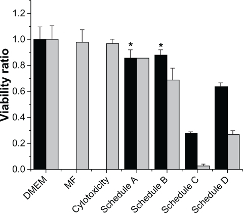
Figure 3 Apoptosis ratio over viability ratio for Caco-2 cells exposed to various modes of hyperthermia.
Notes: ![]()
Abbreviation: DMEM – cells maintained in the incubator; MF – magnetic field application to cells without particles. Cytotoxicity – cells in contact with particle suspension without the application of a magnetic field.
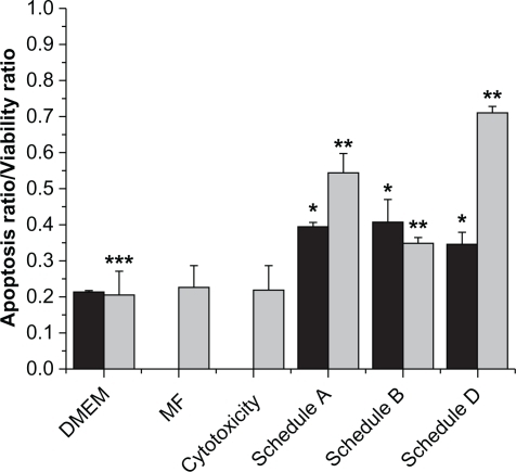
Figure 4 Viability analysis of MCF-7 cells exposed to various modes of hyperthermia.
Notes: ![]()
Abbreviation: DMEM – cells maintained in the incubator; MF – magnetic field application to cells without particles. Cytotoxicity – cells in contact with particle suspension without the application of a magnetic field.
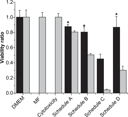
Figure 5 Apoptosis ratio over viability ratio for MCF-7 cells exposed to various modes of hyperthermia.
Notes: ![]()
Abbreviation: DMEM – cells maintained in the incubator; MF – magnetic field application to cells without particles. Cytotoxicity – cells in contact with particle suspension without the application of a magnetic field.
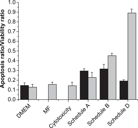
Apoptosis analysis was also performed on Caco-2 cells after each of the treatments (), except for schedule C since most cells were dead at that point. Apoptosis after HWH was statistically similar for all treatments (P = 0.65, α = 0.05) but different from controls, exhibiting an increase to ∼35%–40%. On the other hand, for MFH, schedule A showed an apoptosis of ∼55%, schedule B an apoptosis of ∼35%, and schedule D an apoptosis of ∼70%, which have statistically significant differences from the negative control (P = 0.02, α = 0.05) and HWH (P = 0.008, α = 0.05).
Similar experiments were performed on MCF-7 cells in order to establish whether the observed phenomenon was specific to the Caco-2 cell line. Results for HWH for all of the treatments, except schedule C, demonstrated a reduction in cell viability to ∼85%, with no statistically significant difference between treatments (P = 0.15 for α = 0.05). HWH using schedule C (2 hours of application and a resting time of 48 hours) resulted in a reduction in cell viability to ∼45% (see ). In the case of MFH, there was a significant difference in reduction in cell viability for schedules A, B, C, and D, for which cell viability decreased to ∼80%, ∼50%, ∼5%, and ∼30%, respectively. Apoptosis analysis in MCF-7 cells showed an increase to ∼30% for schedules A and B and to ∼20% for schedule C after HWH (). On the other hand, the resulting apoptosis for MCF-7 after MFH was ∼25% for schedule A, ∼45% for schedule B, and ∼90% for schedule D. Schedule C was not measured since most cells were dead.
Discussion
This work intended to quantify differences between conventional hyperthermia (HWH) and MFH. Cytocompatibility analysis indicated that there was a reduction in cell viability as the amount of iron oxide in suspension increased. This reduction in cell viability may be attributed to loss of colloidal stability as significant precipitation of the particles was observed at higher concentrations. Recently, Miles et alCitation26 examined the colloidal stability of nanoparticles in the presence of phosphate based buffers. Results demonstrated that carboxylic acid groups were displaced by the phosphate ions promoting particle aggregation. Since the DMEM formulation possesses a high concentration of phosphate ions (0.109 g/L of sodium phosphate monobasic), this was most likely the cause for particle aggregation at high concentrations. Loss of viability may be due to potential physical damage caused by precipitated particles. As such, a concentration of 0.6 mg iron oxide/mL was selected for MFH experiments to ensure that the observed effects were solely due to the effects of MFH.
Experimental controls performed on cell viability and apoptosis analysis demonstrated that neither magnetic nanoparticles alone nor the magnetic field alone affected cell viability under the conditions tested. By comparing the 1-hour and 2-hours MFH treatments, it was observed that there was a difference in viability when measured immediately, 24 hours, or 48 hours after treatment. As expected, exposure to hyperthermia for 2 hours resulted in a greater effect on cell viability when compared with exposure for 1 hour. The ability of cells to protect themselves from heat stress by producing heat shock proteins could be the reason for the need to apply a longer period of heat.Citation27 By comparing the results between MFH and HWH, it can be observed that there was a greater decrease in cell viability with MFH, compared with HWH, for both cell types. Jordan et alCitation17,Citation18 reported that WiDr cells treated by MFH and HWH did not present a difference in viability, whereas BT-20 cells did show a significant difference in cell viability after MFH in comparison with HWH. However, in their case two different cell models with two different particles were employed. As a result, direct comparisons are difficult to make. Also, they performed MFH on a cell pellet, where only cells on the surface of the pellet will have direct contact with the particles. Our experiments were conducted with cells in suspension; hence, nanoparticles and cells were in direct contact during the treatment. More importantly, studies in both cell lines were conducted using similarly coated nanoparticles. This allows for a more direct comparison between HWH and MFH. Our results indicated that for both cell models MFH had a greater effect on cell viability when compared with HWH. These results indicate that MFH is an effective tool to target heat to desired areas while potentially overcoming current challenges of traditional hyperthermia treatments.
Conclusion
This work explored differences between conventional hyperthermia (HWH) and MFH. Cytotoxicity experiments demonstrated that as colloidal stability was lost, an increase in cytotoxic effects was observed when either of the cell lines were exposed to IO-CMDX nanoparticles. Subsequent experiments were conducted with particle concentrations which did not result in loss in cell viability in the absence of a magnetic field. HWH experiments suggested that apoptosis was induced in both cell models at 45°C. The apoptotic effect was slow and its effects on viability were not observed until 48 hours after 2 hours of treatment. On the other hand, an effect due to MFH on cell viability was detected 24 hours after 2 hours of treatment, suggesting that MFH had a more significant effect on cell viability than HWH. When cell viability was compared between HWH and MFH, results clearly indicated that there is a significant additional effect on cell viability due to MFH when compared with hot water treatment, providing additional evidence of the promising use of magnetic nanoparticles for localized hyperthermia applications. The mechanisms for the observed differences are still unknown.
Acknowledgements
This work was supported by the US National Science Foundation (Grant No. CBET-0609117).
Disclosure
The authors report no conflicts of interest in this work.
References
- KrishnanKMBiomedical nanomagnetics: a spin through possibilities in imaging, diagnostics, and therapyIEEE Trans Magn20104672523255820930943
- LatorreMRinaldiCApplications of magnetic nanoparticles in medicine: magnetic fluid hyperthermiaP R Health Sci J200928322723819715115
- JainPLeeKEl-SayedIEl-SayedMCalculated absorption and scattering properties of gold nanoparticles of different size, shape, and composition: applications in biological imaging and biomedicineJ Phys Chem B2006110147238724816599493
- MerrittWMLinYGSpannuthWAEffect of interleukin-8 gene silencing with liposome-encapsulated small interfering RNA on ovarian cancer cell growthJ Nat Cancer Inst2008100535937218314475
- BriggerIDubernetCCouvreurPNanoparticles in cancer therapy and diagnosisAdv Drug Deliv Rev200254563165112204596
- FerrariMCancer nanotechnology: opportunities and challengesNat Rev Cancer20055316117115738981
- GuptaAGuptaMSynthesis and surface engineering of iron oxide nanoparticles for biomedical applicationsBiomaterials200526183995402115626447
- RosensweigRHeating magnetic fluid with alternating magnetic fieldJ Magn Magn Mater20022521–3370374
- WadaSYueLTazawaKFurutaINagaeHTakemoriSNew local hyperthermia using dextran magnetite complex (DM) for oral cavity: experimental study in normal hamster tongueOral Dis20017319219511495196
- JordanAScholzRMaier-HauffKThe effect of thermotherapy using magnetic nanoparticles on rat malignant gliomaJ Neurooncol2006578171416314937
- KawaiNFutakuchiMYoshidaTEffect of heat therapy using magnetic nanoparticles conjugated with cationic liposomes on prostate tumor in boneProstate200868778479218302228
- HilgerIHiergeistRHergtRWinnefeldKSchubertHKaiserWAThermal ablation of tumors using magnetic nanoparticles: an in vivo feasibility studyInvest Radiol2002371058058612352168
- Maier-HauffKRotheRScholzRIntracranial thermotherapy using magnetic nanoparticles combined with external beam radiotherapy: results of a feasibility study on patients with glioblastoma multiformeJ Neurooncol2007811536016773216
- JohannsenMGneveckowUEckeltLClinical hyperthermia of prostate cancer using magnetic nanoparticles: presentation of a new interstitial techniqueInt J Hyperthermia200521763764716304715
- JohannsenMGneveckowUThiesenBThermotherapy of prostate cancer using magnetic nanoparticles: feasibility, imaging, and three-dimensional temperature distributionEur Urol20075261653166117125906
- ThiesenBJordanAClinical applications of magnetic nanoparticles for hyperthermiaInt J Hyperthermia200824646747418608593
- JordanAWustPScholzRCellular uptake of magnetic fluid particles and their effects on human adenocarcinoma cells exposed to AC magnetic fields in vitroInt J Hyperthermia19961267057228950152
- JordanAScholzRWustPEndocytosis of dextran and silancoated magnetite nanoparticles and the effect of intracellular hyperthermia on human mammary carcinoma cells in vitroJ Magn Magn Mater1999194185196
- SunXRXingLGLingCCBiological rationales and clinical applications of temperature controlled hyperthermia – implications for multimodal cancer treatmentsCurr Med Chem201017273045305720629627
- HurwitzMDThe effect of mild temperature hyperthermia on tumor hypoxia and blood perfusion: relevance for radiotherapy, vascular targeting and imagingInt J Hyper2010263224231
- RabinYIs intracellular hyperthermia superior to extracellular hyperthermia in the thermal sense?Int J Hyper2002183194202
- LefebureSBuboisECabuilVNeveuSMassartRMonodisperse magnetic nanoparticles: preparation and dispersion in water and oilsJ Mater Res1998131029752981
- FortinJPWilhelmCServaisJMenagerCBacriJCGazeauFSize-sorted anionic iron oxide nanomagnets as colloidal mediators for magnetic hyperthermiaJ Am Chem Soc20071292628263517266310
- CreixellMHerreraAPLatorre-EstevesMAyalaVTorres-LugoMRinaldiCEffect of graft method on the stability and cytotoxicity of carboxymethyl dextran coated magnetic nanoparticlesJ Mater Chem20102085398547
- DeweyWCArrhenius relationships from the molecule and cell to the clinicInt J Hyper2009251320
- MilesWCGoffJDHuffstetlerPPSynthesis and colloidal properties of polyether–magnetite complexes in water and phosphate-buffered salineLangmuir2009252802813
- RylanderMNFengYBassJDillerKRThermally induced injury and heat shock protein expression in cells and tissuesAnn NY Acad Sci20051066122224216533928