
Abstract
Tissue engineering (TE) is an interdisciplinary field integrating engineering, material science and medical biology that aims to develop biological substitutes to repair, replace, retain, or enhance tissue and organ-level functions. Current TE methods face obstacles including a lack of appropriate biomaterials, ineffective cell growth and a lack of techniques for capturing appropriate physiological architectures as well as unstable and insufficient production of growth factors to stimulate cell communication and proper response. In addition, the inability to control cellular functions and their various properties (biological, mechanical, electrochemical and others) and issues of biomolecular detection and biosensors, all add to the current limitations in this field. Nanoparticles are at the forefront of nanotechnology and their distinctive size-dependent properties have shown promise in overcoming many of the obstacles faced by TE today. Despite tremendous progress in the use of nanoparticles over the last 2 decades, the full potential of the applications of nanoparticles in solving TE problems has yet to be realized. This review presents an overview of the diverse applications of various types of nanoparticles in TE applications and challenges that need to be overcome for nanotechnology to reach its full potential.
Introduction
Tissue engineering (TE) is the study of the growth of new tissues and organs, starting from a base of cells and scaffolds.Citation1,Citation2 The scaffolds are used as three-dimensional (3D) structures in which cells grow, proliferate and differentiate into various cell types. Growth factors are introduced into the scaffolds to direct cell behavior toward any desired process where the eventual goal is to produce fully functional organs or tissues capable of growth and regeneration and suitable for implantation.Citation3,Citation4 Despite such promises, TE faces numerous limitations; transcribing these ideas into reality seems like an uphill task. The inability of engineered materials to mimic the natural properties of tissues is one of the roadblocks. Nanotechnology through customized nanoparticle engineering has the potential to solve this challenge.Citation4,Citation5
Nanoparticles are characterized by their nanoscale dimension, enabling them to develop critical physical and chemical characteristics that enhance their performance and therefore make them beneficial for a wide range of applications.Citation6 In the biomedical field, nanoparticles have been used for controlled drug delivery,Citation7,Citation8 imaging of specific sites, probing of DNA structures,Citation9–Citation11 biomolecular sensing, gene delivery, photothermal ablation of cellsCitation12 and, most recently, TE.Citation13,Citation14 Additionally, many therapies utilize nanoparticles for the treatment of cancer,Citation15 diabetes,Citation16 allergy,Citation17 infectionCitation18 and inflammation.Citation19
Very recently, nanoparticles have been used in TE in order to obtain improved mechanical and biological performances.Citation20 The surface conjugation and conducting properties of gold nanoparticles (GNPs), the antimicrobial properties of silver and other metallic nanoparticles and metal oxides, the fluorescence properties of quantum dots and the unique electromechanical properties of carbon nanotubes (CNTs) have made them very useful in numerous TE applications. In addition, magnetic nanoparticles (MNPs) have been applied in the study of cell mechanotransduction, gene delivery, controlling cell patterning and construction of complex 3D tissues.
The advantage of nanoparticles in TE stems from their small size and their associated large surface to volume ratio, which is comparable to peptides and small proteins. They can easily diffuse across membranes and facilitate uptake by cells. Moreover, one is not limited by a predetermined size for nanoparticles, since they can be made in customized sizes and surface characteristics in order to suit any purpose. Nanoparticles also mimic the natural nanometer size scale of extracellular matrix (ECM) components of tissues themselves.
In one sense, nanoscale structures can be considered as an integral part of our body where the components of organs and tissues such as ECM and cells comprise various atoms, molecules, nanostructures, microstructures and macroscale structures hierarchically.
On the other hand, there are numerous external nanoparticles that enter and exit our body frequently through the inhaled air or oral and topical routes, which depending on the toxicity or nature of the particle may or may not be harmful to the body. A third source of nanoparticles in our body could be the release or secretion of elements from biomedical prosthetics and implants transplanted in our body. As reported in several studies,Citation21–Citation23 metal nanoparticles, namely, Ag, Cr, Fe, Mo, Ni, Ta, Cr, Co, Sb and Sc,Citation22 were formed both in tissues adjacent to the implants and distant to the implantation sites after implementation of bioprosthetics in the body. A high accumulation of metallic elements was found in the brain and lungs of patients with hip endoprosthesis, while hip arthroplasty patients were found to exhibit high concentrations of Cr, Co and Mo ions in their hair as well.Citation24 These three types of nanoparticles mentioned earlier are beyond the scope of this review.
In this review, we present an overview of the diverse applications of various types of nanoparticles in TE applications. This review addresses the applications of magnetic, metallic, ceramic and metal oxide nanoparticles in TE. They are advantageous, as they tend to be biocompatible and have low immunogenicity. The specific intrinsic properties of each material may hold the answer to overcoming the current hurdles in TE, where the use of materials engineering has not been exploited to its full potential. The insights provided in this review will be beneficial for material scientists and tissue engineers working with nanoparticles, allowing them to navigate through applications that best fit their need.
Brief background of nanoparticles
Nanoparticles are entities of any shape with a size range of 1–100 nm in any one dimension.Citation25 Such a unique size gives these particles the properties of both bulk materials and molecular structures. As such, nanoparticles are viewed as the “bridge” between the macroscopic and microscopic structures. Their small size gives them one of their most attractive intrinsic properties: a high surface to volume ratio. Nanoparticles are highly mobile when they are in a free state, causing them to have tremendously slow sedimentation rates (). In addition, they are characterized by a wide range of compositions ranging from soft to hard materials depending on their use and may exhibit what is known as the quantum effect. This quantum effect allows one to have extreme control over the surface energy of these particles, which in turn can control initial protein adsorption to dictate cellular interactions.Citation26
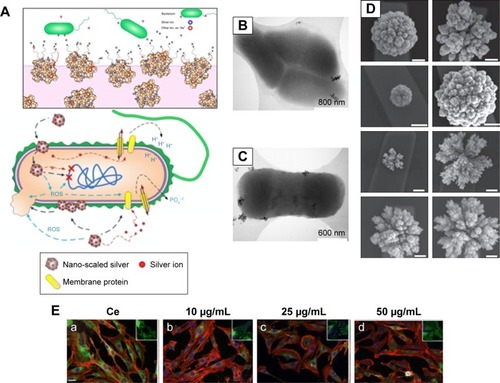
Figure 1 Interaction of bacteria and mammalian cells with nanoparticles.
Notes: (A) Schematics of the effects of nanoparticles on bacterial cells. (B and C) Representative electron microscopy images showing the interaction of E. coli with the samples of cerium oxide nanoparticles at different magnifications. (D) Programmable nanomaterial synthesis using GNPs under different deposition parameters. (E) Effects of CeO2 nanoparticles on cardiac progenitor cell morphology and growth. CeO2-untreated (a) or CeO2-treated cells 10 μg/mL (b), 25 μg/mL (c) and 50 μg/mL (d). (A–C) Republished with permission of American Society for Microbiology, from Effects of engineered cerium oxide nanoparticles on bacterial growth and viability. Pelletier DA, Suresh AK, Holton GA, et al. Appl Environ Microbiol. 76(24):7981–7989. Copyright 2010.Citation167 (D) Reprinted by permission from Springer Nature: Nat Nanotechnol. Programming the detection limits of biosensors through controlled nanostructuring. Soleymani L, Fang Z, Sargent EH, Kelley SO. Copyright 2009.Citation168 (E) Reprinted with permission from Pagliari F, Mandoli C, Forte G, et al. Cerium oxide nanoparticles protect cardiac progenitor cells from oxidative stress. ACS Nano. 2012;6(5):3767–3775. Copyright 2012 American Chemical Society.Citation169
Abbreviations: E. coli, Escherichia coli; GNP, gold nanoparticle; ROS, reactive oxygen species.
Depending on the shape, nanoparticles can be of 0D, 1D, 2D or 3D structure types.Citation27,Citation28 Based on their source or type of materials, they can be divided into different categories such as carbon-based, metal-based, ceramic-based, polymeric-based, semiconductor-based and lipid-based nanoparticles.Citation28 Nanoparticles can be synthesized by using two main methods such as 1) bottom-up and 2) top-down approaches.Citation29 Furthermore, these methods can be divided into mechanical, chemical and biological syntheses,Citation30 and these include chemical vapor deposition, physical vapor deposition, a sol–gel method, radio frequency (RF) plasma method, pulsed laser method, thermolysis and solution combustion method. Various characterization methods used to characterize the nanoparticles depend on the property of the materials. The morphology of the nanoparticles has been analyzed using polarized optical microscopy (POM), scanning electron microscopy (SEM), transmission electron microscopy (TEM), atomic force microscopy (AFM), X-ray photoelectron spectroscopy (XPS), infrared (IR), X-ray diffraction (XRD), Raman spectroscopy, Brunauer–Emmett–Teller (BET) and Zeta size analyzer.Citation30,Citation31
These properties provide unique applications of nanoparticles in a wide variety of research fields, eg, biomedical sciences, electronics, optics and more. While significant progress has been made for numerous nanoparticle applications in these fields, their applications in TE are still in their infancy stage hindered by some significant challenges. Here, we aim to review the application of nanoparticles in TE along with the challenges associated with them and address how such challenges can be tackled widening the application of nanoparticles in a broad range of TE applications.
Applications of nanoparticles in TE
Nanoparticles have been used to serve various functions in TE, ranging from enhancement of biological, electrical and mechanical properties to gene delivery, DNA transfection, viral transduction and patterning of cells, to facilitate the growth of various types of tissues to molecular detection and biosensing. The use of the right type of nanoparticles in TE can significantly enhance the biological, mechanical and electrical properties of scaffolds as well as can serve various functions depending on the applicationsCitation32 as described in the following.
Biological property enhancement: increased cell proliferation rates
In particular, two types of nanoparticles, namely, GNPs and titanium dioxide (TiO2) nanoparticles, have been used to enhance cell proliferation rates for bone and cardiac tissue regeneration, respectively. GNPs have shown superior biocompatibility and the ability for surface modification, which has resulted in interesting biomedical applications.Citation33,Citation34 In bone TE, it was first observed that GNPs promote osteogenic differentiation of an osteoblast precursor cell line, MC3T3-E1.Citation35,Citation36 In addition, these nanoparticles also influenced osteoclast (or bone resorbing cell) formation from hematopoietic cells while providing protective effects on mitochondrial dysfunction in osteoblastic cells.Citation37,Citation38 Another report showed that GNPs can promote osteogenic and adipogenic differentiation of mesenchymal stem cells (MSCs). This happened after their intracellular uptake during the establishment of the p38 mitogen-activated protein kinase (MAPK) pathway.Citation38 It was found, however, that this osteogenic differentiation is dependent on the size of GNPs. One study demonstrated that GNPs of diameters 30–50 nm were favorable and more effective for human adipose-derived stem cell (ADSC) function,Citation37 while another study showed a preference for 20 and 40 nm sized GNPs for MC3T3-E1 osteoblast-like cell function.Citation39
In light of these studies, GNPs present themselves as excellent candidates for bone tissue regeneration. They seem to be perfect candidates to replace bone morphogenetic proteins (BMPs). Although BMPs have beneficial effects on bone regeneration and repair,Citation40,Citation41 they also have many disadvantages. Their high cost and susceptibility to result in unwanted bone formation and local inflammatory reactions are some of the main reasons that have prompted researchers toward the search for alternative bone growth materials, and GNPs may be a promising substitute.Citation42–Citation44 Numerous papers have reported promising in vitro experimental results using GNPs. However, there were considerable variations in the experimental results when carried out in animals.
To overcome this, hybrid scaffolds (GNPs and gelatin) were studied under in vivo conditions for their effectiveness in initiating osteogenic differentiation consequent to human ADSCs. The hydrogels were placed in the parietal bone of rabbits, where bone defects were made prior to the application of the hydrogel. The study showed enhanced proliferation of osteoblasts as the GNP content in the hydrogels increased.Citation44
In other studies, GNPs and gold nanowires placed within a scaffold had a remarkable impact on cell proliferation and synapse formation, which is the key to a functional organ transplant. Additionally, these materials may also direct stem cell differentiation, without using growth factors, thus minimizing/avoiding the numerous side effects of the application of growth factors in the body.Citation45,Citation46
GNPs have also been successfully used to produce a favorable cellular environment for cardiac TE applications. GNP-loaded nanofibers led to the enhancement of cardiomyogenic differentiation to help infarcted myocardium regeneration.Citation47
In another study, TiO2 nanoparticles were added to the scaffold in which human embryonic stem cell-derived cardiomyocytes showed enhanced cell proliferation.Citation48 TiO2 nanoparticles have also been 3D printed with polymers, such as poly(lactic-co-glycolic acid) (PLGA) to exactly match the nanostructured roughness of bone itself and significantly improve bone cell performance.Citation49 Interestingly in this study, the TiO2 nanoparticles were also shown to decrease the harmful effects of acid byproducts released by PLGA during degradation, which are known to kill cells.Citation49 Thus, incorporation of certain types of nanoparticles into scaffolds can significantly alter the biological properties of scaffolds and thus help in TE.
Enhancement of mechanical properties
Nanoparticle-embedded nanocomposite polymers both in the form of hydrogels and electrospun fibers have exhibited superior mechanical properties for TE applications compared to scaffolds without nanoparticle reinforcements.Citation50–Citation52 For example, a TiO2-embedded biodegradable patch showed a higher tensile strength in reinforcing the scar after myocardial infarction.Citation53 In another study, hydrogel microfibers with poly(N-isopropylacrylamide) (PNIPAm) and MNPs increased the mechanical strength of hydrogel microfibers.Citation54 Introducing attapulgite nanoparticles enhanced the mechanical properties of chitosan/β-glycerophosphate disodium salt (CS/GP) injectable hydrogels.Citation55 Attapulgite amplified the cross-links because of the hydrogen bonds that were formed between attapulgite nanoparticles and CS molecules. It showed five times larger tensile strength and elongation compared to the neat CS/GP hydrogel at break.Citation55
For skin TE, 3D nanocomposite scaffolds were prepared using a mixture of type I collagen and polyvinylpyrrolidone (PVP)-coated TiO2 nanoparticles. In this mixture, hydrogen bonds were formed between collagen, PVP and TiO2, which improved the ultimate tensile strength of the scaffolds.Citation56,Citation57 In other studies, GNPs, silver nanoparticles and MNPs were embedded in the scaffolds to improve the scaffold mechanics,Citation58 while in some studies, hydroxyapatite (HA) nanoparticles enhanced the mechanical properties of electro-spun silk fibroin scaffolds by making bonds between HA and silk fibroin fibers.Citation59 Thus, nanoparticles can play a vital role in enhancing the mechanical properties of scaffolds in TE.
CNTs have also been used to enhance the mechanical properties of polymers for TE applications.Citation60 CNTs reinforce the polymers especially due to their remarkable mechanical properties, tensile strength and fiber-like structure.Citation61,Citation62 Recently, CNTs were added into polylactic acid (PLA) blends and were electrospun into nanofiber mats; it was found that they had amazing biocompatibility and osteoinductive potential as compared to PLA/gelatin nanofibers. The CNT-contained PLA nanofibers improved mechanical properties of the hybrid samples, and such a 3D system appears to be interesting for potential applications as a TE scaffold.Citation63 The addition of CNTs in polycaprolactone (PCL), for example, increased the mechanical properties of PCL several folds when applied to enhance the proliferation and differentiation of rat bone marrow stem cells (BMSCs).Citation64 Similarly, graphene-based nanoparticles have also been used in TE for the replacement of damaged connective tissue. For example, composites based on graphene oxide (GO) and silk fibroin showed improved proliferation, adhesion and viability of human periodontal ligament mesenchymal stem cells (PDLSCs) as compared to silk fibroin alone.Citation65 In other studies, graphene-based nanomaterials of stem cells enhanced the proliferation, attachment and osteogenic differentiation for bone regeneration in addition to improvement of mechanical properties.Citation66–Citation68
In one study, composites of a cyclic copolymer (COC) also termed as TOPAS and HA were synthesized for bone repair and regeneration. HA nanoparticles were uniformly dispersed inside the TOPAS copolymer, and improved mechanical properties were reported due to better dispersion and interfacial interactions with HA chains. Preosteoblasts showed improved cell adhesion and proliferation on TOPAS/HA hybrids as compared to TOPAS alone. An improvement in biomedical potential was cited due to improved mechanical properties of TOPAS/HA composites compared to TOPAS alone.Citation69 In another study, silk fibers were incorporated into chitosan/glycerophosphate hydrogels to enhance the mechanical capacity of the constructs that could be applied for hyaline cartilage regeneration. The newly developed chitosan/glycerophosphate reinforced with silk fibers not only showed improved mechanical properties but also manifested enhanced proliferation potential of chondrocytes on these scaffolds.Citation70 Similarly, CaCO3 nanoparticles were incorporated into chitosan/poly(vinyl alcohol) (PVA) blends to improve the mechanical properties of the electrospun nanofiber mats. Chitosan/PVA nanofibers showed increased tensile strength and biocompatibility when the concentration of CaCO3 varied from 1 to 5 wt%. In addition, the reinforcement of chitosan/PVA nanofiber scaffolds with 4% CaCO3 proved to be effective scaffolds for ATDC5 cell proliferation and provided a positive environment for cell growth as compared to chitosan/PVA and chitosan/PVA–apatite nanoformulations. Therefore, chitosan/PVA/CaCO3 nanofibers showed promising artificial leads for cartilage tissue regeneration.Citation71
Enhancement of electrical properties
Various nanoparticles have been used to enhance the electrical properties of scaffolds, which can be highly beneficial in cardiac TE.Citation47 In one study, when depositing GNPs into fibrous decellularized matrices, cardiac cells within the scaffolds exhibited improved morphology, better striation behavior and enhanced electrical coupling proteins. This study provided promising results for engineering cardiac patches with nanoparticles to treat myocardial infarction.Citation72
Similar studies were carried out using gold nanostructures, which were integrated into scaffolds to boost matrix conductivity and develop an electrical signal between different cardiac components. GNP-based electrospun fibrous scaffolds were reported to be functional for cardiac tissue regeneration. GNPs were deposited on the surface of the gelatin and PCL–gelatin fibers, creating nanocomposites with a nominal gold thickness that showed that cardiac tissues engineered within these GNP scaffolds can be used to improve the function of the infarcted heart.Citation73–Citation75
Gold nanowires have also been used as conductive materials alongside scaffolds to enhance the electrical coupling between the cells. With time, cardiac muscle cells started growing within the 3D porous scaffolds and resulted in synapse formation.Citation76 Studies have also highlighted that the use of CNTs in polymer composites can significantly improve conductivity to promote cardiomyocyte functions;Citation77 it is clear that conductive nanomaterials have a promising future in cardiac applications. is designed to show the electrical conductivity of polymers and nanocomposites and their diverse applications in the biomedical field.
Table 1 Electrical conductivity of nanocomposites and their applications in the biomedical field
Antibacterial applications
Importantly, the Centers for Disease Control (CDC), USA, recently predicted more deaths from antibiotic-resistant bacteria than from all cancers combined by 2050.Citation62,Citation74,Citation78,Citation80–Citation83 This has certainly highlighted the growing need to identify alternative means to kill bacteria without using antibiotics, and nanoparticles may provide the answer.Citation84 Some metal oxides, particularly silver nanoparticles, have shown great antimicrobial effects, as well as wound healing capabilities ().Citation85–Citation89 Xing et alCitation83 assessed the antibacterial activity of poly(3-hydroxybutyrate-co-3-hydroxyvalerate) (PHBV) nanofibrous scaffolds containing silver. It was found that silver-containing PHBV nanofibrous scaffolds had high antibacterial properties and they exhibited excellent in vitro cell compatibility. This shows that PHBV nanofibrous scaffolds containing silver nanoparticles have prospects to be used in joint arthroplasty and, thus, should be further studied. Another study dealt with the categorization and antimicrobial activity of a nanosilver-based biocomposite scaffold for bone TE.Citation90 Results suggested that biocomposite scaffolds containing nanosilver could regulate bacterial infection during reconstructive bone surgery and that the presence of silver nanoparticles in the scaffolds acted as an affixed coating for protection against infection, sepsis and malfunctioning of implants.
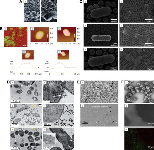
Figure 2 Antimicrobial activity of nanoparticles on bacteria.
Notes: (A) Antibacterial nanoscaffolds with silver nanoparticles. (B) (i) Simultaneous fluorescent and transmission image of E. coli culture mixed with aerogel prepared MgO/fluorescein (the fluorescent areas are green; the nonfluorescent areas are red). (ii and iii) AFM images. (iv and v) Cross sections and their section z-heights below. (C) Photothermal inactivation of heat-resistant bacteria on nanoporous gold disk arrays at different magnifications and array structures (i–vi). Reprinted with permission from Optical Society of America. Santos GM, de Santi Ferrara FI, Zhao G, Rodrigues DF, Shih W. Photothermal inactivation of heat-resistant bacteria on nanoporous gold disk arrays. Opt Mater Express. 2016;6(4):1217–1229.Citation170 (D) Transmission electron micrographs of Bacillus incubated with 5 μg⋅mL−1 mercaptopropionic acid–GNP (i and ii), 5 μg⋅mL−1 cationic 3-mercaptopropylamine–GNP (iii and iv) and 0.5 μg⋅mL−1 cationic polyelectrolyte poly(allylamine hydrochloride)–GNP (v and vi). White arrows in figure D point to binding sites of NPs with cells; yellow arrows denote lysed cells or empty cells; dashed arrows show cell wall-free cytoplasmic content. (E) Novel selenium nanoparticles killing E. coli when coated on polyvinyl chloride. Arrows indicate bacteria. (F) The use of iron oxide MNPs and a magnet to penetrate an E. coli biofilm and kill bacteria (as indicated by the red dead stain; live bacteria are stained green). (i) Iron oxide nanoparticles coated in polymersomes. Scale bar=1 μm. (ii) Bright-field image showing the placement of a black magnet under the well plate (left) and (iii) the same area under fluorescent imaging showing red (dead) bacteria in the exact same area. Scale bar=50 μm. (A–B) Reprinted with permission from Stoimenov PK, Klinger RL, Marchin GL, et al. Metal oxide nanoparticles as bactericidal agents. Langmuir. 2002;18(17):6679–6686. Copyright 2002 American Chemical Society.Citation81 (D) Reproduced from Feng Z, Gunsolus IL, Qiu TA, et al. Impacts of gold nanoparticle charge and ligand type on surface binding and toxicity to Gram-neg ative and Gram-positive bacteria. Chem Sci. 2015;6(9):5186–5196.Citation171
Abbreviations: E. coli, Escherichia coli; GNP, gold nanoparticle; MNP, magnetic nanoparticle.
In another study conducted by Marsich et al,Citation91 antibacterial nanoscaffolds were developed by introducing silver nanoparticles to alginate and HA scaffolds. These scaffolds possessed enhanced antimicrobial properties, beneficial in prosthesis implementation. Patrascu et alCitation92 also fabricated composite scaffolds with silver nanoparticles where collagen–silver nanoparticle scaffolds were produced for skin repair, especially for burn victims, while composite HA–silver nanoparticles and HA/collagen–silver nanoparticle composite scaffolds were developed as potential bone graft materials.Citation92 Silver nanoparticles have also been used with poly(l-lactide-co-caprolactone) (PLCL) scaffolds for skin TE. The scaffolds were developed using electrospinning techniques.Citation82 Similarly, silver nanoparticles were used in PCL composite scaffolds for in vitro tissue-engineered vascular grafts.Citation93 The antimicrobial features of silver nanoparticles enabled their infusion into various composite scaffold materials such as HA, alginate, PLCL, PCL and collagen.Citation82,Citation91–Citation94 However, recent studies have highlighted that bacteria may develop a resistance to silver nanoparticles, so extreme caution and additional studies should be undertaken for all of the abovementioned materials.Citation95
Apart from silver nanoparticles, different metal oxide nanoparticles have also proven to be effective as bactericidal agents. Specifically, magnesium oxide nanoparticles and their halogen adducts with Cl2 and Br2 showed strong antibacterial properties.Citation81 In order to assess the effectiveness of the nanoparticles, a study was performed by Stoimenov et al on three groups of bacteria: Escherichia coli, Bacillus megaterium and Bacillus subtilis. It was shown that the nanoparticulate formulations were able to destroy the populations of E. coli and B. megaterium bacteria in ~20 min.
There has also been excitement over the use of selenium nanoparticles since it has been shown that selenium kills bacteria by different mechanisms than the above mentioned nanoparticles, which rely on reactive oxygen species (ROS).Citation96 It has been speculated that nanoparticles that rely on ROS generation to kill bacteria may lead to mutations of bacteria, thus causing the bacteria to be resistant to such materials. In contrast, selenium nanoparticles, which have been shown to kill both gram-positive and gram-negative bacteria (), alter thiolcontaining protein function inside bacteria, for which bacterial resistance has not been reported.Citation97
Furthermore, iron oxide nanoparticles have demonstrated much promise in killing post-biofilm formation in bacteria.Citation98 Specifically, iron oxide nanoparticles (especially when functionalized with sugars such as fructose and sucrose) can penetrate biofilms, whereas antibiotics cannot, under a magnetic field to disrupt and kill bacteria ().Citation99,Citation100 This has enormous consequences in TE since currently if a biomaterial becomes infected, it needs to be removed and adjacent tissue cleaned. Strategies that do not rely on implant removal and cleaning can have a bright future in TE.
Stimulation of cells for mechanotransduction
It is well known that various bioactive molecules and growth factors regulate cell function in the human body. It has been shown that, in addition to these substances, mechanical forces play a major role in determining cell functions by affecting mechanotransduction pathways.Citation101 Numerous approaches have been used, including the introduction of shear stress by bioreactors and stiffness of patterned substrates to mechanically control cell functions. However, MNPs have proven to be superior to all these methods since they have the ability to be controlled remotely, spatially and temporally through a magnetic field.Citation54,Citation80,Citation102 On a microscopic level, the process happens as follows. First, the MNPs are coated with a certain targeting antibody. Once the magnetic field is applied, the cells are clustered in the direction of the magnetic field. Based on the antibody used, receptor-mediated cell function is affected. Mannix et alCitation103 were able to achieve an increase in intracellular calcium ion levels, while Gopinath et al were able to induce the apoptotic pathway using such approaches.Citation104 These two experiments along with many other studies have shown how the integration of MNPs can provide full details of cell functions and differentiation in regenerative medicine in a remote manner.
In another study, silver nanoparticles were used to induce cell apoptosis in BHK21 and HT29 cells. The introduction of silver nanoparticles at a concentration of 11.0 μg⋅mL−1 triggered extracellular stresses on the membranes, resulting in an upregulation of p53 that triggered the right molecules to induce cell death.Citation104 shows the silver nanoparticle-and MNP-induced apoptotic pathways. Of course, in these approaches, one needs to be concerned about other cellular functions that may be inadvertently changed through nanoparticle–cell membrane attachment or internalization.
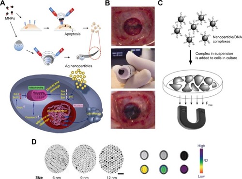
Figure 3 Utilization of magnetic particles in bioengineering applications.
Notes: (A) Upper side of the figure shows MNP-induced apoptosis, whereas the bottom side shows silver nanoparticle-induced apoptosis. (B) Transplantation procedure of MSC sheets using an electromagnet with harvesting, transportation and transplantation. (C) Principles and schematic of magnetofection. (D) Size-dependent contrast agents with their T2-weighted magnetic resonance images and color maps; the scale bar is 50 nm. (A) Reprinted from Colloids Surf B Biointerfaces. 77(2). Gopinath P, Gogoi SK, Sanpui P, Paul A, Chattopadhyay A, Ghosh SS. Signaling gene cascade in silver nanoparticle induced apoptosis. Pages 240–245., Copyright 2010, with permission from Elsevier.Citation104 (B) © 2007 Wiley Periodicals, Inc. J Biomed Mater Res Part B: Appl Biomater 2007. Reproduced from Shimizu K, Ito A, Yoshida T, Yamada Y, Ueda M, Honda H. Bone tis sue engineering with human mesenchymal stem cell sheets constructed using magnetite nanoparticles and magnetic force. Journal of Biomedical Materials Research.Citation172 (D) Reprinted from Springer Nature: Nature Medicine. Lee JH, Huh YM, Jun YW, et al. Artificially engineered magnetic nanoparticles for ultra-sensitive molecular imaging. Copyright 2007.Citation173
Abbreviations: MNP, magnetic nanoparticle; Bcl-2, B-cell lymphoma 2; MSC, mesenchymal stem cells.
Gene delivery
Gene delivery technology targeting matured cells or stem cells has become an important issue within TE. Human mesenchymal stem cells (hMSCs) are multipotent cells that show immunosuppressive properties and have an intrinsic capacity to differentiate into various types of cells, including chondrocytes, osteoblasts, myocytes and adipocytes.Citation105,Citation106 For effective gene therapy applications, it is vital to build up a suitable vector system with a high gene transfection efficiency, low cytotoxicity and high specificity to unhealthy cells. Gene delivery is classified into two categories: viral and nonviral. The promise for gene delivery has been seen through the use of nanoparticles and self-assembled nanomaterials as described in the following.
DNA transfection
The advantages of nonviral methods are their simplicity and the absence of an immune response, while the disadvantage is low efficiency due to low transfection rate.Citation107 Magnetofection is a new method for gene delivery in which gene transfection is accomplished using MNPs (). To achieve magnetofection using plasmid DNA, cationic lipids or polymers with complexes of DNA interact with magnetic beads and then through a magnetic force are attracted onto target cells so that they can accumulate on the surface.Citation108 There has been substantial research into various methods of magnetofection in combination with Lipofectamine, polyethyleneimine or dioleoyltrimethylammounium.Citation108 Furthermore, in endothelial cellsCitation109 and embryonic stem cells,Citation110 which are generally resistant to traditional transfection methods, magnetofection showed high expression levels in target cells. It was found that in fibroblasts and keratinocytes using reporter genes, the transfection efficiencies using the magnetofection technique were 36- and 10-fold higher, respectively, compared to the lipofection technique by cationic liposomes.Citation111
Some studies have shown that iron oxide magnetic particles can attach to the gene and improve transfection efficiency. These particles were distributed within a polymer matrix or internalized in a polymer or metallic case, which binds DNA through charge contact.Citation112,Citation113 Another study looked at the improvement of mesoporous silica nanoparticles to uphold the uptake by the target cells. Coupled with N-{1-(2,3-dioleoyloxy)propyl}-N,N,N trimethylammonium chloride (DOTAP chloride), the structure helped to avoid aggregation and encouraged uptake.Citation114 CNTs, which have shown numerous applicationsCitation115,Citation116 and can be synthesized through several different approaches,Citation117,Citation118 were also shown of remarkable potential as nonviral gene delivery agents. A technique called as nanotube spearing has been used to prepare nickel-embedded, magnetic nanotubes where DNA is attached.Citation119
Viral transduction
Viral transduction methods have also been used in TE applications; however, one problem associated with this method is the difficulty in preparing viral vectors with a high titer.Citation107,Citation120 In one case, retroviral vectors were captured using magnetite cationic liposomes (MCLs) by adding the MCLs to a solution of retroviral vector pseudo typed through the vesicular stomatitis virus glycoprotein.Citation121 The MCLs were then collected through the use of a magnetic force and seeded into mouse neuroblastoma cells, thereby increasing the viral titer by 55-fold. It was noted that physical interactions due to magnetic force enhances cellular uptake of MNPs. When transduction efficiency of magnetofection with MCLs was compared with conventional methods using polybrene, the effectiveness was 6.7-fold higher for the MCLs.Citation122
No coxsackievirus and adenovirus receptor (CAR) were expressed by using adenoviral vectors conjugated with polyethyleneimine. These vectors were coated with MNPs, which enabled transduction of a number of cell lines. Using these particles, magnetofection has been utilized to transfect a number of cell types, including epithelial cells from lungsCitation123 and endothelial cells from blood vessels.Citation109 These particles have also been used to effectively deliver antisense oligonucleotidesCitation124 and siRNA to inhibit gene expression. Further development of magnetically labeled vectors with magnetic fields should provide tools for the effective treatment of different types of diseases.
Magnetic cell patterning
Successful TE requires the fabrication of tissue architecture similar to in vivo conditions. One strategy to accomplish this is to control cell adhesion within a specified design pattern. Current limitations of conventional methods include the need for extensive external equipment or requirement of usage of chemically modified surfaces. The use of MNPs for the purpose of cell patterning may serve to bypass these obstacles by offering an effective and practical alternative. shows the MNPs and the mediated cell patterning on an acryl resin plate. MCLs show high-cell binding capacity. A cell-patterning technique was developed using MCLs where a magnet with a magnetic field concentrator was laid under a cell culture surface. Various cell patterns were successfully fabricated using this technique by manipulating the line patterns of the magnetic field concentrators. When human umbilical vein endothelial cells were used, it was found that for the same number of cells, the cells were connected and formed capillary-like structures when patterned in a line.Citation125 For the construction of vascular grafts, it was found that the tubular geometry did not allow for an effective delivery of the cells onto the scaffold. Another study using porcine decellularized common carotid artery showed that when a cylindrical magnet was placed into the lumen of the artery and then immersed into a suspension of NIH3T3 fibroblasts (3T3s) that had been magnetically labeled using original MCLs with a positive surface charge, almost all the 3T3s were attached to the artery compared to the very low seeding efficiency that was observed in the absence of a magnet. Reseeding the porcine decellularized common carotid artery with both smooth human muscle cells and human dermal fibroblasts successfully constructed the vascular graft using the Mag-seeding technique.Citation126
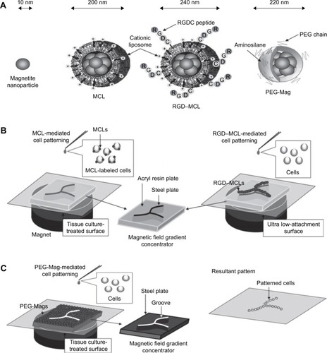
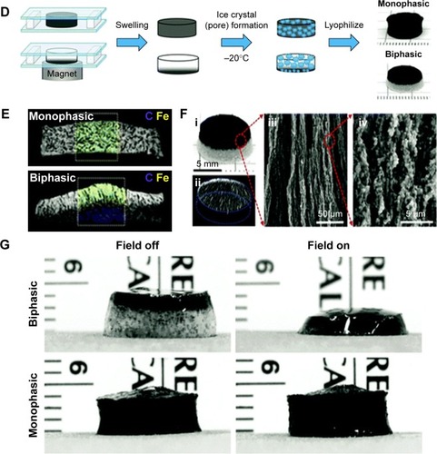
Figure 4 Magnetic cell patterning and ferrogel fabrication.
Notes: (A–C) Magnetic cell patterning using MCLs, RGD–MCLs and PEG-Mags and their resultant pattern of cells. Reprinted from Academic Press, Ito A, Kamihira M. Progress in Molecular Biology and Translational Science/Tissue Engineering Using Magnetite Nanopar ticles, pages 355–395, Copyright 2011.Citation174 (D–G) Schematic of fabrication of monophasic and biphasic ferrogels, their SEM/EDS and micro-CT images and these ferrogels in the presence of no magnetic field (field off) and a moderate vertical magnetic field gradient (field on). Reproduced from Cezar CA, Kennedy SM, Mehta M, et al. Biphasic ferrogels for triggered drug and cell delivery. Adv Healthc Mater. 2014;3(11):1869–1876. With permission from John Wiley and Sons.Citation175
Abbreviations: EDS, energy dispersive spectroscopy; MCL, magnetite cationic liposome; RGD–MCL, arginine–glycine–aspartic acid motif-containing peptide coupled to a phospholipid group on magnetite cationic liposomes; PEG-Mags, poly(ethylene glycol) in combination with a magnetic force; SEM, scanning electron microscopy; micro-CT, micro-computed tomography.
An arginine–glycine–aspartic acid motif-containing peptide coupled to a phospholipid group on magnetite cationic liposomes (RGD–MCLs) has been shown to facilitate cell adhesion and proliferation. The RGD–MCLs were added to an ultralow-attachment plate where the culture surface was modified with a hydrophilic and neutrally charged hydrogel layer. Human keratinocyte (HaCaT) cells were seeded onto the plate. Without either the RGD or MCLs, a few cells were attached to the plate. With MCLs, cells attached but low proliferation rates were noted. However, when RGD–MCLs were used, the cells adhered within a day and proliferated. Of course, one needs to be careful when using RGD since it is not an exclusive cell-binding motif and, thus, if not deactivated or fully covered by the target cells, competitor cells may attach and decrease the efficacy of the TE construct.
Another strategy investigated for cell patterning was to use MNPs coated with aminosilane and modified with poly(ethylene glycol) in combination with a magnetic force (PEG-Mags).Citation122 The PEG-Mags were magnetically decorated onto the surface of a tissue culture dish resulting in two regions, namely, the cell-resistant PEG-Mags covering region and the native substrate surface, which was the cell adhesion region. When HaCaT cells were seeded, they adhered only to the native substrate surface and the PEG-Mags were washed away exposing the surface. When mouse myoblast C2C12 cells were seeded into the dish, they adhered to the substrate surface creating a patterned coculture of heterotypic cells. Furthermore, almost no cytotoxic effect was observed by using PEG-Mags, signifying that this strategy of combing PEG with the MNPs provided particles with biocompatibility and low toxicity, adding to the appeal of this method.Citation122
Constructing 3D tissues
Nanotechnology shows potential perspectives in TE. Nano-scale structures can control cellular activities such as adhesion, propagation and segregation, and most importantly, nanomaterials have unique visual and magnetic properties and thus are suitable agents for monitoring cellular performance in vivo subsequent a transplantation. Nanomaterials were subsequently developed as 3D tissue-engineered scaffolds for skin, bone, vasculature and other tissues;Citation127–Citation131 additionally, nanostructures have also been revealed to control the actions of primary stem cells such as adhesion, growth and differentiation.Citation132–Citation134 Recently, MNPs have gained lots of attention due to their exceptional magnetic properties and unique dimensions.Citation135 MNPs are smaller in size (20–300 nm), and their magnetization directions support thermal fluctuations at room or biological temperature.
Skin
Keratinocyte sheets of five or more cellular layers were constructed, which were adequately strong for improvement and transplantation. A well of ultralow-attachment plated with a covalently bound hydrogel layer and a neodymium magnet under the plate was used to seed magnetically labeled keratinocytes. Without either the magnet or MCLs, the keratinocytes did not stick on to the culture surface.Citation136 It was also observed that sheets consisted of undifferentiated keratinocytes that may be more effective in wound healing. In addition, as soon as the five-layered sheets were cultured in a medium of high calcium, they stratified to produce 10-layer epidermal sheets formed within this high-calcium medium and were substantially stronger. An added advantage of magnetic force was that the sheets labeled with MCLs floated up to the culture surface and stuck to the hydrophilic poly(vinylidene fluoride)-treated membrane placed on a cylindrical magnet, making recovery simpler.
Skeletal muscle
Magnetic force-based scaffolds of free multilayered sheets of skeletal muscle cells were constructed using mouse MCL-labeled myoblast C2C12 cells seeded in ultra-low attachment plates with a magnet below the plates. The cells accumulated uniformly forming dense, strong and multilayered sheets. On the other hand, when the magnet was removed, the cells formed small aggregates instead.Citation137 The method was then adjusted to fabricate string-shaped 3D tissues similar to skeletal muscle fiber bundles. The magnetic field gradient forced the cells to aggregate in string-like lines, and by controlling the magnetic field gradient, the width and thickness could be manipulated.Citation137 To date, a number of new strategies have been established for the manufacturing of skeletal muscle tissues. Saxena et alCitation138 presented vascularized skeletal muscle constructs where myoblasts were immobilized on artificial biodegradable polymers. Another group showed a 3D fabrication with scaffold-free self-assembly of myoblast cells on a silicone surface.Citation139 Moon du et alCitation140 developed skeletal muscle tissue constructs using collagen-based acellular tissue scaffolds with improved contractile force generation. Akiyama et alCitation141 and Sato et alCitation142 developed a TE technique called magnetic force-based tissue engineering (Mag-TE) where a magnetic force was applied and cells labeled with MNPs were used to engineer tissue constructs. Using Mag-TE, they also showed that the B-cell lymphoma 2 (Bcl-2) protein improved the development of synthetic skeletal muscle tissue constructs with an elevated cell density.Citation141,Citation142
Liver
Recent research has shown that Mag-TE can be used as a cell sheet for engineering of liver tissue. An example can be seen in in magnetic cell patterning and Ferro gel fabrication. In case of the liver, it was found that the magnetic force allowed for close and tight cell interactions, which may have caused the deposition of an ECM and cytokines between cell layers, enhancing liver function. Hepatocytes and endothelial cells were cocultured using Mag-TE. Human aortic endothelial cells (HAECs) were labeled using MCLs and placed onto a layer of rat hepatocytes. Multilayers of HAECs were formed where magnets were placed and remained attached to the hepatocyte layer in an even manner. The layered cocultures also managed to maintain an increased amount of albumin secretion for a period of at least 8 days.Citation143 Another study looked at magnetically labeled HepG2 (hepatocyte model) and NIH3T3 (stromal fibroblast model) cells. The cells were seeded in wells of an ultra-low attachment plated with a magnet underneath. The cells were attracted to the magnet at the bottom and formed strong sheet-like structures. When the magnet was removed, the cell sheets detached and were disengaged without disruption with the use of a magnet.Citation144
Figure 5 Magnetic culturing of cells using cell levitation and cocultures.
Notes: (A) Schematic of heterotypic cell cocultures using magnetic force. (B) Schematic of magnetic levitation in a Petri dish (left) and a 24-well plate (right). The cells are levitated to the air–liquid interface by placing a neodymium magnet above the dish or well. Cell culture under magnetic stimuli: (C) schematic drawing of the setup, (D) bright-field image and (E) fluorescence images of the two types of microcryogels (poly(ethyelene glycol) diacryate microcryogels [PM]; and magnetic gelatin microcryogels [MGM]) mixed together and co-cultured in the system. Scale bar is 200 μm. (A) Reprinted from Trends Biotechnol. 27(8). Corchero JL, Villaverde A. Biomedical applications of distally controlled magnetic nanoparticles. Pages 468–476. Copyright 2009, with permission from Elsevier.Citation176 (B) The publisher for this copyrighted material is Mary Ann Liebert, Inc. publishers. Tseng H, Gage JA, Raphael RM, et al. Assembly of a three-dimensional multitype bronchiole coculture model using magnetic levitation. Tissue Eng Part C Methods. 2013;19(9):665–675.Citation177 (C–E) Republished with permission of Royal Society of Chemistry, from Magnetically controllable 3D microtissues based on magnetic microcryogels, Liu W, Li Y, Feng S, et al, Volume 14(15), 2014; permission conveyed through Copyright Clearance Center, Inc.Citation178
![Figure 5 Magnetic culturing of cells using cell levitation and cocultures.Notes: (A) Schematic of heterotypic cell cocultures using magnetic force. (B) Schematic of magnetic levitation in a Petri dish (left) and a 24-well plate (right). The cells are levitated to the air–liquid interface by placing a neodymium magnet above the dish or well. Cell culture under magnetic stimuli: (C) schematic drawing of the setup, (D) bright-field image and (E) fluorescence images of the two types of microcryogels (poly(ethyelene glycol) diacryate microcryogels [PM]; and magnetic gelatin microcryogels [MGM]) mixed together and co-cultured in the system. Scale bar is 200 μm. (A) Reprinted from Trends Biotechnol. 27(8). Corchero JL, Villaverde A. Biomedical applications of distally controlled magnetic nanoparticles. Pages 468–476. Copyright 2009, with permission from Elsevier.Citation176 (B) The publisher for this copyrighted material is Mary Ann Liebert, Inc. publishers. Tseng H, Gage JA, Raphael RM, et al. Assembly of a three-dimensional multitype bronchiole coculture model using magnetic levitation. Tissue Eng Part C Methods. 2013;19(9):665–675.Citation177 (C–E) Republished with permission of Royal Society of Chemistry, from Magnetically controllable 3D microtissues based on magnetic microcryogels, Liu W, Li Y, Feng S, et al, Volume 14(15), 2014; permission conveyed through Copyright Clearance Center, Inc.Citation178](/cms/asset/fa97a2bd-b067-4f6d-a6ba-fe6cf7476373/dijn_a_12193941_f0005_c.jpg)
Nanoparticles in biomolecular detection
The application of biomolecular detection and biosensors is rapidly gathering pace in the field of TE.Citation145 Biosensors are biological devices that recognize a change in molecules through physiochemical detection (). In TE, various types of biosensors have been used, such as electrochemical, optical, magnetic and acoustic, for the detection of functional protein molecules, glucose and pathogenic microbes.Citation146 Most importantly, however, is the rise in the application of nanoparticles in biomolecular detection. Nanoparticles in biosensors have been used for DNA, nucleic acid and protein detection, among other applications, due to their high reactivity and their advantageous chemical properties.Citation147,Citation148 The rising prominence of nanoparticle applications in biomolecular sensors and the importance of biosensors in TE highlight the potential of nanoparticles in TE. Hopefully in the near future, researchers can incorporate intelligent nanoscale biosensors inside scaffolds, cell sheets or associated locations to track the development of engineered tissues after transplantation and grafting. We also believe that incorporating nanomaterials in 3D-engineered tissues could serve to reduce inflammatory responses to transplanted engineered tissues by modulating immune cell adhesion and cell viability.
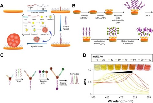
Figure 6 Biosensing principles using nanoparticles and nanomaterials.
Notes: (A) Electromechanical biodetection schematic for silver nanoparticles. (B) Quantum dot electromechanical detection of DNA strands. (C) Electrochemical detection schematic for silver nanoparticles. (D) Example of localized surface plasmon resonance modulation by altering nanoparticle compositions. (A) Reprinted by permission from Springer Nature: Anal Bioanal Chem. Aptamer-based electrochemical approach to the detection of thrombin by modification of gold nanoparticles. Li L, Zhao H, Chen Z, Mu X, Guo L. Copyright 2010.Citation179 (B) Reprinted with permission from Wang J, Liu G, Merkoçi A. Electrochemical coding technology for simultaneous detection of multiple DNA targets. J Am Chem Soc. 2003;125(11):3214–3215. Copyright 2003 American Chemical Society.Citation180 (C) Reprinted with permission from Li H, Sun Z, Zhong W, Hao N, Xu D, Chen HY. Ultrasensitive electrochemical detection for DNA arrays based on silver nanoparticle aggregates. Anal Chem. 2010;82(13):5477–5483. Copyright 2010 American Chemical Society.Citation165 (D) Reprinted with permission from Doria G, Conde J, Veigas B, et al. Noble metal nanoparticles for biosensing applications. Sensors (Basel). 2012;12(2):1657–1687.Citation181
Abbreviation: Abs, absolute.
Optical detection
One of the prime examples of the use of nanoparticles as biosensors is through optical sensors and optical imaging in which semiconductors (ie, quantum dots) as well as GNPs and silica nanoparticles have been used.Citation148 Quantum dots have been used due to their high stability, brightness and lasting luminescence.Citation149 Goldman et alCitation150 used luminescent quantum dots for protein detection in immunoassays.Citation149 Quantum dots were also used for DNA sensing in a study conducted by Sharon et al.Citation151 In this study, the hemin structure in CdSe and ZnS quantum dots was used in order to develop new optical sensing techniques. Owing to the risk of toxicity during in vivo applications associated with heavy quantum dots,Citation152 carbon quantum dots have been developed by Ding et al.Citation153 These quantum dots have been found to have low cytotoxicity and have been used for molecule detection. Metal nanoparticles, on the other hand, are used in optical detection due to their tunable strength, bandwidth and frequency. These characteristics, attributed to the collective oscillation mode, make metal nanoparticles perfect for optical applications.Citation154 Newman et alCitation155 developed optical sensors based on GNPs. In their study, the GNPs with silica microparticles were used to detect organophosphate molecules.
Electrical detection
Nanoparticles have also been used as electrical detectors for DNA molecules through electric signals. With nanoparticles acting as intermediaries, the action of the DNA is converted to electrical signals.Citation156 Park et alCitation157 developed an electrical DNA detection method through binding of oligonucleotides with GNPs with a high accuracy. GNPs have also been implemented by Noor et alCitation158 for DNA detection. In their study, DNA was mounted on a silicon chip between gold nanoelectrodes. The GNPs were found to increase conductivity between the electrodes, thus enhancing the process of DNA detection. MNPs and CNTs have also been used for electrical detection. One example is an experimentCitation159 that was conducted by encapsulating MNP in CNTs. The results showed enhanced electrical and magnetic properties.
Electrochemical detection
Electrochemical sensors utilize the selective detection of biological elements with the sensitivity of electrical sensors for element recognition. The sensors chemically react with the analyte to produce an electrical signal.Citation160 Nanoparticles have been used as electrochemical biosensors due to the greater surface area they offer, along with their greater chemical convenience and superior electrical conductivity.Citation161 Wang et alCitation162 used GNPs to enhance the immobilization of an antigen. The study showed that the use of GNPs also improved antigen and antibody detection. GNPs have also been used as electrochemical sensors for DNA detection. One example is a study conducted by Li et al.Citation163 Apart from GNPs, various types of nanoparticles have been used in electrochemical sensors. One example is a study conducted by Cui et alCitation164 in which CdTe nanoparticles were used in order to develop a two-fold immunosensor as an electrochemical sensor and fluorescent label. GNPs were also used to swathe the chip upon which the sensor was constructed. Another study was conducted by Li et alCitation165 in which oligonucleotide-functionalized silver nanoparticles were developed for DNA detection. Silver nanoparticles were shown to have excellent properties that are yet to be properly utilized.
Another example of electrochemical detection is a sensor that was developed for an orthopedic hip implant. This titanium-based implant was first anodized to possess nanotubes from which CNTs were grown using chemical vapor deposition.Citation166 The CNTs could then measure the resistance of the cells that attach to the implant, send such information via RF to a hand-held device and even be remotely activated to release bone growth factors, anti-inflammatory agents or antibiotics to the site of the need to ensure implant success. Such electrochemical detection of biological events may be the future of nanomaterial use in medical devices as they can sense in real time biological events depending on the individual’s response to the implant to promote success, perhaps the true definition of personalized medicine.
Challenges and future perspectives
While nanoparticles have demonstrated promising potential in TE applications such as enhancement of biological, mechanical and electrical properties; antimicrobial effects; gene delivery and construction of engineered tissues, many challenges still lie ahead to introduce them into widespread clinical applications. For example, a compelling need exists, at first, for better assessment tools and methods of nanoparticle toxicity, carcinogenicity and teratogenicity. Second, the toxicity, carcinogenicity and teratogenicity of nanoparticles are all highly dose-dependent and exposure-dependent. In many applications, the nanoparticles are used below their threshold concentrations at which they are considered not harmful. However, bioaccumulation of nanoparticles inside the body over a large period of time is well known. Thus, any nanoparticle used in the human body has the potential to accumulate over a long period of time to reach a concentration that can cause toxicity to cells, cancers or harmful effects on reproductive systems and fetuses before their birth. In addition, even though there are numerous products containing nanoparticles/nanomaterials already in the market, there are still some scientific and methodological gaps in the knowledge on specific hazards of nanomaterials. Currently, to the best of our knowledge, there are no international standards yet for nano-specific risk assessments, including specific data requirements and testing strategies. The risk assessments of nanomaterials are laborious and costly. Currently, manufacturers are committed to assess the safety of their nanoparticle-based products and to implement the necessary safety measures (self-supervision). To date, the regulatory tools are not nano-specific; eg, the data requirements for notification of chemicals, criteria for classification and labeling requirements for safety data sheets are still not widely available. Thus, there is a need for precautionary measures for applications of nanoparticles wherever there is a possibility of chronic bioaccumulation.
Conclusion
Nanoparticles exhibit superior biocompatibility and well-established strategies for surface modification, which have made them highly effective in numerous biomedical applications. The electric coupling between decellularized cells and proliferation rates upon several tissues have also been enhanced using nanoparticles. The validity of nanoparticles, when it comes to antibacterial growth, has also been studied with much promise. These nanoparticles have been deposited on biocomposite scaffolds, thus regulating bacterial infection during reconstructive bone surgery. Induction of cell mechanotransduction, which is responsible for many physiological processes in the body, was also stimulated by remotely controlled nanoparticles. This review has mentioned a new method for gene delivery. Specifically, magnetofection, which was accomplished through the use of plasmid DNA cationic lipids with complexes of DNA as they interacted through a magnetic force, thus increasing transfection efficiency. Related to this is the use of nanoparticles for the purpose of cell patterning. Three strategies were investigated for cell patterning: the use of MCLs, RGD motif-containing peptide coupled to the phospholipid of magnetite cationic liposomes and aminosilane modified with PEG and magnetic force (PEG-Mags).
The numerous existing applications in the literature reiterate the great potential nanoparticles can have on TE through biosensors. Nanoparticles have been shown to develop optical, electrical and electrochemical biosensors for molecules, proteins and DNA detection with highly accurate results. By optimizing these functions, these sensors could have a great influence on medical use. Although nanoparticles show a promising future in TE applications, there is still a lack of in vivo experimentation that needs to be done in order to verify the wide variety of successful results from in vitro studies.
We believe that in the near future, we could visualize the use of smart nanoparticles that can engage and direct stem cells to preferred sites in the body and dictate the formation of tissues in vivo.
Acknowledgments
This article was supported by the GCC research program (No GCC-2017-005), Qatar University, Qatar, and by NPRP grant (NPRP 9-144-03-021) from QNRF (Qatar National Research Funding), a part of Qatar Foundation. The Statements made herein are solely the responsibility of the authors. Shabir H acknowledges funding from Swiss National Science Foundation.
Disclosure
The authors report no conflicts of interest in this work.
References
- HasanAMemicAAnnabiNElectrospun scaffolds for tissue engineering of vascular graftsActa Biomater2014101112523973391
- HasanAPaulAMemicAKhademhosseiniAA multilayered microfluidic blood vessel-like structureBiomed Microdevices20151758826256481
- PaulAManoharanVKrafftDNanoengineered biomimetic hydrogels for guiding human stem cell osteogenesis in three dimensional microenvironmentsJ Mater Chem B2016420521528
- HasanAPaulAVranaNEMicrofluidic techniques for development of 3D vascularized tissueBiomaterials201435267308732524906345
- HasanARagaertKSwieszkowskiWBiomechanical properties of native and tissue engineered heart valve constructsJ Biomech20144791949196324290137
- KestellAEDeLoreyGTNanoparticles: Properties, Classification, Characterization, and FabricationHauppauge, NYNova Science Publishers Inc2009
- WilsonDSDalmassoGWangLSitaramanSVMerlinDMurthyNOrally delivered thioketal nanoparticles loaded with TNF-alpha-siRNA target inflammation and inhibit gene expression in the intestinesNat Mater201091192392820935658
- ShiJVotrubaARFarokhzadOCLangerRNanotechnology in drug delivery and tissue engineering: from discovery to applicationsNano Lett20101093223323020726522
- MironovVKasyanovVMarkwaldRRNanotechnology in vascular tissue engineering: from nanoscaffolding towards rapid vessel biofabricationTrends Biotechnol200826633834418423666
- KooOMRubinsteinIOnyukselHRole of nanotechnology in targeted drug delivery and imaging: a concise reviewNanomedicine20051319321217292079
- FurnoFMorleyKSWongBSilver nanoparticles and polymeric medical devices: a new approach to prevention of infection?J Antimicrob Chemother20045461019102415537697
- PrasadGLBiomedical applications of nanoparticlesWebsterTJSafety of NanoparticlesNew YorkSpringer200989109
- BasarkarASinghJPoly (lactide-co-glycolide)-polymethacrylate nanoparticles for intramuscular delivery of plasmid encoding interleukin-10 to prevent autoimmune diabetes in micePharm Res2009261728118779928
- WangXYangLChenZGShinDMApplication of nanotechnology in cancer therapy and imagingCA Cancer J Clin20085829711018227410
- PanyamJLabhasetwarVBiodegradable nanoparticles for drug and gene delivery to cells and tissueAdv Drug Deliv Rev200355332934712628320
- BriggerIDubernetCCouvreurPNanoparticles in cancer therapy and diagnosisAdv Drug Deliv Rev200254563165112204596
- KataokaKHaradaANagasakiYBlock copolymer micelles for drug delivery: design, characterization and biological significanceAdv Drug Deliv Rev200147111313111251249
- HansmaHGSurface biology of DNA by atomic force microscopyAnnu Rev Phys Chem200152719211326059
- RoyKMaoHQHuangSKLeongKWOral gene delivery with chitosan-DNA nanoparticles generates immunologic protection in a murine model of peanut allergyNat Med19995438739110202926
- SensenigRSapirYMacDonaldCCohenSPolyakBMagnetic nanoparticle-based approaches to locally target therapy and enhance tissue regeneration in vivoNanomedicine (Lond)2012791425144222994959
- LuxFZeislerRInvestigations of the corrosive deposition of components of metal implants and of the behaviour of biological trace elements in metallosis tissue by means of instrumental multi-element activation analysisJ Radioanal Nucl Chem1974192289297
- SchnabelCHerpersUMichelRLöerFBuchhornGWillertHGChanges of concentrations of the elements Co, Cr, Sb, and Sc in tissues of persons with joint implantsKučeraJObrusníkISabbioniENuclear Analytical Methods in the Life Sciences 1994Springer1994389395
- AginsHAlcockNWBansalMMetallic wear in failed titanium-alloy total hip replacementsJ Bone Joint Surg19887033473563279037
- Rodríguez de la FlorMHernández-VaqueroDFernández-CarreiraJMMetal presence in hair after metal-on-metal resurfacing arthroplastyJ Orthop Res201331122025203123893786
- RadomskaALeszczyszynJRadomskiMWThe nanopharmacol-ogy and nanotoxicology of nanomaterials: new opportunities and challengesAdv Clin Exp Med201625115116226935510
- KhangDKimSYLiu-SnyderPPalmoreGTDurbinSMWebsterTJEnhanced fibronectin adsorption on carbon nanotube/poly(carbonate) urethane: independent role of surface nano-roughness and associated surface energyBiomaterials200728324756476817706277
- MostofizadehALiYSongBHuangYSynthesis, properties, and applications of low-dimensional carbon-related nanomaterialsJ Nanomater2011201116
- KhanISaeedKKhanINanoparticles: properties, applications and toxicitiesArabian J Chem2017
- AliAZafarHZiaMSynthesis, characterization, applications, and challenges of iron oxide nanoparticlesNanotechnol Sci Appl201694927578966
- GawandeMBGoswamiAFelpinFXCu and Cu-based nanoparticles: synthesis and applications in catalysisChem Rev201611663722381126935812
- LiuC-JBurghausUBesenbacherFWangZLPreparation and characterization of nanomaterials for sustainable energy productionACS Nano20104105517552620973572
- MemicAAlhadramiHAHussainMAHydrogels 2.0: improved properties with nanomaterial composites for biomedical applicationsBiomed Mater201511101410426694229
- GiljohannDASeferosDSDanielWLMassichMDPatelPCMirkinCAGold nanoparticles for biology and medicineAngew Chem Int Ed Engl201049193280329420401880
- DykmanLKhlebtsovNGold nanoparticles in biomedical applications: recent advances and perspectivesChem Soc Rev20124162256228222130549
- BoisselierEAstrucDGold nanoparticles in nanomedicine: preparations, imaging, diagnostics, therapies and toxicityChem Soc Rev20093861759178219587967
- ChenYLiNYangYLiuYA dual targeting cyclodextrin/gold nanoparticle conjugate as a scaffold for solubilization and delivery of paclitaxelRSC Adv201551289388941
- KoWKHeoDNMoonHJThe effect of gold nanoparticle size on osteogenic differentiation of adipose-derived stem cellsJ Colloid Interface Sci2015438687625454427
- SuhKSLeeYSSeoSHKimYSChoiEMGold nanoparticles attenuates antimycin A-induced mitochondrial dysfunction in MC3T3-E1 osteoblastic cellsBiol Trace Elem Res20131531–342843623645457
- LiuDZhangJCYiCQYangMSThe effects of gold nanoparticles on the proliferation, differentiation, and mineralization function of MC3T3-E1 cells in vitroChin Sci Bull2010551110131019
- BessaPCCasalMReisRLBone morphogenetic proteins in tissue engineering: the road from laboratory to clinic, part II (BMP delivery)J Tissue Eng Regen Med200822–3819618383454
- LimJSKookMSJungSPlasma treated high-density polyethylene (HDPE) medpor implant immobilized with rhBMP-2 for improving the bone regenerationJ Nanomater201420147
- BenglisDWangMYLeviADA comprehensive review of the safety profile of bone morphogenetic protein in spine surgeryNeurosurgery2008625 suppl 2ONS423ONS431 discussion ONS43118596525
- LinZFatehASalemDMIntiniGPeriosteum: biology and applications in craniofacial bone regenerationJ Dent Res201493210911624088412
- HeoDNKoWKBaeMSEnhanced bone regeneration with a gold nanoparticle-hydrogel complexJ Mater Chem B201421115841593
- SubbiahRVeerapandianMYunKSNanoparticles: functionalization and multifunctional applications in biomedical sciencesCurr Med Chem201017364559457721062250
- JaconiMNanomedicine: gold nanowires to mend a heartNat Nanotechnol201161169269322051743
- RavichandranRSridharRVenugopalJRSundarrajanSMukherjeeSRamakrishnaSGold nanoparticle loaded hybrid nanofibers for cardiogenic differentiation of stem cells for infarcted myocardium regenerationMacromol Biosci201414451552524327549
- JawadHLyonARHardingSEAliNNBoccacciniARMyocardial tissue engineeringBr Med Bull200887314718790825
- LiuHSlamovichEBWebsterTJLess harmful acidic degradation of poly(lactic-co-glycolic acid) bone tissue engineering scaffolds through titania nanoparticle additionInt J Nanomed200614541545
- WanjaleSDJogJPCrystallization and phase transformation kinetics of poly (1-butene)/MWCNT nanocompositesPolymer2006471864146421
- El FrayMBoccacciniARNovel hybrid PET/DFA–TiO2 nanocomposites by in situ polycondensationMater Lett2005591823002304
- LiuAHongZZhuangXSurface modification of bioactive glass nanoparticles and the mechanical and biological properties of poly (L-lactide) compositesActa Biomater2008441005101518359672
- JawadHBoccacciniARAliNNHardingSEAssessment of cellular toxicity of TiO2 nanoparticles for cardiac tissue engineering applicationsNanotoxicology20115337238020858044
- LimDLeeEKimHParkSBaekSYoonJMulti stimuli-responsive hydrogel microfibers containing magnetite nanoparticles prepared using microcapillary devicesSoft Matter20151181606161325594916
- WangQChenDSynthesis and characterization of a chitosan based nanocomposite injectable hydrogelCarbohydr Polym20161361228123726572466
- KubotaYShuinTKawasakiCPhotokilling of T-24 human bladder cancer cells with titanium dioxideBr J Cancer1994706110711117981061
- LiNFanXTangKZhengXLiuJWangBNanocomposite scaffold with enhanced stability by hydrogen bonds between collagen, polyvinyl pyrrolidone and titanium dioxideColloids Surf B Biointerfaces201614028729626764111
- LudererFBegerowISchmidtWEnhanced visualization of biodegradable polymeric vascular scaffolds by incorporation of gold, silver and magnetite nanoparticlesJ Biomater Appl201328221923122492201
- KimHCheLHaYRyuWMechanically-reinforced electrospun composite silk fibroin nanofibers containing hydroxyapatite nanoparticlesMater Sci Eng C201440324335
- GonçalvesEMOliveiraFJSilvaRFThree-dimensional printed PCL-hydroxyapatite scaffolds filled with CNTs for bone cell growth stimulationJ Biomed Mater Res B Appl Biomater201610461210121926089195
- JuYMYuBKoobTJA novel porous collagen scaffold around an implantable biosensor for improving biocompatibility. I. In vitro/in vivo stability of the scaffold and in vitro sensitivity of the glucose sensor with scaffoldJ Biomed Mater Res A200887113614618085651
- HanZJRiderAEIshaqMCarbon nanostructures for hard tissue engineeringRSC Adv20133281105811072
- MagieraAMarkowskiJMenaszekEPLA-based hybrid and composite electrospun fibrous scaffolds as potential materials for tissue engineeringJ Nanomater2017201711
- PanLPeiXHeRWanQWangJMultiwall carbon nanotubes/polycaprolactone composites for bone tissue engineering applicationColloids Surf B Biointerfaces20129322623422305638
- Rodríguez-LozanoFJGarcía-BernalDAznar-CervantesSPotential of graphene for tissue engineering applicationsTransl Res2015166439940025936761
- FengLLiuZGraphene in biomedicine: opportunities and challengesNanomedicine20116231732421385134
- KimJKimYRKimYJGraphene-incorporated chitosan substrata for adhesion and differentiation of human mesenchymal stem cellsJ Mater Chem B201317933938
- ChenGYPangDWHwangSMTuanHYHuYCA graphene-based platform for induced pluripotent stem cells culture and differentiationBiomaterials201233241842722014460
- AinQUKhanANNabaviniaMMujahidMEnhanced mechanical properties and biocompatibility of novel hydroxyapatite/TOPAS hybrid composite for bone tissue engineering applicationsMater Sci Eng C201775807815
- MirahmadiFTafazzoli-ShadpourMShokrgozarMABonakdarSEnhanced mechanical properties of thermosensitive chitosan hydrogel by silk fibers for cartilage tissue engineeringMater Sci Eng C201333847864794
- SambudiNSSathyamurthyMLeeGMElectrospun chitosan/poly (vinyl alcohol) reinforced with CaCO3 nanoparticles with enhanced mechanical properties and biocompatibility for cartilage tissue engineeringCompos Sci Technol20151067684
- ShevachMFleischerSShapiraADvirTGold nanoparticle- decellularized matrix hybrids for cardiac tissue engineeringNano Lett201414105792579625176294
- ShevachMMaozBMFeinerRNanoengineering gold particle composite fibers for cardiac tissue engineeringJ Mater Chem B201313952105217
- NavaeiASainiHChristensonWSullivanRTRosRNikkhahMGold nanorod-incorporated gelatin-based conductive hydrogels for engineering cardiac tissue constructsActa Biomater20164113314627212425
- KharazihaMMemicAAkbariMBrafmanDANikkhahMNano-enabled approaches for stem cell-based cardiac tissue engineeringAdv Healthc Mater20165131533155327199266
- ShinSRShinCMemicAAligned carbon nanotube–based flexible gel substrates for engineering biohybrid tissue actuatorsAdv Funct Mater201525284486449527134620
- StoutDABasuBWebsterTJPoly (lactic-co-glycolic acid): carbon nanofiber composites for myocardial tissue engineering applicationsActa Biomater2011783101311221571106
- CollierJHCampJPHudsonTWSchmidtCESynthesis and characterization of polypyrrole-hyaluronic acid composite biomaterials for tissue engineering applicationsJ Biomed Mater Res A2000504574584
- KimHSHobbsHLWangLBiocompatible composites of polyaniline nanofibers and collagenSynth Met20091591313131318
- SniadeckiNJAnguelouchAYangMTMagnetic microposts as an approach to apply forces to living cellsProc Natl Acad Sci U S A200710437145531455817804810
- StoimenovPKKlingerRLMarchinGLMetal oxide nanoparticles as bactericidal agentsLangmuir2002181766796686
- SambergMEMentePHeTKingMWMonteiro-RiviereNAIn vitro biocompatibility and antibacterial efficacy of a degradable poly (L-lactide-co-epsilon-caprolactone) copolymer incorporated with silver nanoparticlesAnn Biomed Eng20144271482149324150238
- XingZCChaeWPBaekJYChoiMJJungYKangIKIn vitro assessment of antibacterial activity and cytocompatibility of silver-containing PHBV nanofibrous scaffolds for tissue engineeringBiomacromolecules20101151248125320415469
- El HajjFHasanANakhlehJNanosilver loaded GelMA hydrogel for antimicrobial coating of biomedical implantsAdvances in Biomedical Engineering (ICABME), 2015 International Conference onIEEE2015
- KimJSKukEYuKNAntimicrobial effects of silver nanoparticlesNanomedicine2007319510117379174
- SalahNHabibSSKhanZHHigh-energy ball milling technique for ZnO nanoparticles as antibacterial materialInt J Nanomed20116863
- Al-AmriSAnsariMSRafiqueSNi doped CuO nanoparticles: structural and optical characterizationsCurr Nanosci2015112191197
- AzamAAhmedASOvesMKhanMSMemicASize-dependent antimicrobial properties of CuO nanoparticles against Gram-positive and-negative bacterial strainsInt J Nanomed201273527
- AzamAAhmedASOvesMKhanMSHabibSSMemicAAntimicrobial activity of metal oxide nanoparticles against Gram-positive and Gram-negative bacteria: a comparative studyInt J Nanomed201276003
- SaravananSNethalaSPattnaikSTripathiAMoorthiASelvamuruganNPreparation, characterization and antimicrobial activity of a bio-composite scaffold containing chitosan/nano-hydroxyapatite/nano-silver for bone tissue engineeringInt J Biol Macromol201149218819321549747
- MarsichEBellomoFTurcoGTravanADonatiIPaolettiSNano-composite scaffolds for bone tissue engineering containing silver nanoparticles: preparation, characterization and biological propertiesJ Mater Sci Mater Med20132471799180723553569
- PatrascuJMNedelcuIASonmezMComposite scaffolds based on silver nanoparticles for biomedical applicationsJ Nanomater201520153
- MadhavanRVRosemaryMJNandkumarMAKrishnanKVKrishnanLKSilver nanoparticle impregnated poly (ε-caprolactone) scaffolds: optimization of antimicrobial and noncytotoxic concentrationsTissue Eng A2010173–4439449
- DuraipandyNLakraRSrivatsanKVPlumbagin caged silver nanoparticle stabilized collagen scaffold for wound dressingJ Mater Chem B20153714151425
- SilverSBacterial silver resistance: molecular biology and uses and misuses of silver compoundsFEMS Microbiol Rev2003272–334135312829274
- TranPAWebsterTJSelenium nanoparticles inhibit Staphylococcus aureus growthInt J Nanomed201161553
- RamosJTranPWebsterTSelenium nanoparticles for the prevention of PVC-related medical infectionsBioengineering Conference (NEBEC), 2012 38th Annual NortheastPhiladelphia, PAIEEE2012
- TaylorENWebsterTJMultifunctional magnetic nanoparticles for orthopedic and biofilm infectionsInt J Nanotechnol201081–22135
- MahmoudiMSerpooshanVSilver-coated engineered magnetic nanoparticles are promising for the success in the fight against antibacterial resistance threatACS Nano2012632656266422397679
- ParkHParkHJKimJAInactivation of Pseudomonas aeruginosa PA01 biofilms by hyperthermia using superparamagnetic nanoparticlesJ Microbiol Methods2011841414520971135
- OrrAWHelmkeBPBlackmanBRSchwartzMAMechanisms of mechanotransductionDev Cell2006101112016399074
- TsengPJudyJWDi CarloDMagnetic nanoparticle-mediated massively parallel mechanical modulation of single-cell behaviorNat Methods20129111113111923064517
- MannixRJKumarSCassiolaFNanomagnetic actuation of receptor-mediated signal transductionNat Nanotechnol200831364018654448
- GopinathPGogoiSKSanpuiPPaulAChattopadhyayAGhoshSSSignaling gene cascade in silver nanoparticle induced apoptosisColloids Surf B Biointerfaces201077224024520197232
- PittengerMFMackayAMBeckSCMultilineage potential of adult human mesenchymal stem cellsScience1999284541114314710102814
- WangSCastroRAnXElectrospun laponite-doped poly (lactic-co-glycolic acid) nanofibers for osteogenic differentiation of human mesenchymal stem cellsJ Mater Chem201222442335723367
- ItoAKamihiraMTissue engineering using magnetite nanoparticlesProg Mol Biol Transl Sci201110435539522093224
- SchererFAntonMSchillingerUMagnetofection: enhancing and targeting gene delivery by magnetic force in vitro and in vivoGene Ther20029210211857068
- KrötzFSohnHYGloeTPlankCPohlUMagnetofection potentiates gene delivery to cultured endothelial cellsJ Vasc Res200340542543414530599
- LeeCHKimEYJeonKSimple, efficient, and reproducible gene transfection of mouse embryonic stem cells by magnetofectionStem Cells Dev200817113314218271700
- InoKKawasumiTItoAHondaHPlasmid DNA transfection using magnetite cationic liposomes for construction of multilayered gene-engineered cell sheetBiotechnol Bioeng2008100116817618078300
- NeubergerTSchöpfBHofmannHSuperparamagnetic nanoparticles for biomedical applications: possibilities and limitations of a new drug delivery systemJ Magn Magn Mater20052931483496
- HarrisLGoffJDCarmichaelAYMagnetite nanoparticle dispersions stabilized with triblock copolymersChem Mater200315613671377
- DobsonJGene therapy progress and prospects: magnetic nanoparticle-based gene deliveryGene Ther200613428328716462855
- KhanZHAnsariMSSalahNACOBALT catalyzed-multi-walled carbon nanotubes film sensor for carbon mono-oxide gasDig J Nanomater Biostruct2011641947
- AlshehriRIlyasAMHasanACarbon nanotubes in biomedical applications: factors, mechanisms, and remedies of toxicity: miniperspectiveJ Med Chem201659188149816727142556
- SalahNAl-GhamdiAMemicAHabibSSKhanZHFormation of carbon nanotubes from carbon-rich fly ash: growth parameters and mechanismMater Manuf Processes2016312146156
- SalahNHabibSSKhanZHMemicANahasMNGrowth of carbon nanotubes on catalysts obtained from carbon rich fly ashDig J Nanomater Biostructures20127312791288
- CaiDMatarazaJMQinZHHighly efficient molecular delivery into mammalian cells using carbon nanotube spearingNat Methods20052644945415908924
- PaulAHasanARodesLSangaralingamMPrakashSBioengineered baculoviruses as new class of therapeutics using micro and nanotechnologies: principles, prospects and challengesAdv Drug Deliv Rev20147111513024503281
- ItoATakahashiTKameyamaYKawabeYKamihiraMMagnetic concentration of a retroviral vector using magnetite cationic liposomesTissue Eng C Methods20081515764
- AkiyamaHItoAKawabeYKamihiraMCell-patterning using poly (ethylene glycol)-modified magnetite nanoparticlesJ Biomed Mater Res A20109231123113019306262
- GerstingSWSchillingerULausierJGene delivery to respiratory epithelial cells by magnetofectionJ Gene Med20046891392215293350
- KrötzFde WitCSohnHYMagnetofection – a highly efficient tool for antisense oligonucleotide delivery in vitro and in vivoMol Ther20037570071012718913
- InoKItoAHondaHCell patterning using magnetite nanoparticles and magnetic forceBiotechnol Bioeng20079751309131717216656
- ShimizuKItoAArinobeMEffective cell-seeding technique using magnetite nanoparticles and magnetic force onto decellularized blood vessels for vascular tissue engineeringJ Biosci Bioeng2007103547247817609164
- XuCYInaiRKotakiMRamakrishnaSAligned biodegradable nanofibrous structure: a potential scaffold for blood vessel engineeringBiomaterials200425587788614609676
- LiWJTuliROkaforCA three-dimensional nanofibrous scaffold for cartilage tissue engineering using human mesenchymal stem cellsBiomaterials200526659960915282138
- LiCVepariCJinHJKimHJKaplanDLElectrospun silk-BMP-2 scaffolds for bone tissue engineeringBiomaterials200627163115312416458961
- SillTJvon RecumHAElectrospinning: applications in drug delivery and tissue engineeringBiomaterials200829131989200618281090
- FengZQChuXHuangNPThe effect of nanofibrous galacto-sylated chitosan scaffolds on the formation of rat primary hepatocyte aggregates and the maintenance of liver functionBiomaterials200930142753276319232710
- NamgungSBaikKYParkJHongSControlling the growth and differentiation of human mesenchymal stem cells by the arrangement of individual carbon nanotubesACS Nano2011597383739021819114
- TeoBKWongSTLimCKNanotopography modulates mechanotransduction of stem cells and induces differentiation through focal adhesion kinaseACS Nano2013764785479823672596
- LavenusSTrichetVLe ChevalierSHoornaertALouarnGLayrollePCell differentiation and osseointegration influenced by nanoscale anodized titanium surfacesNanomedicine20127796798022394187
- XuCSunSNew forms of superparamagnetic nanoparticles for biomedical applicationsAdv Drug Deliv Rev201365573274323123295
- RheinwaldJGreenHSerial cultivation of strains of human epidermal keratinocytes in defined clonal and serum-free cultureJ Invest Dermatol19756331342
- YamamotoYItoAKatoMPreparation of artificial skeletal muscle tissues by a magnetic force-based tissue engineering techniqueJ Biosci Bioeng2009108653854319914590
- SaxenaAKMarlerJBenvenutoMWillitalGHVacantiJPSkeletal muscle tissue engineering using isolated myoblasts on synthetic biodegradable polymers: preliminary studiesTissue Eng19995652553110611544
- DennisRGKosnikPE2ndGilbertMEFaulknerJAExcitability and contractility of skeletal muscle engineered from primary cultures and cell linesAm J Physiol Cell Physiol20012802C288C29511208523
- Moon duGChristGStitzelJDAtalaAYooJJCyclic mechanical preconditioning improves engineered muscle contractionTissue Eng Part A200814447348218399787
- AkiyamaHItoAKawabeYKamihiraMGenetically engineered angiogenic cell sheets using magnetic force-based gene delivery and tissue fabrication techniquesBiomaterials20103161251125919942286
- SatoMItoAAkiyamaHKawabeYKamihiraMEffects of B-cell lymphoma 2 gene transfer to myoblast cells on skeletal muscle tissue formation using magnetic force-based tissue engineeringTissue Eng A2012191–2307315
- ItoATakizawaYHondaHTissue engineering using magnetite nanoparticles and magnetic force: heterotypic layers of cocultured hepatocytes and endothelial cellsTissue Eng2004105–683384015265301
- ItoAJitsunobuHKawabeYKamihiraMConstruction of heterotypic cell sheets by magnetic force-based 3-D coculture of HepG2 and NIH3T3 cellsJ Biosci Bioeng2007104537137818086436
- BarthesJÖzçelikHHindiéMNdreu-HaliliAHasanAVranaNECell microenvironment engineering and monitoring for tissue engineering and regenerative medicine: the recent advancesBiomed Res Int2014201492190525143954
- HasanANurunnabiMMorshedMRecent advances in application of biosensors in tissue engineeringBiomed Res Int2014201430751925165697
- ThaxtonCSRosiNLMirkinCAOptically and chemically encoded nanoparticle materials for DNA and protein detectionMRS Bull2005305376380
- TansilNCGaoZNanoparticles in biomolecular detectionNano Today2006112837
- RosenthalSJChangJCKovtunOMcBrideJRTomlinsonIDBio-compatible quantum dots for biological applicationsChem Biol2011181102421276935
- GoldmanERMedintzILMattoussiHLuminescent quantum dots in immunoassaysAnal Bioanal Chem2006384356056316344927
- SharonEFreemanRWillnerICdSe/ZnS quantum dots-G-quadruplex/hemin hybrids as optical DNA sensors and aptasensorsAnal Chem201082177073707720695436
- HardmanRA toxicologic review of quantum dots: toxicity depends on physicochemical and environmental factorsEnviron Health Perspect2006114216516451849
- DingHChengLWMaYYKongJLXiongHMLuminescent carbon quantum dots and their application in cell imagingNew J Chem201337825152520
- ZijlstraPOrritMSingle metal nanoparticles: optical detection, spectroscopy and applicationsRep Prog Phys20117410106401
- NewmanJRobertsJBlanchardGOptical organophosphate sensor based upon gold nanoparticle functionalized fumed silica gelAnal Chem20077993448345417378539
- AgastiSSRanaSParkMHKimCKYouCCRotelloVMNano-particles for detection and diagnosisAdv Drug Deliv Rev201062331632819913581
- ParkSJTatonTAMirkinCAArray-based electrical detection of DNA with nanoparticle probesScience200229555591503150611859188
- NoorMRGoyalSChristensenSMIqbalSMElectrical detection of single-base DNA mutation using functionalized nanoparticlesAppl Phys Lett2009957073703
- CleuziouJPWernsdorferWOndarçuhuTMonthiouxMElectrical detection of individual magnetic nanoparticles encapsulated in carbon nanotubesACS Nano2011532348235521344889
- RonkainenNJHalsallHBHeinemanWRElectrochemical biosensorsChem Soc Rev20103951747176320419217
- SuniIIImpedance methods for electrochemical sensors using nanomaterialsTrends Anal Chem2008277604611
- WangMWangLWangGApplication of impedance spectroscopy for monitoring colloid Au-enhanced antibody immobilization and antibody-antigen reactionsBiosens Bioelectron200419657558214683641
- LiC-ZLiuYLuongJHImpedance sensing of DNA binding drugs using gold substrates modified with gold nanoparticlesAnal Chem200577247848515649043
- CuiRPanHCZhuJJChenHYVersatile immunosensor using CdTe quantum dots as electrochemical and fluorescent labelsAnal Chem200779228494850117927140
- LiHSunZZhongWHaoNXuDChenHYUltrasensitive electrochemical detection for DNA arrays based on silver nanoparticle aggregatesAnal Chem201082135477548320550213
- SirivisootSWebsterTJMultiwalled carbon nanotubes enhance electrochemical properties of titanium to determine in situ bone formationNanotechnology2008192929510121730595
- PelletierDASureshAKHoltonGAEffects of engineered cerium oxide nanoparticles on bacterial growth and viabilityAppl Environ Microbiol201076247981798920952651
- SoleymaniLFangZSargentEHKelleySOProgramming the detection limits of biosensors through controlled nanostructuringNat Nanotechnol200941284484819893517
- PagliariFMandoliCForteGCerium oxide nanoparticles protect cardiac progenitor cells from oxidative stressACS Nano2012653767377522524692
- SantosGMde Santi FerraraFIZhaoGRodriguesDFShihWPhotothermal inactivation of heat-resistant bacteria on nanoporous gold disk arraysOpt Mater Express20166412171229
- FengZGunsolusILQiuTAImpacts of gold nanoparticle charge and ligand type on surface binding and toxicity to Gram-negative and Gram-positive bacteriaChem Sci2015695186519629449924
- ShimizuKItoAYoshidaTYamadaYUedaMHondaHBone tissue engineering with human mesenchymal stem cell sheets constructed using magnetite nanoparticles and magnetic forceJ Biomed Mater Res B Appl Biomater200782247148017279563
- LeeJHHuhYMJunYWArtificially engineered magnetic nanoparticles for ultra-sensitive molecular imagingNat Med2007131959917187073
- ItoAKamihiraMTissue Engineering Using Magnetite NanoparticlesAntonioVProgress in Molecular Biology and Translational ScienceAcademic Press2011355395
- CezarCAKennedySMMehtaMBiphasic ferrogels for triggered drug and cell deliveryAdv Healthc Mater20143111869187624862232
- CorcheroJLVillaverdeABiomedical applications of distally controlled magnetic nanoparticlesTrends Biotechnol200927846847619564057
- TsengHGageJARaphaelRMAssembly of a three-dimensional multitype bronchiole coculture model using magnetic levitationTissue Eng Part C Methods201319966567523301612
- LiuWLiYFengSMagnetically controllable 3D micro-tissues based on magnetic microcryogelsLab Chip201414152614262524736804
- LiLZhaoHChenZMuXGuoLAptamer-based electrochemical approach to the detection of thrombin by modification of gold nanoparticlesAnal Bioanal Chem2010398156357020607523
- WangJLiuGMerkoçiAElectrochemical coding technology for simultaneous detection of multiple DNA targetsJ Am Chem Soc2003125113214321512630867
- DoriaGCondeJVeigasBNoble metal nanoparticles for biosensing applicationsSensors (Basel)20121221657168722438731