
Abstract
Introduction
Inorganic materials are widely used in medical devices, such as artificial hearts, vessels, and joints, in stents, and as nanocarriers for drug-delivery systems. Carbon nanomaterials are of particular interest due to their biological inertness and their capability to accommodate molecules. Several attempts have been proposed, in which carbon nanomaterials are used as nanocarriers for the systemic delivery of drugs.
Materials and methods
We developed a drug-delivery system in which oxidized single-walled carbon nanohorns (oxSWNHs) were immobilized on a titanium (Ti) surface using material-binding peptides to enable localized drug delivery. For this purpose, we utilized a bispecific peptidic aptamer comprising a core sequence of a Ti-binding peptide and a SWNH-binding peptide to immobilize oxSWNHs on Ti.
Results
Scanning electron microscopy was used to confirm the presence of oxSWNHs adsorbed onto the Ti surface, and a quartz crystal microbalance was used to evaluate the binding process during oxSWNH adsorption. The oxSWNHs-ornamented Ti substrate was nontoxic to cells and released biologically active dexamethasone over a sustained period.
Conclusion
This oxSWNHs-immobilized system can be used to modify the surface of Ti in implants and be loaded with drugs that stimulate osteogenesis and bone regeneration.
Introduction
Controlled surface modification of substrates is critical for the development of biomaterials that can interact with cells.Citation1–Citation4 Surface modifications can be classified as physical, chemical, or biological.Citation5 In the chemical approach, various types of drug compounds are used as components of modifiers, which induce highly specific reactions in the biological system. To develop a functionalized surface that can release drugs into the microenvironment adjacent to the materials, we must first establish a system to load the drug and slowly release it from the carrier.
Titanium (Ti) is widely used as a biomaterial for dental implants, artificial joints, and stents.Citation6 For developing next-generation titanium implants, several modification methods have been proposed to reduce the time required for osseointegration. These methods include the use of sintered metal beads, surface blasting, acid or alkaline treatment, anodization, and ion implantation.Citation7–Citation10 Surface chemistry is a pivotal factor in the regulation of interactions between the biomaterial and cells/tissue.Citation11–Citation14 In addition to these physical and chemical methods, modification using natural proteins, such as collagen, cell-adhesive molecules, or growth factors, has also been attempted.Citation5 For example, Kashiwagi et alCitation3 ornamented Ti surfaces with modified bone-morphogenetic protein (BMP) that had an affinity for Ti. Different methodologies have been designed to control drug release from Ti.Citation15,Citation16 One strategy relies on surface modification. Usually, the rough Ti surface contributes to enhanced drug loading as compared with plane Ti. For example, a Ti plate containing nanopores shows enhanced drug-loading capability.Citation17 Another approach involves attachment of therapeutic molecules directly to the Ti surface. Therapeutic molecules are chemically attached to a pure Ti surfaceCitation18 or to self-assembled monolayers, which act as a glue.Citation19 Additionally, drug coatings comprising drugs and hydroxyapatiteCitation20 or polymerCitation21 show controlled drug-release profiles in vitro and in vivo.
Carbon nanomaterials have potential applications as carriers in drug-delivery systemsCitation22–Citation29 because of their favorable characteristics, including stability, inertness, and a large surface area for drug loading.Citation30–Citation33 A single-walled carbon nanohorn (SWNH) is a spherical aggregate with a diameter of ~50 nm to ~100 nm and comprising ~2,000 single-walled graphene tubules with a nonuniform diameter of 2 nm to 5 nm and a length of 40 nm to 50 nm.Citation34 SWNHs have an extensive surface area and intertubule interstices that can accommodate large amounts of adsorbed molecules.Citation35 Moreover, the surface area can be enlarged by oxidization, which causes the formation of nanowindows in the SWNH walls,Citation36 through which a variety of small molecules can infiltrate into the inner space.Citation37 Oxidized SWNHs (oxSWNHs) can open two to four holes that allow the passage of various materials, including drugs, which can then be stored inside the aggregate.Citation37,Citation38 The ability of oxSWNHs to load and release drugs, such as dexamethasone (DEX),Citation28 doxorubicin,Citation39 and cisplatin,Citation40,Citation41 has also been shown, and no acute toxicity has been reported for SWNHs in animals,Citation33,Citation42,Citation43 highlighting their utility for biological applications.
In this study, we used a Ti-binding peptide (TBP-1)Citation44 and SWNH-binding peptide (NHBP-1)Citation45 to immobilize a drug-delivery system on a Ti surface. TBP-1 is a material-binding peptide isolated as an aptamer against Ti. Although originally isolated as a 12-mer peptide (RKLPDAPGMHTW), further mutational analyses revealed that a hexapeptide sequence (RKLPDA; named minTBP-1) is sufficient to mediate binding. The arginine, proline, and aspartate in this sequence (R1, P4, and D5, respectively) are critical for Ti recognition, suggesting that electrostatic interactions constitute the predominant binding force.Citation46 NHBP-1 is a 12-amino acid aptamer (DYFSSPYYEQLF) isolated as a specific SWNH binder using a peptide phage system. Non-covalent modification with a conjugate of polyethylene glycol and NHBP-1 enhances the dispersibility of hydrophobic oxSWNHs under aqueous conditions.Citation41
Recent studies of peptide aptamers have highlighted the importance of valency on the part of the displayed peptide for target affinity with the conjugate. Multivalent display of peptide aptamers increases the affinity of the conjugate for the target,Citation47,Citation48 and in one instance, multivalency increased conjugate adsorption.Citation2 We speculated that TBP–NHBP generated by fusing minTBP-1 and NHBP-1 would be adsorbed onto oxSWNHs, enabling a multivalent display of minTBP-1 on oxSWNHs and allowing the latter to be adsorbed onto the Ti surface. To test this possibility, we synthesized TBP–NHBP and developed a method for immobilizing oxSWNHs on the Ti surface using this peptide. We also tested the ability of DEX-loaded oxSWNHs immobilized on a Ti surface to release the drug in a sustained fashion and examined how this affects the activation of preosteoblasts grown on the functionalized Ti surface.
Materials and methods
Adsorption of oxSWNHs onto Ti plates
oxSWNHs were prepared by oxidation of as-grown SWNHs in dry air, with the temperature increased from room temperature to 550°C at a rate of 1°C/min.Citation49 oxSWNHs were dispersed in dimethylformamide (DMF) at 10 mg/mL, and after sonication at 130 W (Bioruptor® UCD-200; Cosmo Bio Co. Ltd, Tokyo, Japan), 5 µL of the oxSWNH suspension was diluted by adding 175 µL of 10% DMF in water (purified with a Biocel A10 filter; Merck Millipore, Billerica, MA, USA), followed by brief sonication. A 20-µL volume of TBP–NHBP (RKLPDAGGDYFSSPYYEQLF; 1 mM in DMF) was added to the solution, which was vortexed for 10 min at room temperature. TBP–NHBP comprised the peptide aptamers minTBP-1 (RKLPDA) and NHBP-1 (DYFSSPYYEQLF) connected via a GG linker. The custom peptide was constructed via solid-phase peptide synthesis by Anygen Co. Ltd (Gwangju, Korea). The final concentrations of oxSWNHs and TBP–NHBP in the mixture were 0.25 mg/mL and 100 µM, respectively. Ti plates (Japan Industrial Specification H4600; 99.9 mass % Ti, 10×10×1 mm; Shinkinzoku Co. Ltd., Osaka, Japan) were incubated for 1 h at room temperature in 500 µL of the oxSWNHs/TBP–NHBP mixture in 24-well culture plates (BD Biosciences, Sparks, MD, USA), washed three times with water filtered through a 0.22-µm membrane (Millipore), and then dried under vacuum for 8 h. The ratio of TBP–NHBP to oxSWNHs was evaluated in pilot experiments under various conditions. oxSWNHs adsorbed onto Ti plates were quantified using an electronic balance (UMX2; Mettler-Toledo, Billerica, MA, USA) and observed by scanning electron microscopy (SEM; VE-8800, Keyence, Osaka, Japan).
Quartz crystal microbalance (QCM) measurements
The binding of oxSWNHs to the Ti surface was evaluated with a QCM (QCM-D300; Q-Sense AB, Götenborg, Sweden) and a Ti sputter-coated QCM sensor that was first cleaned for 10 min using an ultraviolet/ozone cleaner surface-treatment system (ProCleaner™, BioForce Nanosciences, Inc., Salt Lake City, UT, USA). Measurements were performed at 25°C, and data were collected at 14.8 MHz. The sensor was first equilibrated with the mixture containing oxSWNHs and TBP–NHBP1 peptide (100 µM) in 10% DMF, followed by washing with 0.5 mL of 10% DMF. The second and subsequent layers were formed in the same manner. Data were analyzed using Q-tools software (https://qtools.com).
Cell viability
Cell viability was qualitatively analyzed by fluorescence microscopy. MC3T3-E1 cells (ATCC) were seeded on Ti plates (10×10×1 mm) in 24-well plates (BD Biosciences) at a density of 1×10⁵ cells/well and cultured for 48 h. MC3T3E-1 cells were maintained in α-minimal essential medium (α-MEM; Gibco®, Thermo Fisher Scientific, Grand Island, NY, USA) supplemented with 10% fetal bovine serum (FBS; JRH Bioscience, Lenexa, KS, USA), 100 µg/mL penicillin (Banyu Pharmaceutical Co., Tokyo, Japan), and 100 U/mL streptomycin (Meiji Seika Kaisha, Tokyo, Japan) in continuous culture at 37°C under a humidified atmosphere with 5% CO2 and 95% air. After three washes with phosphate-buffered saline (PBS; Gibco-BRL, Carlsbad, CA, USA), adherent cells were stained with 2 µM calcein-AM and 4 µM propidium iodide (Dojindo Laboratories, Kumamoto, Japan) in PBS and washed twice with PBS. The stained cells were photographed under a DMLP reflection microscope (Leica Microsystems, Bensheim, Germany) equipped with a DC480 or DFC350 FX digital camera.
Adsorption of DEX-oxSWNHs onto Ti plates
oxSWNHs (500 µg/mL) were dispersed by sonication in 10% DMF, and cell-culture-grade DEX (1,000 µg/mL; Sigma-Aldrich, St Louis, MO, USA) in 10% DMF was added at a volumetric ratio of 1:1. The mixture was vortexed for 10 min and incubated overnight at room temperature. DEX-oxSWNHs were mixed with TBP–NHBP and vortexed for 10 min. Ti plates (10×10×1 mm) in 24-well culture plates were incubated for 1 h at room temperature in 500 µL of the above mixture, washed three times with water filtered through a 0.22-µm membrane (Millex-GS; Millipore), and dried under vacuum for 8 h. The amount of DEX was quantified with [3H]-DEX (1.48 TBq/mmol; Amersham Bioscience, Piscataway, NJ, USA) using an LS6500 scintillation counter (Beckman Coulter, Fullerton, CA, USA).
Analysis of in vitro release of DEX from DEX-oxSWNHs on a Ti surface
DEX-oxSWNHs were prepared as described using [3H]-DEX and incubated at 37°C in PBS, Roswell Park Memorial Institute (RPMI) 1640 medium (Gibco), or α-MEM supplemented with 5% FBS, 100 µg/mL penicillin, and 100 U/mL streptomycin. Samples were collected at predetermined time points, and the amount of [3H]-DEX released into the supernatant was quantified using a scintillation counter. For cumulative-release experiments, the supernatant was replaced with fresh medium at each time point.
DEX-responsive promoter assay
An expression vector containing the luciferase-reporter gene under the control of a DEX-responsive element (pBV2-MMTV-LUC) was gifted by Drs H Noguchi and I Abe of the University of Shizuoka (Shizuoka, Japan).Citation50 ST2 cells (RIKEN BioResource Center) were seeded at a density of 2.5×10⁴ cells/well on Ti plates (10×10×1 mm) in 24-well plates. ST2 cells were maintained in RPMI 1640 supplemented with 10% FBS, 100 µg/mL penicillin, and 100 U/mL streptomycin in continuous culture at 37°C under a humidified atmosphere with 5% CO2 and 95% air. After incubating for 1 day, cells were co-transfected with pBV2-MMTV-LUC and thymidine kinase promoter-driven Renilla luciferase DNA (Promega Corporation, Fitchburg, WI, USA) using FuGENE6 (Promega). After an additional 24 h, luciferase activity was measured using the dual-luciferase reporter assay system (Promega) and an AutoLumat LB953 luminometer (Berthold Technologies, Oak Ridge, TN, USA) according to the manufacturer’s instructions.
Alkaline phosphatase (ALP) assay
MC3T3-E1 cells were seeded at a density of 1.0×10⁴ cells/well on Ti plates (10×10×1 mm) in 24-well plates and grown until they reached 70%–80% confluence. The culture medium was changed to a differentiation-inducing medium consisting of α-MEM supplemented with 5% FBS, 100 U/mL penicillin, 100 µg/mL streptomycin, 50 µM ascorbic acid, 10 mM β-glycerophosphate, and 20 ng/mL recombinant human bone morphogenetic protein-4 (Genzyme/TECHNE, Cambridge, MA, USA). The cells were incubated for 5, 7, 10, or 14 days, during which time the medium was refreshed every 3 days. Medium changes were performed carefully to ensure that oxSWNHs or DEX-oxSWNHs adhering to the Ti plates were not disturbed and removed along with the medium. To evaluate ALP activity, the medium was removed, and cells were washed three times with Tris-buffered saline (TBS) comprising 20 mM Tris (pH 7.4) and 150 mM NaCl. The cells were harvested by scraping them into 200 µL TBS containing 1% Triton X-100 on ice and sonicated on ice for 5 min. The resulting cell lysates were centrifuged at 16,100 g for 20 min at 4°C, and the supernatant (50 µL) was mixed with p-nitrophosphate. The amount of p-nitrophenol released after 45 min was measured by spectrophotometry (405 nm) using a Model 550 microplate reader (Bio-Rad Laboratories Inc., Hercules, CA, USA). The protein content of the supernatant was assayed by the Lowry method using a bicinchoninic acid protein assay kit (Pierce, Rockford, IL, USA). ALP activity was expressed as the amount of released p-nitrophenol normalized to total protein concentration.
Statistical analysis
Differences between groups were evaluated by one-way analysis of variance. Any P-values <0.05 and <0.01 were considered significant.
Results
Immobilization of oxSWNHs/TBP–NHBP on a Ti surface
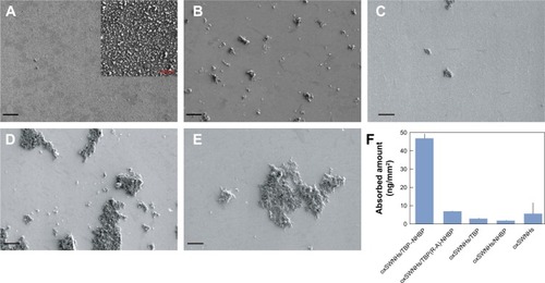
We generated TBP–NHBP (RKLPDAGGDYFSSPYYEQLF), a fusion of minTBP-1 and NHBP-1 connected via a GG linker, to mediate the binding of oxSWNHs to the Ti surface (). TBP–NHBP was mixed with oxSWNHs in 10% DMF to obtain the dispersed oxSWNHs/TBP–NHBP complex, which was adsorbed onto the Ti surface by incubating Ti plates in dispersed solutions of the complex. Three samples [oxSWNHs without peptide (oxSWNHs), a mixture of minTBP-1 and oxSWNHs (oxSWNHs/TBP), and a mixture of NHBP and oxSWNHs (oxSWNHs/NHBP)] were adsorbed onto the Ti plates and served as the controls. shows the SEM image of the oxSWNHs/TBP-NHBP-adsorbed particles on the Ti surface. When oxSWNHs were adsorbed onto Ti plates with TBP–NHBP, the weight of the plate increased relative to the controls (). By contrast, the adsorption of oxSWNHs without TBP–NHBP onto the Ti surface was negligible (). When oxSWNHs alone were incubated with the Ti plates, aggregates formed that were nonuniformly adsorbed on the plate surface (). Additionally, we used a TBP–NHBP variant containing an arginine (R) to alanine (A) mutation in the minTBP-1 sequence AKLPDA [TBP(R-A)-NHBP] to determine whether the minTBP-1 sequence in TBP–NHBP was effective for Ti binding. oxSWNHs did not uniformly adsorb to Ti in the presence of TBP(R-A)-NHBP (), consistent with a previous report showing that loss of the charged R residue reduced the binding of minTBP-1 to Ti.Citation44,Citation47 Additionally, the dispersibility of oxSWNHs might have been reduced by the R-to-A substitution. These results indicated that the minTBP-1 sequence promoted binding of oxSWNHs to Ti. Additionally, oxSWNHs did not uniformly adsorb to the Ti surface when mixed with TBP or NHBP alone (), indicating that TBP–NHBP conferred the binding characteristics of minTBP-1 on oxSWNHs.
Figure 1 Schematic representation of oxSWNHs containing the TBP–NHBP peptide complex (oxSWNHs/TBP–NHBP) on the Ti surface. oxSWNHs were modified by artificial peptide aptamers against SWNH (NHBP-1) and Ti (minTBP-1).
Abbreviations: oxSWNHs, oxidized single-walled carbon nanohorns; TBP, Ti-binding peptide; NHBP-1, SWNH-binding peptide.
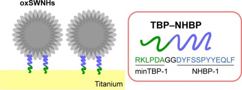
Figure 2 Scanning electron micrographs of Ti plates functionalized with oxSWNHs/TBP–NHBP. (A–E) Ti plates were functionalized with oxSWNHs/TBP–NHBP (A), oxSWNHs (B), oxSWNHs/TBP(R-A)-NHBP (C), oxSWNHs/TBP (D), or oxSWNHs/NHBP (E). The inset shows magnified sections. Black scale: 20 µm; red scale: 2 µm. (F) Adsorption amounts measured using an electronic balance.
Abbreviations: oxSWNHs, oxidized single-walled carbon nanohorns; TBP, Ti-binding peptide; NHBP-1, SWNH-binding peptide.
To investigate the ability of TBP–NHBP to promote adsorption of oxSWNHs onto the Ti surface, we used a QCM to monitor changes in the resonance frequency (f) and dissipation (D) of the Ti plate. Initial infusion of an oxSWNHs/TBP–NHBP solution into the QCM measurement cell reduced f bŷ65 Hz, with this response plateauing within 1 min and ameliorated by subsequent infusion of a wash buffer, indicating that oxSWNHs/TBP-NHBP were bound to the Ti surface (). When the cell chamber was refilled with an oxSWNHs/TBP–NHBP solution, f declined, whereas D increased. By contrast, injection of oxSWNHs alone reduced f by only 9 Hz, indicating a weak association with the Ti sensor ().
Figure 3 Time-dependent changes in f and D determined using a QCM. (A–F) oxSWNHs/TBP–NHBP (A), oxSWNHs (B), TBP–NHBP (C), oxSWNHs/TBP–NHBP (D), TBP–NHBP (E), and TBP–NHBP and oxSWNHs/TBP–NHBP (F) deposited on a Ti-coated sensor. Values on the longitudinal axis represent negative Δf and positive ΔD values. Changes associated with oxSWNHs/TBP–NHBP, oxSWNHs, and TBP–NHBP adsorption are indicated by green, gray, and purple lines, respectively. Green, gray, and purple arrowheads indicate the time points at which oxSWNHs/TBP–NHBP, oxSWNHs, and TBP–NHBP, respectively, were infused into the measurement chamber. Vs represent the time points at which the wash solution (10% DMF) was infused.
Abbreviations: oxSWNHs, oxidized single-walled carbon nanohorns; TBP, Ti-binding peptide; NHBP-1, SWNH-binding peptide; DMF, dimethylformamide.
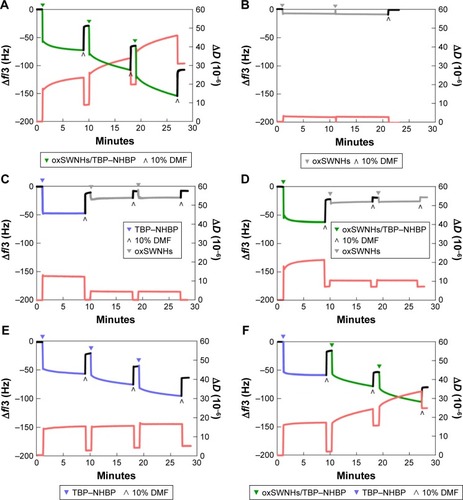
We then investigated whether oxSWNHs must be premixed with TBP–NHBP for efficient adsorption to Ti by testing whether pre-adsorption of TBP–NHBP alone to Ti facilitated oxSWNH immobilization through interaction of the latter with the NHBP part of the peptide on the solid phase. To this end, we injected TBP–NHBP alone onto Ti plates, followed by injection of oxSWNHs. This did not result in adsorption of oxSWNHs to Ti (), and moreover, there was no adsorption by injection of oxSWNHs after oxSWNHs/TBP–NHBP were injected and allowed to adsorb to the Ti sensor (). These results demonstrated the necessity of pre-mixing TBP–NHBP and oxSWNHs for adsorption of oxSWNHs onto the Ti surface. When TBP–NHBP alone was used, adsorption onto Ti plates was observed (); however, use of oxSWNHs/TBP–NHBP resulted in an increase in D (). Even if cleaning was performed prior to each flow of oxSWNHs/TBP-NHBP, an increase in D was observed, although when TBP-NHBP only was used, a minimal increase in D was observed. It is possible that this resulted due to the unstable adsorption of some of the oxSWNHs to Ti in the chamber. Therefore, the minTBP-1 portion of TBP–NHBP also contributed to the immobilization of oxSWNHs on the Ti surface.
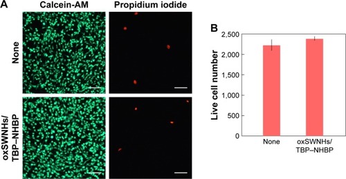
We then investigated whether oxSWNHs/TBP–NHBP on Ti was toxic to cells by culturing MC3T3-E1 cells on oxSWNHs/TBP–NHBP on the Ti plates. The live/dead assay using calcein/propidium-iodide staining revealed good cell viability after 2 days of culture (). These results demonstrated that uncoated Ti and oxSWNHs/TBP–NHBP on Ti exhibited comparable effects on cell viability. SWNHs do not contain contaminating metals, as they are prepared by laser ablation from pure graphite without a metallic catalyst, which reduces the risk of toxicity.Citation33
Figure 4 Viability of MC3T3-E1 cells cultured for 2 days on oxSWNHs/TBP–NHBP on Ti. Viable cell counts were determined relative to cells grown on uncoated Ti (none). (A) Live (green, left panel) and dead (red, right panel) cells were stained with calcein-AM and propidium iodide, respectively. White scale: 100 µm. (B) Counts of live cells on uncoated Ti and oxSWNHs/TBP–NHBP-conjugated Ti. Error bars indicate standard deviation (n=4).
Abbreviations: oxSWNHs, oxidized single-walled carbon nanohorns; TBP, Ti-binding peptide; NHBP-1, SWNH-binding peptide.
In vitro release of DEX from DEX-oxSWNHs/TBP–NHBP on Ti
Controlled release from a drug-carrier complex is an important property of drug-delivery systems. In vitro binding and release of the anti-inflammatory glucocorticoid DEX by oxSWNHs was previously reported.Citation28 Here, we evaluated the in vitro drug-release potential of oxSWNHs on Ti according to the cumulative doses of [3H]-DEX released from DEX-oxSWNHs on Ti plates in PBS, α-MEM, and RPMI medium at 37°C (). The initial release of DEX from Ti was slower in α-MEM or RPMI medium than in PBS. Additionally, DEX from Ti in α-MEM or RPMI medium was released in a sustained fashion for up to 2 weeks.
Figure 5 In vitro release of DEX from DEX-oxSWNHs/TBP–NHBP on Ti. Time course of cumulative release of [3H]-DEX from DEX-oxSWNHs/TBP–NHBP on Ti plates in PBS (blue line), RPMI medium/5% FBS (green line), and α-MEM/5% FBS (red line) at 37°C over 14 days. The inset shows cumulative release of DEX from DEX-oxSWNHs/TBP–NHBP on Ti plates in PBS (blue line), RPMI medium/5% FBS (green line), and α-MEM/5% FBS (red line) at 37°C for 24 hours. Error bars indicate standard deviation (n=3).
Abbreviations: DEX, dexamethasone; PBS, phosphate-buffered saline; RPMI, Roswell Park Memorial Institute; α-MEM, α-minimal essential medium; oxSWNHs, oxidized single-walled carbon nanohorns; TBP, Ti-binding peptide; NHBP-1, SWNH-binding peptide.
![Figure 5 In vitro release of DEX from DEX-oxSWNHs/TBP–NHBP on Ti. Time course of cumulative release of [3H]-DEX from DEX-oxSWNHs/TBP–NHBP on Ti plates in PBS (blue line), RPMI medium/5% FBS (green line), and α-MEM/5% FBS (red line) at 37°C over 14 days. The inset shows cumulative release of DEX from DEX-oxSWNHs/TBP–NHBP on Ti plates in PBS (blue line), RPMI medium/5% FBS (green line), and α-MEM/5% FBS (red line) at 37°C for 24 hours. Error bars indicate standard deviation (n=3).Abbreviations: DEX, dexamethasone; PBS, phosphate-buffered saline; RPMI, Roswell Park Memorial Institute; α-MEM, α-minimal essential medium; oxSWNHs, oxidized single-walled carbon nanohorns; TBP, Ti-binding peptide; NHBP-1, SWNH-binding peptide.](/cms/asset/0f5658c7-f9d9-44f7-adf9-3d9bce5e073d/dijn_a_12193961_f0005_c.jpg)
Biological activity of DEX released from DEX-oxSWNHs/TBP–NHBP on Ti
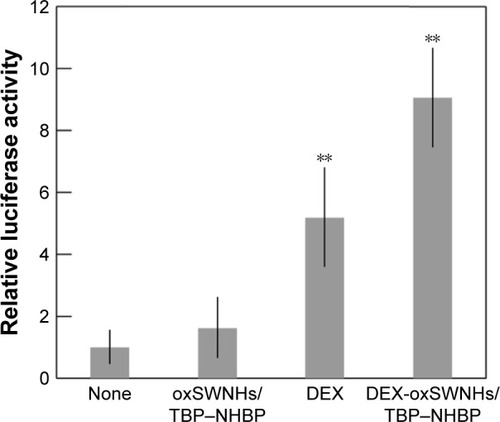
DEX is a synthetic agonist of glucocorticoid receptor (GR), which is a nuclear receptor that binds to glucocorticoid response elements (GREs) to activate target-gene transcription.Citation51 We examined the responsiveness of GREs to DEX released from DEX-oxSWNHs/TBP–NHBP on Ti using the reporter plasmid pBV2-MMTV-LUC containing a luciferase gene under the control of GREs.Citation50 To this end, we transfected ST2 cells with pBV2-MMTV-LUC, followed by incubation with DEX-oxSWNHs/TBP–NHBP, DEX, or oxSWNHs/TBP–NHBP on Ti. DEX and DEX-oxSWNHs/TBP–NHBP activated luciferase expression, with the latter having a greater effect, whereas the cells alone and oxSWNHs/TBP–NHBP had no effect (). Additionally, DEX-oxSWNHs/TBP-NHBP showed an even greater activation than DEX alone on Ti.
Figure 6 Effects of DEX-oxSWNHs on GR transcriptional activity. ST2 cells were transfected with pBV2-MMTV-LUC and incubated with oxSWNHs/TBP–NHBP, DEX, or DEX-oxSWNHs/TBP–NHBP on Ti plates. Error bars indicate standard deviation (n=5). **P<0.01.
Abbreviations: oxSWNHs, oxidized single-walled carbon nanohorns; TBP, Ti-binding peptide; NHBP-1, SWNH-binding peptide; DEX, dexamethasone.
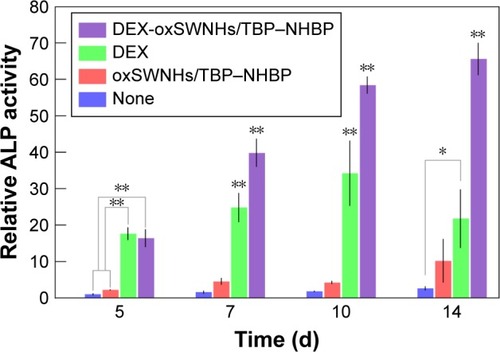
DEX promotes the differentiation of preosteoblasts into mature osteoblasts, a process characterized by ALP expression.Citation52 Additionally, preosteoblastic MC3T3-E1 cells treated with DEX show upregulated expression of ALP, as well as BMP-4, which is a potent inducer of osteoblast differentiation and bone formation.Citation53,Citation54 Here, we found that treatment of MC3T3-E1 cells with DEX-oxSWNHs/TBP–NHBP adsorbed on Ti in the presence of BMP-4 increased ALP expression from day 5 to day 14, whereas oxSWNHs/TBP–NHBP had no effect (). Moreover, on day 14, the activity was higher for DEX-oxSWNHs/TBP–NHBP adsorbed on Ti as compared with that observed in control samples (P<0.05). Our results indicated that DEX released from DEX-oxSWNHs on Ti plates was biologically active.
Figure 7 Effects of DEX-oxSWNHs on ALP activity. MC3TS-E1 cells were cultured with oxSWNHs/TBP–NHBP, DEX, or DEX-oxSWNHs/TBP–NHBP on Ti plates for 5, 7, 10, and 14 days. Error bars indicate standard deviation (n=5). **P<0.01; *P<0.05.
Abbreviations: ALP, alkaline phosphatase; DEX, dexamethasone; oxSWNHs, oxidized single-walled carbon nanohorns; TBP, Ti-binding peptide; NHBP-1, SWNH-binding peptide.
Discussion
Ti and its alloys are frequently used for bone and dental implants, and their junction with adjacent bone tissue is paramount. Ti-based materials are also used as scaffolds for biomolecules that control cellular activities, such as adhesion, differentiation, and proliferation.Citation5 Ti implants should ideally have osteoinduction capacity, representing the ability to stimulate the formation of new bone from progenitor cells. Various nanomaterials, including carbon nanotubes (CNTs), multi-walled CNTs, and SWNHs, promote bone formation by interacting with osteoblasts.Citation55–Citation57 In the present work, we combined the TBP–NHBP peptide with oxSWNHs to obtain dispersed oxSWNHs/TBP–NHBP complexes, which were adsorbed onto the Ti surface. Incorporation of the TBP–NHBP peptide conferred the binding characteristics of minTBP-1 to oxSWNHs and mediated their uniform bidimensional adsorption onto the Ti surface. NHBP-1 binds to oxSWNHs, but has low solubility, whereas fusion with highly soluble minTBP-1 enhances the dispersibility of oxSWNHs. This is the likely reason for the uniform adsorption of oxSWNHs onto the Ti surface without aggregation. We also showed that oxSWNHs/TBP–NHBP were adsorbed onto the Ti surface by monitoring the changes in f and D in a Ti plate, observing that pre-mixing TBP–NHBP and oxSWNHs was important for oxSWNHs adsorption. The decrease in f and increase in D were not saturated within the observation range, suggesting that surface treatment can be readily controlled. Furthermore, oxSWNHs/TBP–NHBP immobilized on Ti were nontoxic to cultured cells, even after 2 days of culture and in contrast with CNTs.Citation58 SWNHs contain no metal impurities, because SWNHs are synthesized through laser ablation or arc discharge using pure graphite rods without any metal catalysts.Citation59,Citation60 Therefore, SWNHs have negligible toxicity both in vitro and in vivo,Citation33,Citation42,Citation43,Citation61 which facilitates fabrication of the functionalized nanoplatforms for biomedical applications with low toxicity.
To assess the potential of oxSWNHs on Ti for drug delivery, we compared the release of the anti-inflammatory glucocorticoid DEX in PBS, α-MEM, and RPMI medium at 37°C. DEX was released from Ti at a slower rate in α-MEM and RPMI than in PBS; moreover, in cell-culture media, the release was sustained for up to 14 days. Variable solubility of DEX in PBS and cell-culture media could account for the differences in the release profiles. Additionally, GREs were activated by DEX and especially DEX-oxSWNHs/TBP–NHBP, with adsorption of the latter onto Ti increasing ALP activity in MC3T3-E1 cells relative to control samples on day 14. Therefore, DEX was not delivered in a single burst at the implantation site, but was steadily released for a sustained effect.
The results of the in vitro assays indicated that DEX released from DEX-oxSWNHs on Ti plates was biologically active. To date, there have been no studies on the effectiveness of DEX delivered by oxSWNHs/TBP–NHBP on Ti for inducing osteogenesis. The majority of studies on Ti-implant-surface treatments have focused on physicochemical surface modifications to improve osseointegration and interactions between surfaces and tissues. In the Ti-surface-modification method that has been reported, a special apparatus, such as a plasma spray apparatus, was required. However, the method described in this study constitutes a simple surface-reforming method that mixes oxSWNHs, TBP-NHBP, and a drug together, followed by incubation of Ti with the mixed solution, which enables it to transport the function of the drug. Because oxSWNHs are capable of transporting various drugs, we believe that it will be possible to perform surface modifications of Ti by enabling the drug to support oxSWNHs/TBP–NHBP according to the clinical purpose. Our findings have provided a basis for the development of new biomaterials for use in dental implants and as osteoblast scaffolds for bone regeneration.
Conclusion
We have successfully developed a solid-phase drug-delivery system where oxSWNHs were immobilized on a base material using TBP-NHBP, a bispecific material-binding peptide combining functions of minTBP-1 and NHBP-1. Through the use of TBP-NHBP, oxSWNHs, which otherwise could not be attached to Ti, were successfully immobilized on the Ti surface. SEM was used to confirm the presence of oxSWNHs adsorbed onto the Ti surface, and a QCM was used to evaluate the binding process during oxSWNH adsorption. Moreover, we were able to impart the function of sustained drug release to a substrate by using oxSWNHs. The oxSWNHs/TBP-NHBP-immobilized Ti surface was nontoxic to cells and could be used to deliver DEX, an anti-inflammatory agent, over a sustained period. The released DEX was confirmed as biologically active, suggesting the potential use of oxSWNHs/TBP-NHBP as a drug-delivery system to stimulate osteogenesis and bone regeneration. Therefore, our findings suggested that surface modification is a useful approach for designing medical biomaterials with desirable properties and possible practical applications.
Acknowledgments
We thank Dr T Imamura for kindly providing the MC3T3-E1 cells and ST2 cells, Dr K-I Sano for valuable discussion, and Ms T Minamisawa for technical assistance.
This work was supported by a JSPS KAKENHI (grant no 21791959, 16K11524).
Disclosure
The authors report no conflicts of interest in this work.
References
- JennyGJauernikJBierbaumSA systematic review and meta-analysis on the influence of biological implant surface coatings on periimplant bone formationJ Biomed Mater Res A2016104112898291027301790
- KokubunKKashiwagiKYoshinariMInoueTShibaKMotif-programmed artificial extracellular matrixBiomacromolecules20089113098310518826322
- KashiwagiKTsujiTShibaKDirectional BMP-2 for functionalization of titanium surfacesBiomaterials20093061166117519022501
- YuasaKKokubuEKokubunKAn artificial fusion protein between bone morphogenetic protein 2 and titanium-binding peptide is functional in vivoJ Biomed Mater Res A201410241180118623625448
- SchulerMTrentinDTextorMTosattiSGPBiomedical interfaces: titanium surface technology for implants and cell carriersNanomedicine20061444946317716147
- BrunetteDMTengvallPTextorMThomsenPTitanium in Medicine: Material Science, Surface Science, Engineering, Biological Responses and Medical ApplicationsBerlinSpringer Verlag2001
- AmigóVSalvadorMDRomeroFSolvesCMorenoJFMicrostructural evolution of Ti–6Al–4V during the sintering of microspheres of Ti for orthopedic implantsJ Mater Process Technol20031411117122
- KimHMMiyajiFKokuboTNakamuraTEffect of heat treatment on apatite-forming ability of Ti metal induced by alkali treatmentJ Mater Sci Mater Med19978634134715348733
- PopaCSimonVVida-SimitiIBatinGCandeaVSimonSTitanium- hydroxyapatite porous structures for endosseous applicationsJ Mater Sci Mater Med200516121165117116362217
- RautrayTRNarayananRKwonT-YKimK-HSurface modification of titanium and titanium alloys by ion implantationJ Biomed Mater Res B Appl Biomater201093258159120127988
- NayabSNJonesFHOlsenIEffects of calcium ion-implantation of titanium on bone cell function in vitroJ Biomed Mater Res A200783229630217437306
- ShenXHuYXuGRegulation of the biological functions of osteoblasts and bone formation by Zn-incorporated coating on microrough titaniumACS Appl Mater Interfaces2014618164261644025148131
- QiaoYZhangWTianPStimulation of bone growth following zinc incorporation into biomaterialsBiomaterials201435256882689724862443
- LiangCWangHYangJFemtosecond laser-induced micropattern and Ca/P deposition on Ti implant surface and its acceleration on early osseointegrationACS Appl Mater Interfaces20135168179818623927373
- Kazemzadeh-NarbatMKindrachukJDuanKJenssenHHancockREWWangRAntimicrobial peptides on calcium phosphate-coated titanium for the prevention of implant-associated infectionsBiomaterials201031369519952620970848
- RadinSDucheynePControlled release of vancomycin from thin sol–gel films on titanium alloy fracture plate materialBiomaterials20072891721172917184835
- AninweneGE2ndYaoCWebsterTJEnhanced osteoblast adhesion to drug-coated anodized nanotubular titanium surfacesInt J Nano-medicine200832257264
- AntociVJrKingSBJoseBVancomycin covalently bonded to titanium alloy prevents bacterial colonizationJ Orthop Res200725785886617415753
- ManiGJohnsonDMMartonDDrug delivery from gold and titanium surfaces using self-assembled monolayersBiomaterials200829344561457318790530
- ZavgorodniyAVBorrero-LópezOHoffmanMLeGerosRZRohanizadehRCharacterization of the chemically deposited hydroxy-apatite coating on a titanium substrateJ Mater Sci Mater Med20112211921052792
- ChoiJKonnoTTakaiMIshiharaKControlled drug release from multilayered phospholipid polymer hydrogel on titanium alloy surfaceBiomaterials200930285201520819560818
- PantarottoDPartidosCDGraffRSynthesis, structural characterization, and immunological properties of carbon nanotubes functionalized with peptidesJ Am Chem Soc2003125206160616412785847
- MattsonMPHaddonRCRaoAMMolecular functionalization of carbon nanotubes and use as substrates for neuronal growthJ Mol Neurosci200014317518210984193
- ChenRJBangsaruntipSDrouvalakisKANoncovalent functionalization of carbon nanotubes for highly specific electronic biosensorsProc Natl Acad Sci U S A200310094984498912697899
- PantarottoDPartidosCDHoebekeJImmunization with peptide-functionalized carbon nanotubes enhances virus-specific neutralizing antibody responsesChem Biol2003101096196614583262
- MiyakoEDeguchiTNakajimaYPhotothermic regulation of gene expression triggered by laser-induced carbon nanohornsProc Natl Acad Sci U S A2012109197523752822529368
- MiyakoEKonoKYubaEHosokawaCNagaiHHagiharaYCarbon nanotube-liposome supramolecular nanotrains for intelligent molecular-transport systemsNat Commun20123122623187626
- MurakamiTAjimaKMiyawakiJYudasakaMIijimaSShibaKDrug-loaded carbon nanohorns: adsorption and release of dexamethasone in vitroMol Pharm20041639940516028351
- HirataEMiyakoEHanagataNCarbon nanohorns allow acceleration of osteoblast differentiation via macrophage activationNanoscale2016830145141452227412794
- KasaiTMatsumuraSIizukaTCarbon nanohorns accelerate bone regeneration in rat calvarial bone defectNanotechnology201122606510221212475
- DepanDMisraRDKThe interplay between nanostructured carbon-grafted chitosan scaffolds and protein adsorption on the cellular response of osteoblasts: structure-function property relationshipActa Biomater2013946084609423261921
- MurakamiTSawadaHTamuraGYudasakaMIijimaSTsuchidaKWater-dispersed single-wall carbon nanohorns as drug carriers for local cancer chemotherapyNanomedicine (Lond)20083445346318694307
- MiyawakiJYudasakaMAzamiTKuboYIijimaSToxicity of single-walled carbon nanohornsACS Nano20082221322619206621
- IijimaSYudasakaMYamadaRNano-aggregates of single-walled graphitic carbon nano-hornsChem Phys Lett19993093–4165170
- MurataKKanekoKKanohHAdsorption mechanism of supercritical hydrogen in internal and interstitial nanospaces of single-wall carbon nanohorn assemblyJ Phys Chem B2002106431113211138
- MurataKKanekoKSteeleWAMolecular potential structures of heat-treated single-wall carbon nanohorn assembliesJ Phys Chem B2001105421021010216
- MurataKHiraharaKYudasakaMIijimaSKasuyaDKanekoKNanowindow-induced molecular sieving effect in a single-wall carbon nanohornJ Phys Chem B2002106491266812669
- YugeRIchihashiTMiyawakiJYoshitakeTIijimaSYudasakaMHidden caves in an aggregate of single-wall carbon nanohorns found by using Gd2O3 probesJ Phys Chem C2009113727412744
- MurakamiTFanJYudasakaMIijimaSShibaKSolubilization of single-wall carbon nanohorns using a PEG-doxorubicin conjugateMol Pharm20063440741416889434
- AjimaKYudasakaMMurakamiTMaignéAShibaKIijimaSCarbon nanohorns as anticancer drug carriersMol Pharm20052647548016323954
- MatsumuraSAjimaKYudasakaMIijimaSShibaKDispersion of cisplatin-loaded carbon nanohorns with a conjugate comprised of an artificial peptide aptamer and polyethylene glycolMol Pharm20074572372917685580
- TaharaYMiyawakiJZhangMHistological assessments for toxicity and functionalization-dependent biodistribution of carbon nanohornsNanotechnology2011222626510621586808
- ZhangMYamaguchiTIijimaSYudasakaMSize-dependent biodistribution of carbon nanohorns in vivoNanomed Nanotechnol Biol Med201395657664
- SanoK-IShibaKA hexapeptide motif that electrostatically binds to the surface of titaniumJ Am Chem Soc200312547142341423514624545
- KaseDKulpJLYudasakaMEvansJSIijimaSShibaKAffinity selection of peptide phage libraries against single-wall carbon nanohorns identifies a peptide aptamer with conformational variabilityLangmuir200420208939894115379530
- HayashiTSanoK-IShibaKIwahoriKYamashitaIHaraMCritical amino acid residues for the specific binding of the Ti-recognizing recombinant ferritin with oxide surfaces of titanium and siliconLangmuir20092518109011090619735142
- SanoK-IAjimaKIwahoriKEndowing a ferritin-like cage protein with high affinity and selectivity for certain inorganic materialsSmall200518–982683217193533
- SanoK-ISasakiHShibaKUtilization of the pleiotropy of a peptidic aptamer to fabricate heterogeneous nanodot-containing multilayer nanostructuresJ Am Chem Soc200612851717172216448147
- FanJYudasakaMMiyawakiJAjimaKMurataKIijimaSControl of hole opening in single-wall carbon nanotubes and single-wall carbon nanohorns using oxygenJ Phys Chem B200611041587159116471720
- AbeIUmeharaKMoritaRNemotoKDegawaMNoguchiHGreen tea polyphenols as potent enhancers of glucocorticoid-induced mouse mammary tumor virus gene expressionBiochem Biophys Res Commun2001281112212511178969
- YamamotoKRSteroid receptor regulated transcription of specific genes and gene networksAnnu Rev Genet1985192092523909942
- DiefenderferDLOsyczkaAMGarinoJPLeboyPSRegulation of BMP-induced transcription in cultured human bone marrow stromal cellsJ Bone Joint Surg Am200385-ASuppl 3S19S28
- LuppenCASmithESpevakLBoskeyALFrenkelBBone morphogenetic protein-2 restores mineralization in glucocorticoid-inhibited MC3T3-E1 osteoblast culturesJ Bone Miner Res20031871186119712854828
- LuppenCALeclercNNohTBrief bone morphogenetic protein 2 treatment of glucocorticoid-inhibited MC3T3-E1 osteoblasts rescues commitment-associated cell cycle and mineralization without alteration of Runx2J Biol Chem200327845449954500312933820
- ShimizuMKobayashiYMizoguchiTCarbon nanotubes induce bone calcification by bidirectional interaction with osteoblastsAdv Mater201224162176218522447724
- SaitoNUsuiYAokiKCarbon nanotubes for biomaterials in contact with boneCurr Med Chem200815552352718289008
- MisraRDChaudhariPMOsteoblasts response to nylon 6,6 blended with single-walled carbon nanohornJ Biomed Mater Res A201310141059106822965545
- ShvedovaAACastranovaVKisinERExposure to carbon nanotube material: assessment of nanotube cytotoxicity using human keratinocyte cellsJ Toxicol Environ Health A200366201909192614514433
- KasuyaDYudasakaMTakahashiKKokaiFIijimaSSelective production of single-wall carbon nanohorn aggregates and their formation mechanismJ Phys Chem B20021061949474951
- LiNWangZZhaoKShiZGuZXuSSynthesis of single-wall carbon nanohorns by arc-discharge in air and their formation mechanismCarbon201048515801585
- JiangB-PHuL-FShenX-COne-step preparation of a water-soluble carbon nanohorn/phthalocyanine hybrid for dual-modality photothermal and photodynamic therapyACS Appl Mater Interfaces2014620180081801725248075