
Abstract
Traditional supplements of selenium generally have a low degree of absorption and increased toxicity. Therefore, it is imperative to develop innovative systems as transporters of selenium compounds, which would raise the bioavailability of this element and allow its controlled release in the organism. Nanoscale selenium has attracted a great interest as a food additive especially in individuals with selenium deficiency, but also as a therapeutic agent without significant side effects in medicine. This review is focused on the incorporation of nanotechnological applications, in particular exploring the possibilities of a more effective way of administration, especially in selenium-deficient organisms. In addition, this review summarizes the survey of knowledge on selenium nanoparticles, their biological effects in the organism, advantages, absorption mechanisms, and nanotechnological applications for peroral administration.
Methodology of the review
To carry out this study, different databases such as Web of Science, PubMed, MEDLINE, and Google Scholar were employed. The findings of research studies (original articles, experimental studies) chosen from more than 1,000 viewed scientific publications were compared. The emphasis was put on original articles, while more than 100 review articles were not included. The search was based on phrases such as selenium nanoparticles and antioxidant activity/antimicrobial properties/antiviral effect/antibacterial activity/anticancer agent, selenium nanoparticles and synthesis, nano-selenium and bioavailability/toxicity/safety/drug delivery/orthopedic implants/protective effects/oxidative stress/metal intoxication/immunomodulation/reproduction/growth/gastrointestinal tract/hair, selenium nanoparticles and animals, advantages of nanoparticles, and effects of nano-selenium. The review includes research findings from the years 2000–2017.
Nanotechnological modifications
Nanoparticle (NP) systems appear to be a promising alternative to peroral drug deliveryCitation1 as well as nutritional supplements.Citation2 The effects of bioactive compound supplements – omega-3 and omega-6 fatty acids, probiotics, prebiotics, vitamins, and minerals – in nanoparticulate preparations have already been dealt by numerous studies.Citation3–Citation16 Some applications of NPs in nutrition and medicine have already been approved for clinical use, and many others are at different stages of their development.Citation17
The incentive to incorporate nanotechnology in nutrition has the following advantages: taste and smell, administration and solubility, protection against oxidation and enzymatic degradation, prolongation of residence time, and effective passage through the gastrointestinal tract enhancing the bioavailability of supplemented substances.Citation18 The use of most perorally administered supplements carries a number of drawbacks such as insufficient residence time, low permeability and solubility, and instability during the production process (temperature, presence of oxygen, light) or in the gastrointestinal tract environment (pH, enzymes, presence of other nutrients), which can reduce the activity and potential health benefits of supplements.Citation19 Thus, it is necessary to develop more appropriate products to ensure their physiological and therapeutic effect.Citation20
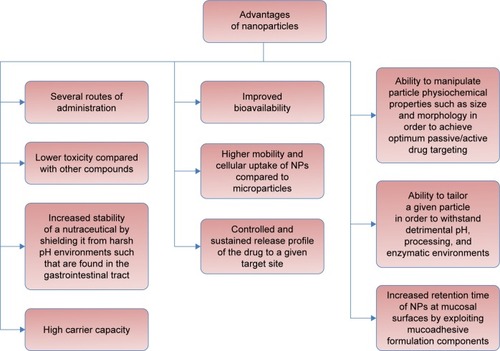
The use of NPs is a promising alternative for the peroral application of supplements that could solve the mentioned shortcomings of traditional forms of supplements and preserve their health benefits.Citation1 The main advantages of NPs are shown in .
Figure 1 Diagram showing the main advantages of nanoparticles (NPs).
Note: Data from various studies.Citation2,Citation22,Citation28,Citation201–Citation204
Selenium NPs as a food additive
The nanoform of selenium attracts even more attention, thanks to its high bioavailability and lower toxicity than inorganic and organic forms,Citation21–Citation23 where inorganic compounds are more toxic than organic ones.Citation24
The biological properties of selenium nanoparticles (SeNPs) depend on their size: smaller particles have a greater activity.Citation25 Particle size affects the cellular intake of NPs; for example, in vitro absorption of 0.1 μm particles was found to be 2.5 and 6 times higher compared to 1 and 10 μm particles, respectively.Citation26 With respect to this fact, in the preparation of dietary supplements, an appropriate particle size and morphology, as well as encapsulation material, should be chosen.Citation27 A system based on the administration of food supplements encapsulated into NPs represents an important potential to improve the bioavailability of supplements with the possibility of modifying their properties, such as resistance to adverse pH, digestion, and enzymatic cleavage.Citation28
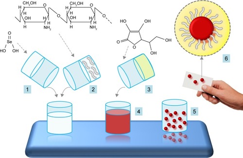
The advantage of nano-selenium (Nano-Se) is the possibility of using selenium in zero oxidation state (Se0), which presents low toxicity and excellent bioavailability compared to other oxidation states (Se+IV, Se+VI);Citation22,Citation25 however, it is very unstable and easily transformed into an inactive form. Although, its stabilization can be achieved by encapsulation into suitable nano-vehicles, for example, chitosan (CS)Citation15 ().
Figure 2 Scheme of preparation of selenium nanoparticles (SeNPs).
Notes: For the synthesis of SeNPs, the selenious acid solution (aqueous solution of SeO2) (1) is mixed with an aqueous solution of a polysaccharide, for example, chitosan (2), and ascorbic acid solution (3), which gradually transforms the initially colorless solution into a red color solution (4). In the next step, elemental SeNPs are coated with chitosan, and the result is encapsulated nano-selenium form (5, in detail 6).Citation50,Citation213
Nanoscale selenium has a very wide range of biomedical applications. Its effect on the reduction of oxidative stress is very well known.Citation12,Citation29 Gao et alCitation30 demonstrated the antioxidant properties of hollow spherical SeNPs reducing the risk of selenium toxicity. Besides its use as an antioxidant with reduced risk of toxicity, Nano-Se also possesses potential as a chemopreventive agent.Citation22 The results of numerous studies indicate that Nano-Se can be more helpful in cancer chemoprevention as a potential anticancer drug,Citation9,Citation31–Citation34 as well as an anticancer drug delivery carrier.Citation32,Citation35,Citation36 Many studies have shown the antimicrobial effectCitation37–Citation39 and antifungal activity of Nano-Se.Citation40 In addition, its protective effects against metal intoxication were well documented.Citation41–Citation43 Moreover, the immunostimulatory effect of nanoscale selenium was confirmed.Citation8,Citation44 Last but not least, Nano-Se has beneficial effects on a number of physiological functions.Citation13,Citation45–Citation47
Besides these unique abilities of Nano-Se, its antiprotozoal effect was described. Based on in vitro and in vivo studies, biogenic SeNPs can be considered as a novel therapeutic agent for the treatment of localized lesions typical of cutaneous leishmaniasis caused by Leishmania major.Citation48 The anti-leishmanial activities of SeNPs against Leishmania infantum were also described. SeNPs have more growth-inhibitory effect on promastigotes than selenium dioxide (SeO2), while the IC50 (half minimal [50%] inhibitory concentration) was determined to be 25 and 50 μg·mL−1, respectively.Citation49
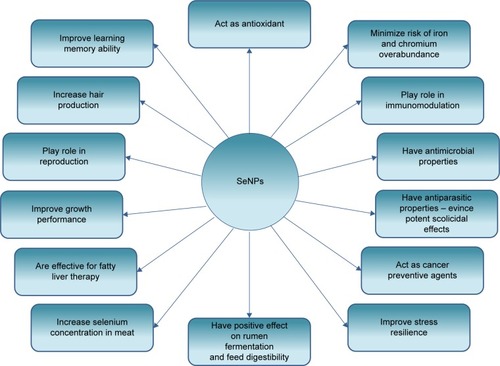
The main effects of the SeNPs found in various scientific studies are summarized in .
Figure 3 Diagram showing the main effects of selenium nanoparticles (SeNPs).
Note: Data from various sources.Citation7,Citation10,Citation12–Citation14,Citation21,Citation29,Citation43,Citation46,Citation47,Citation52,Citation155,Citation176,Citation180,Citation205–Citation212
Synthesis of SeNPs
SeNPs can be synthesized chemically,Citation50 or using physical procedures,Citation51 or can even be obtained by biological way – using microorganisms or plant extracts, the so-called green synthesis.Citation52,Citation53
Chemical synthesis
In terms of chemical synthesis, SeNPs are usually prepared by reduction of selenious acid solution by ascorbic acid in the presence of polysaccharides () such as CS, glucomannan, acacia gum, or carboxymethylcellulose.Citation50 CS is positively charged, biocompatible, non-immunogenic, nontoxic, pH sensitive, and biodegradable, and is therefore a suitable component for oral administration for a wide range of biomedical and nutritional applications.Citation54–Citation56 It has been extensively examined in the pharmaceutical industry due to its potential in the development of medication delivery systems.Citation57 In the molecular structure of polysaccharides, reactive amino, hydroxyl, or carboxyl groups are present, which have a substantial effect on the formation, stabilization, and growth of SeNPs.Citation50 After the synthesis of NPs, their ex situ characterization is performed using different biophysical methods including electron microscopy. The obtained monodisperse spherical selenium particles are very stable in solutionCitation16 and can be used as dietary additive.Citation14
The effects of selenium encapsulation into CS NPs were investigated by Zhang et al.Citation2 The study showed that encapsulation of selenium compounds into CS NPs is an effective way of delivering selenium to cells in which selenium retention increases, while reducing the risk of DNA damage. The development of NP–selenium compound systems may, in the case of low selenium level, significantly improve the bioavailability of selenium and facilitate the expression of selenoproteins.
Another example for chemical preparation of SeNPs is ionic liquid-induced synthesis with sodium selenosulfate as selenium precursor, in the presence of polyvinyl alcohol stabilizer, which can produce spherical SeNPs in the size range of 76–150 nm.Citation58
Physical synthesis
Among the physical techniques for the preparation of SeNPs, synthesis by pulsed laser ablation (PLA) or deposition was described in a study.Citation51 Generally, physical methods have distinct advantages over the chemical ones, since these often require a final calcination step, which makes them unsuitable for certain applications. Furthermore, with sputtering and laser ablation, the stoichiometry of the material is maintained.Citation59 In PLA, the size of the nanoclusters can be controlled by laser parameters, such as fluence, wavelength, and pulse duration, as well as by ambient gas conditions, such as pressure and flow parameters.Citation60 However, physical techniques are not widely applied for producing SeNPs.
Biological synthesis
Originally, SeNPs, like other NPs, were synthesized by various chemical methods. However, the high cost of production and the presence of toxic by-products led to the development of novel methods of NPs synthesis.Citation61 Biological organisms such as plants, fungi, or bacteria have the ability to convert some toxic metal ions to less toxic forms including metal precipitants or NPs.Citation62–Citation64 Thanks to this advantage, researches focused on their use for synthesizing NPs with an ecofriendly approach.Citation65–Citation68
As for green synthesis, SeNPs were synthesized, for example, using the following plant extracts: aqueous extract of Allium sativum,Citation69 tea extract,Citation70 Clausena dentata plant leaf extract,Citation71 Undaria pinnatifida polysaccharide solutions,Citation31 and Terminalia arjuna leaf extract.Citation42
Biosynthesis of nanomaterials using plant extracts has more advantages than other biological methods because it is inexpensive and does not require any special conditions.Citation52
NPs synthesis using bacteria is more effective than chemical synthesis, thanks to the following advantages: 1) high purity of selenium spheres (which are relatively regular and uniform, and their size depends on the bacterium), 2) cheaper and faster production process, and 3) better possibility to control the parameters.Citation72
Microorganisms are capable of synthesizing metal NPs.Citation53 Different bacterial strains are able to reduce Se+IV (selenite) and (or) Se+VI (selenate) to less toxic Se0 with the formation of SeNPs. The biogenic SeNPs have exhibited promising application perspectives in the field of medicine, biosensors, and environmental remediation.Citation73 Interestingly, two halotolerant Bacillus megaterium strains (BSB6 and BSB12), isolated from saline mangrove habitat without selenium contamination, were found to be capable of reducing Se+IV to elemental selenium, even in the presence of high salt concentrations.Citation74
The synthesis of SeNPs by macro- or microorganisms, due to the diversity of reducing enzymes in organisms, involves morphological and shape changes of the particles. By changing the redox state, the reducing enzymes of microorganisms convert metal ions (Se−II) to SeNPs without charge (Se0). The biological activity of SeNPs includes their protective role against DNA oxidation.Citation53 It was found that certain anaerobic bacteria respire toxic selenium oxyanions and as a result cause extracellular accumulation of elemental selenium (Se0). The spectral properties differ considerably from those of amorphous Se0 formed by chemical oxidation of hydrogen selenide (H2Se) and of black, vitreous Se0 formed chemically by reduction of selenite with ascorbate. The microbial synthesis of Se0 nanospheres results in unique, complex, with compact nanostructural arrangement of Se atoms. This probably reflects a wide diversity of enzymes involved in the dissimilatory reduction that are slightly different in various bacteria. Remarkably, these conditions cannot be achieved by current methods of chemical synthesis.Citation66
Different types of bacteria have been used for the biosynthesis of SeNPs, such as the species of phylum Proteobacteria (Escherichia coli ATCC 35218,Citation75 recombinant E. coli,Citation76 Ralstonia eutropha,Citation77 Enterobacter cloacae Z0206,Citation78 Pseudomonas aeruginosa ATCC 27853,Citation79 Klebsiella pneumoniae,Citation80,Citation81 Pantoea agglomerans,Citation25 Zooglea ramigera,Citation82 Rhodopseudomonas palustris strain N,Citation83 Shewanella sp. HN-41,Citation84 Azoarcus sp. CIB,Citation85 Burkholderia fungorum,Citation86 Stenotrophomonas maltophiliaCitation39), Firmicutes (Lactobacillus casei,Citation72,Citation81,Citation87 Lactobacillus acidophilus [LA-5],Citation72 Lactobacillus helveticus [LH-B02],Citation72 Enterococcus faecalis,Citation53 Streptococcus thermophilus,Citation72 Staphylococcus carnosus,Citation88 Bacillus sp. MSh-1,Citation89 Bacillus subtilis,Citation65 Bacillus mycoides SelTE01,Citation38 Bacillus licheniformis JS2Citation90), Actinobacteria (Streptomyces sp. ES2-5,Citation73 Bifidobacterium BB-12Citation72), and Cyanobacteria (Arthrospira [Spirulina] platensisCitation91).
For in vivo synthesis of Nano-Se, unicellular eukaryotic organisms, such as the protozoa Tetrahymena thermophila SB210,Citation92 yeast Saccharomyces cerevisiae,Citation93 and a genetically modified Pichia pastoris,Citation94 or even multicellular organisms, such as a fungus from the phylum of Ascomycota (Aspergillus terreusCitation95), were used.
Problems with traditional forms of oral supplementation of selenium and potential benefits of SeNPs
The bioavailability of various chemical forms of selenium has been the subject of many studies. For instance, Mahan et al stated that the degree of selenium absorption is much lower in ruminants than in nonruminants; after oral supplementation, the selenium uptake in sheep was only 34%, whereas in pigs 85%.Citation96 In ruminants, the microbial digestion in the rumen and reticulum precedes its assimilation in the abomasum and microbial digestion in the small intestine.Citation97,Citation98 Ruminal microorganisms partially transform selenium supplements into insoluble forms, such as elemental selenium and selenides, which are unavailable to animals, and thus, the absorption of selenium from the digestive tract is reduced.Citation99
To avoid this negative phenomenon of reduced selenium absorption due to microbial digestion in ruminants and improve the availability of this microelement, polymer NP systems with in vivo potential have been proposed.
This technology was implemented using the Eudragit® RL and Eudragit® RS polymers.Citation100 These nonbiodegradable polymer NPs were already used in 2005 to encapsulate the lipophilic cyclic undecapeptide cyclosporin A (CyA) for oral administration in rabbits for the prevention of graft rejection and the treatment of autoimmune diseases. CyA was encapsulated by nanoprecipitation in mentioned NP polymers, and its relative bioavailability from both preparations ranged from 20% to 35%.Citation101 The design of selenium in the nanoform for oral administration to ruminants was proposed as late as 2010 by Romero-Pérez et al,Citation100 who monitored the in vitro effect of sodium selenite (Na2SeO3) encapsulated in polymeric NPs by nanoprecipitation and emulsion-evaporation methods using two different solvents – ethanol and acetone. Nanoprecipitated NPs were spherical and had a greater particle size variability; on the contrary, the NPs produced by the emulsion-evaporation technique were both spherical and irregular in shape and homogeneous in size. The size of NPs ranged from 30 to 200 nm. Higher particle size, zeta potential, and polydispersity index were found for the NPs produced by the nanoprecipitation method using ethanol as the solvent of polymers. The release of selenium from NPs was monitored in vitro at different pH values and was higher in the strongly acidic environment (pH less than 4), which is a prerequisite for providing better accessibility of this element in the abomasum. At pH values lower than 4, selenium release increases by 62%Citation100 compared to pH 6. The pH in the rumen ranges from 5.5 to 6.5. Thus, the sheep rumen essentially does not absorb selenium.Citation97 On the contrary, the pH in the abomasum is less than 4; under this condition, the largest amount of selenium is released from the particles. NPs increase the bioavailability of this element because at pH 6 only a small amount of selenium is released (by rumen microorganisms) and the remaining, bigger part, is released then in the more acidic environment of the gastrointestinal tract, which guarantees its better availability.Citation100
SeNPs evince, in addition to better selenium availability, much lower toxicity compared to other selenium compounds.Citation22,Citation23 For example, in mice, SeNPs showed much lower toxicity measured by median lethal dose (50%, LD50), liver impairment, and short-term toxicity.Citation23
Mechanism of passage of NPs through intestinal mucosa
Oral administration of NPs is considered to be the most appropriate and cost-effective method of supplementation. However, absorption of NPs may also be made more difficult by the presence of absorption barriers in the digestive tract. It is necessary to overcome two barriers in the gastrointestinal tract for the absorption of NPs, which are mucus covering the intestinal mucosa and the intestinal mucosa.Citation1
The intestinal epithelium is composed of a series of specialized cells, primarily enterocytes, goblet cells, and M cells. One of the main functions of enterocytes is to control the transition of macromolecules and allow the absorption of nutrients. The goblet cells secrete mucus consisting of a high-molecular-weight glycoprotein (mainly mucin) suspended in the electrolyte solution,Citation102 which covers with the adherent layer the mucous membrane of the intestine. Their primary function is to protect the intestinal mucosa from potential pathogens or chemicals and maintain a different pH between the lumen and the mucosal surface of the intestine.Citation1
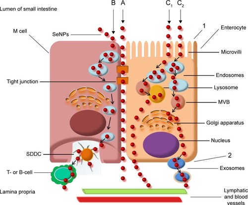
NPs can theoretically pass through the intestinal epithelium in two ways: paracellular (between adjacent cells) or transcellular (through the cells) ().Citation102 Under physiological conditions, the first way is restricted by a narrow region of intercellular spaces and by the tightness of the junctions between the epithelial cells (pore diameter is between 0.3 and 1.0 nm).Citation103 Transcellular transport of NPs takes place through a process called transcytosis, which starts with endocytosis in the apical membrane of the cells. Subsequently, the NPs are transported through the cells and released on the basolateral pole.Citation104
Figure 4 Diagram of nanoparticle transport across the intestinal mucosa of the small intestine. Particles pass either paracellularly, that is, between adjacent cells, or by a transcellular pathway that has been more explored. The transcellular transition takes place via normal enterocytes or M cells. A, paracellular transport; B, transcellular transport: transcytosis through M cell; C1, transcytosis through enterocyte, first mechanism; C2, transcytosis through enterocyte, second mechanism; 1, apical membrane of enterocyte; 2, the basolateral membrane of the enterocyte. M cells form a row of vesicles before transfer from subepithelial dome dendritic cells (SDDCs) to T- or B-lymphocytes (B). Uptake of nanoparticles by microvilli of enterocytes is often followed by endosome formation, microvesicular bodies (MVBs) genesis, and their fusion with lysosomes, and then the particles are transported to lamina propria (C1). Another mechanism of enterocyte utilization for transepithelial transport involves uptake into endosomes, MVB formation, fusion with Golgi apparatus, and exosomal transfer to lamina propria (C2). This is followed by transport into the bloodstream and the lymphatic system.Citation102,Citation214
Intestinal epithelial cells are capable of transferring NPs with mineral elements, although their capacity is limited. Transcellular transport begins with endocytosis (pinocytosis or macropinocytosis). It is an active process requiring energy for the internalization of NPs. Macropinocytosis is actin-dependent but is not regulated by receptors, and a large amount of fluid with particles smaller than 5 μm can be internalized through it.Citation105 The absorption of NPs is influenced by electric charge, surface hydrophobicity, and size.Citation106 The epithelial cell membrane of the gastrointestinal tract is composed of lipids, so that hydrophobic NPs have a higher absorption efficiency than hydrophilic particles. The absorption of NPs of 100 nm in the gastrointestinal tract is 15–250 times higher than that of larger-sized NPs.Citation107
Application of SeNPs through oral administration
Nano-Se as an antioxidant
Nano-Se possesses better antioxidant capability than other chemical forms of selenium while reducing the risk of selenium toxicity. Wang et alCitation22 demonstrated the antioxidant properties of SeNPs that evinced lower toxicity than selenomethionine (SeMet).
Zhang et alCitation108 studied the effect of elemental Nano-Se on the activity of glutathione peroxidase (GPx) in the liver of weanling pigs (Duroc × Landrace × Yorkshire) in comparison to inorganic form of selenium. The animals had significantly higher activities of GPx at a concentration range of 0.50 and 1.0 mg Se·kg−1 diet in the form of Nano-Se than Na2SeO3.
Effect of SeNPs on reproductive performance
Oxidative stress affects the fertility potential of spermatozoa by lipid peroxidation which can lead to sperm dysfunction.Citation109 Selenium deficiency leads to the occurrence of abnormal mitochondria in goat spermatozoa, and Nano-Se supplementation increases selenium concentration in testes, and testicular and semen GPx activity. In addition, it has a protective effect on membrane integrity and the tight arrayment of the mid-piece of the mitochondria. To study the effects of elemental Nano-Se in the diet on the testes ultrastructure, ejaculate quality and GPx activity in goats were investigated. The animals were administered with Nano-Se in the declared selenium dose of 0.3 mg·kg−1 of the feed dry matter (DM) for 12 weeks from weaning to sexual maturity. The results showed that the level of selenium in testes and the activity of GPx and ATPase in the ejaculate significantly increased in the group supplemented with nanoscale selenium compared to the control group. The quality of the ejaculate (volume, density, motility, and pH) was not affected by the addition of selenium, but the percentage of abnormal spermatozoa in the control group of goats was significantly higher compared to the SeNPs group. Using transmission electron microscopy, it was found that in selenium-deficient goats the sperm plasma membrane was damaged, and there were abnormalities in the mitochondria midpiece of spermatozoa.Citation13
Use of Nano-Se for increasing hair follicle development and fetal growth
The study of Wu et alCitation46 demonstrated the importance of maternal administration of selenium at nano size for improving the hair follicle development and promoting the growth of fetus. This was attributed to the influencing antioxidant status in the fetal skin. An increase in the antioxidant defence results in a decrease of reactive oxygen species (ROS) generation, leading to upregulation of IGF-1 and its receptor (IGF-1R) which are crucial for the improvement of both properties.
In cashmere goats, SeNPs have a positive effect on the amount of wool. The administration of 0.5 mg·kg−1 diet (containing 1,500 mg Se·kg−1) in the period from 30 days before conception to 110 days of gestation was manifested in their fetuses with significantly higher expression levels of the GPx, IGF-1, and IGF-1R genes in skin. In addition, increased GPx and superoxide dismutase (SOD) activities, and IGF-1 and selenium concentrations in both skin and blood serum at 110 days were observed. Moreover, a significantly lower malondialdehyde (MDA) production in both skin and serum which affected the antioxidant status in skin of the fetuses, and significantly increased the number of their secondary hair follicles, was observed. The low level of ROS probably upregulates IGF-1 and IGF-1R, which favorably influences the development of hair follicles in the fetus. In addition, the weights of fetus and placenta were significantly higher in SeNPs-receiving group than those in control group.Citation46
Antiviral and antibacterial effects of SeNPs
SeNPs have attracted substantial attention due to their unique antimicrobial activity.Citation53,Citation110–Citation114 Selenium is an essential trace element regulated by cellular redox homeostasisCitation115–Citation117 and is an integral component of selenoproteins controlling some crucial biological processes, such as ROS elimination and specific enzyme modulation.Citation118,Citation119 Selenium deficiency can result in susceptibility to viral infections. The antiviral capability of SeNPs, together with other advantages such as low toxicity and excellent activity, has recently attracted increased attention. The mortality of the H1N1 influenza virus-infected selenium-deficient mice was 3 times higher compared to those receiving Na2SeO3 at the dose of 0.5 mg Se·kg−1, and mice with low serum selenium concentrations showed a marked reduction in body weight (BW) and lower levels of TNF-α and IFN-γ.Citation120 For improving the immune response in the body, the administration of SeNPs can also be an efficient realizable approach.
Li et alCitation121 synthesized oseltamivir (OTV) surface-modified SeNPs (Se@OTV) with superior antiviral properties and restriction on drug resistance. Although the clinical application of OTV itself as an effective antiviral agent is ordinarily limited by the appearance of drug-resistant viruses, OTV decoration of SeNPs evidently inhibited H1N1 influenza virus infection and showed less toxicity. Se@OTV interfered with the entry of H1N1 into host cells through inhibiting the activity of influenza virus glycoproteins – hemagglutinin and neuraminidase. Modified NPs were able to prevent H1N1 from infecting Madin-Darby canine kidney cells and block chromatin condensation and DNA fragmentation. In addition, they inhibited the generation of ROS as well as the activation of phosphorylation of cellular tumor antigen p53 and Akt. Thus, ROS play a pivotal role in the antiviral action. The presence of H1N1 virus increased the intracellular ROS generation from 100% (control) to 380%, and OTV and SeNPs slightly inhibited their production to 270% and 210%, respectively, whereas Se@OTV markedly decreased the ROS generation (120%). It follows that Se@OTV are promising efficient antiviral pharmaceuticals against highly infectious respiratory disease caused by H1N1,Citation121 which belongs to influenza A-type viruses.Citation122
SeNPs also show excellent antibacterial activity. With SeNPs biosynthesized by R. eutropha, 99% inhibition of growth of P. aeruginosa, S. aureus, E. coli, and Streptococcus pyogenes at the concentrations of 100, 100, 250, and 100 μg·mL−1, respectively, was observed. Moreover, the concentration of 500 μg·mL−1 of SeNPs was found to inhibit the growth of pathogenic fungi Aspergillus clavatus. The antimicrobial efficacy of SeNPs can be comparable with commercially available antibiotic ampicillin.Citation77 SeNPs synthesized by another bacterial species E. faecalis can be used as an antistaphylococcal element to effectively prevent and treat S. aureus infections.Citation53 SeNPs with application of bioactive glass scaffolds (45S5Bioglass®) and poly(lactic-co-glycolic acid) (PLGA) (45S5Bioglass®/SeNPs and 45S5Bioglass®/PLGA/SeNPs) showed a considerable antibacterial activity against Gram-positive bacteria, S. aureus and Staphylococcus epidermidis.Citation114 In another study, Yang et alCitation123 introduced the antibacterial effect of Qe/CdSe/ZnS (quercetin/cadmium selenide/zinc sulfide) nanoparticles (QCZNPs) against drug-resistant E. coli and B. subtilis in vitro. QCZNPs showed markedly more effective antibacterial activities than Qe or CdSe NPs. The in vitro study of Wang and WebsterCitation37 showed that the selenium coatings on polycarbonate medical devices significantly inhibited S. aureus growth to 27% compared with an uncoated polycarbonate surface after 72 hours.
The antimicrobial activity of SeNPs depends on the way they are synthesized. Piacenza et alCitation38 evaluated the antimicrobial efficacy of spherical biogenic selenium nanostructures embedded in organic material produced by B. mycoides SelTE01 in comparison with two different chemical SeNP classes produced using L-cysteine or ascorbic acid. The antimicrobial activity was tested on P. aeruginosa and S. aureus biofilms grown onto hydroxyapatite-coated clinical devices and surfaces. After 6 or 24 hours of Na2SeO3 exposure, biogenic SeNPs evinced the same effective antibiofilm activity against both tested strains at 0.078 and 0.3125 mg·mL−1, respectively. On the contrary, chemically synthesized SeNPs at the highest tested concentration (2.5 mg·mL−1) showed only moderate antimicrobial activity. Cremonini et alCitation39 reported that SeNPs synthesized by Gram-negative S. maltophilia and Gram-positive B. mycoides achieved much stronger antimicrobial effects compared with synthetically prepared SeNPs. Both biogenic types of NPs were active at low minimum inhibitory concentrations against a number of clinical isolates of P. aeruginosa. It was interesting that after an exposure of human dendritic cells and fibroblasts to all three SeNPs, the following phenomena were not observed: 1) loss of cell viability, 2) increased release of ROS, and 3) significant increase in the secretion of pro-inflammatory and immunostimulatory cytokines. Therefore, SeNPs appear to be reliable candidates for safe medical applications, either alone or in combination with traditional antibiotics, to inhibit the growth of P. aeruginosa clinical isolates.
Anticancer effects of SeNPs
SeNPs have also showed remarkable anticancer activityCitation52,Citation123–Citation128 and exhibit high potential in cancer chemotherapy and as drug carriers.Citation129–Citation131 The anticancer effects of SeNPs are mediated through their ability to inhibit the growth of cancer cells through induction of cell cycle arrest at S phase.Citation32 The induction of the cell cycle arrest at the S phase is mediated by deregulation of the eIF3 protein complex.Citation132 A recent study revealed that cell membrane plays an important role in SeNPs-induced toxicity in cancer cells. SeNPs treatment changes the biomechanical properties of cancer cells, in particular remarkably decreases the adhesion force and Young’s modulus.Citation133 Besides unique anticancer efficacy, SeNPs have been proved to present a better selectivity between normal and cancer cells than Se+IV at similar concentrations.Citation31 SeNPs can be internalized selectively by cancer cells through endocytosis and induce cell apoptosis by triggering apoptotic signal transduction pathways.Citation134–Citation136
Nano-Se as an anticancer drug
Nano-Se possesses higher anticancer efficacy than other selenium compounds.Citation119 A key mechanism for the chemopreventive effect is the induction of glutathione S-transferase (GST) by selenium.Citation22 The activity of GST in the case of Nano-Se administration increased much earlier and more markedly than in the case of SeMet and selenite.Citation22,Citation137
Yanhua et alCitation127 suggested that novel selenium-substituted hydroxyapatite NPs (SeHAN) could be a new promising anticancer agent to provide both survival advantage and lower toxicity in nude mice model of human hepatocellular carcinoma. The overall survival rate of nude mice in the control, pure hydroxyapatite, and SeHAN groups was 50.00%, 76.92%, and 100.00%, respectively. Blood biochemical studies showed that SeHAN group had significantly lower toxicity on the liver and kidney functions.
In highly metastatic breast cancer mice model, a better prognosis could be achieved by oral administration of SeNP-enriched Lactobacillus brevis. Both components are immunostimulators, and enhanced immune response in cancer-affected mice. Moreover, lactic acid bacteria (LAB) can reduce selenium ions to elemental SeNPs and deposit them in intracellular spaces. When LAB in combination with SeNPs were administered to cancer-bearing mice, a significant increase in natural killer cell cytotoxicity and delayed-type hypersensitivity responses, as well as a high level of IFN-γ and IL-17, compared to the control mice or mice that received L. brevis alone, was observed. In addition, an extended life span and a decline in the tumor metastasis to the liver were recorded in this group compared to the other two groups of mice.Citation44
Chen et alCitation31 reported that SeNPs fabricated in U. pinnatifida polysaccharide solutions induced mitochondria-mediated apoptosis in A375 human melanoma cells. Treatment of this cancer cell line with Nano-Se resulted in a dose-dependent cell apoptosis manifested by DNA fragmentation and phosphatidylserine translocation.
Sonkusre et alCitation34 introduced biologically synthesized SeNPs by B. licheniformis JS2 and developed a method for extraction and purification of intracellular NPs. These neutral-charged, non-agglomerating SeNPs at a concentration as low as 2 μg Se·mL−1 were efficacious in inhibiting proliferation and inducing caspase-independent necrosis to human prostate adenocarcinoma cells (PC3) without causing any significant toxicity to human peripheral blood mononuclear cells.
Luo et alCitation32 examined the in vitro antiproliferative effects of SeNPs (Nano-Se0, 10–40 μmol·L−1) on HeLa (human cervical carcinoma) cells and MDA-MB-231 (human breast carcinoma) cells by optical microscopic inspection and MTT assay. Nano-Se0 effectively inhibited the growth of cells of both the cancer cell lines in a dose-dependent manner. The morphology analysis with atomic force microscopy showed that the HeLa cells treated with 10 μmol·L−1 Nano-Se0 were rough and shrunken with truncated lamellipodia at the terminal part of the cells. Flow cytometric analysis demonstrated that HeLa cells were arrested at S phase of the cell cycle after exposed to mentioned amount of Nano-Se0 (10 μmol·L−1).
It was found that anticancer activity of SeNPs correlates with the level of ERα in breast cancer cells. SeNP-induced cell death and expression of apoptotic markers (pp38, Bax, and cytochrome c) were significantly higher in ERα-positive cells (MCF-7) but not in ERα-negative cells (MDA-MB-231).Citation33
Nano-Se as an anticancer drug delivery carrier
Besides their direct anticancer effects, SeNPs have been appointed as potential anticancer drug delivery carriers.Citation32,Citation35 A key factor that usually contributes to nanomaterial-based drug cytotoxicity is cellular uptake.Citation35 The nanosize of these materials allows an efficient uptake by various cell types and selective drug accumulation at target sites.Citation26 Like other chemotherapeutics, the effective cytotoxicity of nanomaterial-based drugs usually requires a high level of accumulation within the cancer cells.Citation138 Nanomaterials tend to accumulate in cancer cells through a passive targeting processCitation128 and often serve as “nanocarriers” for chemotherapeutics.Citation35,Citation139–Citation147 Moreover, the usage of various surface decorators enhances the cellular uptake and anticancer efficacy of nanomaterials;Citation35,Citation128,Citation135 for example, Yang et alCitation128 developed a simple and solution-phase method for functionalization of SeNPs with Spirulina polysaccharides (SPS). In addition, they found that SPS surface decoration significantly enhanced the cellular uptake and cytotoxicity of SeNPs against several cancer cell lines. The action mechanism of SPS-SeNPs was grounded in inhibition of cancer cell growth through induction of apoptosis, as evidenced by an increase in sub-G1 cell population, DNA fragmentation, chromatin condensation, and phosphatidylserine translocation. In another study, Yang et alCitation148 designed and synthesized folic acid-conjugated SeNPs (FA@SeNPs) as cancer-targeting agents; the anticancer efficacy of FA@ SeNPs was synergistically enhanced by radioactive 125I seeds, and these NPs inhibited colony formation ability, which showed that functionalized SeNPs can be used as a radiation sensitizer for 125I seeds for cancer therapy.
Chan et alCitation129 reported that, based on the Auger-electron effect and Compton effect of Se atoms, cancer-targeted SeNPs in combination with 125I seeds achieve synergetic effects for inhibition of cancer cell growth and colony formation by inducing cell apoptosis and cell cycle arrest. In addition, these could be used as a safe and effective agent for next-generation cancer chemoradiotherapy in clinical applications. The action mechanism showed the activation of intracellular ROS overproduction to regulate p53-mediated DNA damage apoptotic signaling pathways and MAPKs phosphorylation as well as preventing the self-repair of cancer cells simultaneously.
SeNPs can be used as a carrier of 5-fluorouracil to achieve anticancer synergism, as introduced by Liu et al.Citation35 A panel of five human cancer cell lines (A375, MCF-7, HepG2, Colo201, and PC3) evinced susceptibility to 5-fluorouracil surface-functionalized SeNPs (5-FU-SeNPs), with IC50 values ranging from 6.2 to 14.4 μM. Remarkably, despite this potency, the 5-FU-SeNPs possess great selectivity between cancer and normal cells. Induction of apoptosis in A375 human melanoma cells by 5-FU-SeNPs was evidenced by accumulation of sub-G1 cell population, DNA fragmentation, and nuclear condensation.
By comparing the effect of chemically prepared SeNPs on the behavior of cancer cells with that of other inorganic and organic selenospecies, similar alterations in terms of cell viability, proliferation, migration, and cell cycle arrest at the S-G2/M phase after exposure of hepatocarcinoma (HepG2) cells to SeNPs and selenocystine (SeCys2) were observed. In contrast, cells exposed to Se+IV showed evident signs of toxicity such as strong induction of apoptosis and a significant population of cells in the sub-G1 phase compared to control cells. Se+IV, SeMet, and seleno-methylselenocysteine (Se-MetSeCys) did not evince any significant differences in comparison to non-treated cells. While SeNPs only partially inhibited the Cdk1 expression, Se+IV and SeCys2 reduced drastically its expression, which can be toxic for healthy cells. Cdk1 plays an essential role in cell cycle progression and mitosis entry, and its inhibition induces cell cycle arrest. These findings may pave the way for use of Cdk1-targeting SeNPs in mitotic cell death for cancer therapy.Citation36
Nano-Se as a promising orthopedic implant material and an agent reducing bone cancer cell functions
Currently used metallic orthopedic implants possess certain problems such as poor prolonged osseointegration, stress shielding,Citation149 and wear debris-associated bone cell death.Citation150 However, the greatest concern is corrosion as a result of continuous tissue exposure to metal. This limits orthopedic implant efficacy, especially in patients receiving implants due to bone cancer.Citation151 Unfortunately, current orthopedic materials are not designed to prevent the occurrence or reoccurrence of cancer.Citation150 Selenium, due to its chemopreventive and chemotherapeutic properties,Citation150,Citation152–Citation154 appears to be a promising anticancer biocompatible orthopedic implant material.Citation151 Moreover, SeNPs may be used to effectively prevent and treat S. aureusCitation155 and S. epidermidisCitation114 infections, which are one of the leading causes of implant failure.Citation155 In addition, selenium did not evince inhibitory effect on the osteoblastic cell growth.Citation156 Perla and WebsterCitation151 reported increased osteoblast adhesion on particulate surfaces of the compacts made from selenium compared with nonparticulate wrought titanium sheets. Moreover, osteoblast density was further increased on the surfaces of the selenium compacts with nanometer particles. Tran et alCitation150 fabricated titanium orthopedic material coated with a high-density selenium nanoclusters-doped surface. This novel biomaterial inhibited, compared to traditional untreated titanium, cancerous bone cell proliferation while promoting healthy bone cell functions (including adhesion, proliferation, alkaline phosphatase activity, and calcium deposition).
SeNPs precipitated on a common orthopedic tissue engineering material poly-L-lactic acid evinced, without simultaneous use of chemotherapeutics or pharmaceutical agents, selectively decreased long-term osteosarcoma cell density while promoting healthy, noncancerous, osteoblast functions (for instance, up to 2 times higher alkaline phosphatase activity on selenium-coated tissue culture plates compared to osteoblasts grown on typical tissue culture plates), which is an impetus for follow-up studies to replace tumorous bone tissue with healthy bone tissue.Citation157
Effect of SeNPs on oxidative stress parameters
The effect of Nano-Se on oxidative stress parameters was compared with the effect of organically bound selenium in a study. Nano-Se was found to have a comparable efficiency in increasing plasma GPx activity in mice as SeMet but exhibited much lower toxicity assessed on the basis of LD50, acute liver injury, and short-term toxicity. The results of the study showed that the nanoscale selenium can be administered as an antioxidant with a reduced risk of selenium toxicity.Citation22 Upregulation of selenoenzymes by elemental Nano-Se is also comparable to the effect of selenite and Se-MetSeCys, again with a significant reduction of acute toxicity.Citation23,Citation158,Citation159
When comparing the effect of SeNPs (red selenium) (orally 1 mg·kg−1 BW) and inorganic selenium (Na2SeO3) (orally 1 mg Se·kg−1 BW) on antioxidant activities of neutrophils and hematological parameters in sheep for 30 days, it was found that on the 30th day thiobarbituric acid-reactive substance levels were significantly higher in both groups than in control animals, in contrary to the expected decrease in their levels. There were no significant differences between the packed cell volume and red blood cell count between the experimental groups and control group. The white blood cell count in the Nano-Se group showed a significant increase on the 20th and 30th days, and in the Na2SeO3 group on day 20, compared to the control group. There was also a significant increase in neutrophil count and a significant decrease in lymphocyte count on day 10 in the Nano-Se group compared to the second experimental group and the control group, and on the 20th and 30th days in both the experimental groups compared to the control group.Citation12
The effect of SeNPs on heat shock proteins (HSPs) and HSP90 gene expression as additional oxidative stress parameters was investigated. Increased oxygen metabolism induces the formation of ROS.Citation160 Intensive training of trotter horses can lead to oxidative stress, the formation of ROS, and consequently lipid, protein, and DNA damage.Citation161 In addition to adaptive changes in protective enzymes (SOD, catalase [CAT],Citation162 GPxCitation163), oxidative stress to cells is known to induce increased production of stress or HSPs.Citation162 Expression of HSPs is an adaptive mechanism against the disruption of cellular homeostasisCitation164 and integrityCitation165 during physical exercise.
A study of the effect of oral administration of SeNPs at 0.5 mg·kg−1 concentration for 10 days on the expression of genes encoding HSP90 during intensive training in donkeys was performed.Citation160
To assess the expression of the HSP90 gene in gluteus medius muscle, the total RNA was amplified by semiquantitative reverse transcription polymerase chain reaction. The results showed that serum concentration of selenium increased and the expression of the HSP90 gene decreased during rest after workload in the control group. As a response to intense exercise, in the experimental group with the 10-day administration of the SeNPs, both HSP90 expression and serum selenium concentration significantly increased. The induction of HSP resulting from SeNPs administration protected the cells from otherwise lethal stress levels. The result may explain the positive effect of short-term oral supplementation of SeNPs to donkeys on cell stability under stress conditions such as intensive training.Citation160 Excessive dose of the antioxidants in combination with exercise may result in increased oxidative stress and impair exercise-induced adaptation, including direct induction of HSPs.Citation166 Kojouri et alCitation160 showed that oral supplementation of SeNPs in donkeys increased the expression of HSP90 in the gluteal muscle after 24 hours of rest compared to the control group. Expression of HSP90 induced by supplementation of SeNPs can alter the buffering capacity of the cells in response to stressful conditions and protects them from further damage. The epigenetic effect of SeNPs and mechanism of their action on gene expression remains misunderstood.
In addition, the effect of SeNPs on blood urea nitrogen (BUN) in donkeys was investigated. It is known that urea in high concentrations causes oxidative stress and DNA lesions in cells.Citation167 A study of the effect of oral administration of SeNPs on changes in BUN, creatinine, and total protein during intensive training in donkeys revealed that serum selenium concentration was significantly increased after SeNPs supplementation.Citation29 The creatinine concentration in the experimental and control groups was significantly increased in 2 hours of rest after training and rapidly declined in 72 hours of rest after workout in the experimental group. A similar pattern was obtained with changes in BUN in the control group: its concentration was significantly increased in 2 hours of rest after training compared to the group which was dosed with SeNPs. These findings can explain the positive effects of SeNPs supplementation on serum changes in BUN levels and creatinine in response to intense training of donkeys. The positive effect of SeNPs could be related to the incorporation of selenium into proteins, such as selenocysteine (SeCys) and its preventive role in oxidative tissue damage.Citation29
Protective effects of Nano-Se
SeNPs in prevention of cisplatin (CIS)-induced reproductive toxicity
CIS, an anticancer alkylating agent,Citation168 is widely used for cancer treatment.Citation127,Citation169–Citation172 Despite its abundant clinical use, CIS possesses many side effects. It is known that CIS induces DNA adductsCitation173 and forms DNA cross-linksCitation168 which interfere with cellular metabolism, such as DNA replication and transcription, triggering cell death.
Rezvanfar et alCitation168 demonstrated that Nano-Se can be, due to its strong antioxidant potential, useful to prevent CIS-induced gonadotoxicity. Coadministration of SeNPs significantly improved the serum testosterone, sperm quality, and spermatogenesis and reduced CIS-induced free radical toxic stress and spermatic DNA damage in male rats.
Protective effect of Nano-Se against polycyclic aromatic hydrocarbons
In mice exposed to oxidative stress induced by polycyclic aromatic hydrocarbon – 7,12-dimethylbenz(a)anthracene (DMBA), a known immunotoxin and carcinogen,Citation174,Citation175 which were fed with lamb meat supplemented with selenium in the nanoform, the protective effect of Nano-Se against DMBA-induced immunotoxicity was observed. Compared to the control group of mice, also exposed to DMBA, survival of 2 times higher amount of leucocytes (of which 3 times higher amount of phagocytes) was found, and their recovery in the bone marrow was twice higher, and the regenerative capacity of granulopoiesis was 4 times higher. The study showed the functional dietary benefits of lamb meat enriched with selenium obtained by feeding lambs SeNPs.Citation176
Use of SeNPs for minimization of risk of iron overabundance
An in vivo study compared the effect of SeNPs and Na2SeO3 (both were administered at a dose of 1 mg·kg−1 BW for 30 days) on iron homeostasis and expression of genes coding for transferrin and transferrin receptor in sheep.Citation7 The study showed that at the beginning of the administration selenium decreased the serum iron concentration and increased the total iron-binding capacity (TIBC). In addition, after 30 days, a reduction of the iron level in blood serum was observed. The group of animals dosed with SeNPs showed a significantly increased TIBC level after 20 days compared to the Na2SeO3 and control groups, and significantly reduced iron serum level in comparison to the control group. At the beginning, the selenium administration stimulated the expression of the transferrin gene as well as the transferrin receptor gene, but at the end it suppressed their expression. This particular phenomenon was especially observed in animals that received Na2SeO3. This effect could be related to hypoferremia and cellular internalization of iron. Expression of both genes was significantly increased in the SeNPs group after 20 days compared to the Na2SeO3 group.
SeNPs in treatment of heavy metal intoxication
SeNPs were found to be a promising agent to check the chronic toxicity caused due to heavy metals exposure. Prasad and SelvarajCitation42 studied the effect of SeNPs, synthesized using T. arjuna leaf extract, on human lymphocytes treated with arsenite (As+III). Studies on cell viability using MTT assay and DNA damage using comet assay revealed protective effect of SeNPs against As+III-induced cell death and DNA damage. This approach could be used in future for minimizing arsenic-induced ROS-mediated toxic hazard, especially in an area with arsenic-contaminated groundwater and prevalence of arsenicosis.
The protective ability of SeNPs was also found against hexavalent chromium-induced thyrotoxicity.Citation43 Toxic effect as a result of oxidative damage provoked by an intraperitoneal administration of single dose of potassium dichromate (K2Cr2O7; 60 μg·kg−1 BW) to rats was manifested by significant decrease in free T3 (triiodothyronine) and T4 (thyroxine) and glutathione levels, and significant increases in CAT, SOD, and MDA in the chromium-treated group compared to the controls. SeNPs administration resulted in correcting the hormonal levels as well as oxidative stress biomarkers compared to the K2Cr2O7-treated group.
Selenium also evinces protective effect against cadmium-induced nephrotoxicity.Citation41 Two-week oral administration of Na2SeO3 to rats treated with repeated intraperitoneal injection of Cd at sublethal dose (1.50 mg Cd·kg−1 BW for 14 days) reduced lipid peroxidation and restored GPx and SOD activities in the kidneys. Selenium supplementation facilitated renal Cd accumulation in this group compared to Cd-exposed rats without selenium intake. Interestingly, X-ray diffraction analysis carried out on kidney fractions revealed CdSe and/or cadmium sulfide (CdS) NPs (about 62 nm in size). This indicates that Cd may induce the biosynthesis of red fluorescent CdSe and CdS NPs in the kidneys. The reduction of Cd-induced renal toxicity through selenium administration seems to lie in its ability to bind Cd in nanosized insoluble and fluorescent complexes. This implies that Cd complexation with Se or S at a nanoscale level could reduce oxidative stress induced by cadmium in the kidneys.
Nano-Se as an immunostimulator
Chronic oxidative stress reduces the respiratory burst response of neutrophils.Citation177 Nano-Se, possessing an immunostimulating potential,Citation44 evinces, in comparison with Na2SeO3, a stronger and faster effect in supporting the antioxidant defense system, based on increased chemotactic activity and respiratory burst activities of neutrophils.Citation8 The effect of nanoscale selenium and Na2SeO3 (both supplements were administered at 1 mg·kg−1 BW, per os for 30 days) on neutrophils’ characteristics in sheep was compared. To determine the chemotactic activity and respiratory burst activities of the neutrophils, the leading front assay and the nitroblue tetrazolium (NBT) test (it is based on the reduction of the oxidant substances inside the phagocytic cells by NBT revealed by color change) were carried out on heparinized blood samples. It was found that in the Nano-Se group and the Na2SeO3 group, on the 10th, 20th, and 30th days, the neutrophil chemotactic activity increased significantly compared to its basal value. In contrast to the Nano-Se group, in animals supplemented with an inorganic form of selenium, chemotactic activity at day 30 compared to day 20 decreased significantly. In the SeNPs group, a significant increase was observed after 10, 20, and 30 days compared to the control group and at 10 days as compared to the Na2SeO3 group (increase on day 10 was 24% in Nano-Se group and 5% in the group administered with Na2SeO3, compared to basal levels). In the Na2SeO3 group, a significant increase in chemotactic activity was observed after 20 days compared to the control group. Respiratory burst activity value showed a significant increase in both groups from day 0 to day 30.
Effect of Nano-Se on microbial fermentation, nutrients digestibility, and probiotics support
The nanoscale selenium has also shown positive effects on rumen fermentation and increased nutrient conversion. A study of the effect of Nano-Se and selenium yeast (Se-yeast) supplements (both at a dose of 4 g·kg−1 feed DM to meet the need for 4 mg Se) on food digestibility, ruminant fermentation, and urine purine derivatives in sheepCitation14 demonstrated a decrease in ruminal pH, ammonia nitrogen concentration, molar amount of propionate, and acetate/propionate ratio. In addition, an increase in total ruminal volatile fatty acids in the Nano-Se and Se-yeast groups was also observed. Selenium intake significantly improved the in situ degradation of ruminal amylase-treated neutral detergent fiber (aNDF) from Leymus chinensis and crude protein (CP) from soybean meal. The digestibility of DM, organic matter, CP, ethereal extract, aNDF, and acid detergent fiber throughout the digestive tract and urinary excretion of purine derivatives were also significantly affected by the addition of selenium. Ruminal fermentation was improved and feed conversion efficiency increased with Nano-Se compared to Se-yeast. Nano-Se can be considered as a preferentially available source of selenium for ruminants.Citation14
Kheradmand et alCitation40 investigated the effect of Nano-Se on antifungal activity of probiotics on Candida albicans, usually a commensal organism in humans, which can become pathogenic especially in immunocompromised individuals. After exposure to SeNPs-enriched Lactobacillus spp. (Lactobacillus plantarum and Lactobacillus johnsonii), a greater decline in viability of C. albicans than after its exposure to non-Se-enriched LAB was observed. This indicates an increase in the antifungal activity of both bacterial strains by means of selenium. This phenomenon could potentially be used in anti-Candida probiotic formulations in future.
Nano-Se in treatment of metabolic disorders
Recently, promising nanocarriers for oral delivery of antidiabetic supplement to potentiate its curative effect were developed. Yin et alCitation45 designed selenium-coated nanostructured lipid carriers (SeNLCs) for enhancing the oral bioavailability and strengthening the hypoglycemic action of berberine, an antidiabetic phytomedicine. Berberine-loaded SeNLCs administered orally to rats greatly enhanced the bioavailability of berberine 6.6 times and evinced significantly higher hypoglycemic effect than berberine solution as well as berberine-loaded nanostructured lipid carriers. Both properties were improved due to better sustained drug release and intestinal absorption of selenium nanocarriers.
Nano-Se has also been shown to be efficacious in the treatment of fatty liver disease (FLD). Hepatic steatosis (FLD) or fatty liver is a sign of a metabolic disorder affecting 50% of dairy cows immediately after calving, caused by ectopic fat deposition in the liver.Citation178 Fatty liver can cause an inflammatory response in the liver that can lead to permanent liver damage and even to death. Without proper treatment, the mortality may reach up to 25%.Citation179 Apart from changes in dietetic arrangement, the application of Nano-Se may be effective for the therapy of this disease.Citation180 Hegedüs et alCitation47 reported a study of the effect of Nano-Se on FLD therapy in male Wistar rats which showed a low level of inflammation and free radical release in sick animals compared to the control group. This was confirmed by transmethylation ability and histological analysis of samples.Citation47 Bioactive nanoscale selenium appears to be effective, but confirmation of its therapeutic effect requires further experiments.Citation180
Safety and toxicity concerns of orally delivered SeNPs for use as food additives and drug carriers
Despite the growing interest of scientists on SeNPs and reporting of their wide range of positive effects, there are some concerns about their toxicity and objections to their use in clinical practice.
Many studies have demonstrated that the administration of selenium can prevent cancer and reduce its incidence.Citation35,Citation181,Citation182 Moreover, another potential of selenium was conclusively showed in the fight against cancer – through combination with chemotherapeutic and hormonal agents.Citation183,Citation184 Numerous investigations have shown that both tissue and cell distribution profiles of anticancer drugs could be improved by nanotechnology.Citation35,Citation185,Citation186 Nanosized anticancer drugs displayed increased antitumor efficiency and reduced serious side effects.Citation187 It was revealed that selenium can sensitize cancer cells to conventionally used anticancer drugs. For example, Hu et alCitation183 reported that selenium sensitized hormone-refractory prostate cancer cells to apoptosis induced by anticancer drug paclitaxel (Taxol) through enhancing multiple caspases. Li et alCitation184 showed that the combination of doxorubicin with selenium enhanced apoptosis in the MCF-7 human breast cancer cell line.
However, selenium evinces a narrow margin between beneficial and toxic effects. An effective dose of selenium as an anticancer agent approaches the toxicity limit, which substantially limits its clinical application. Nevertheless, the beneficial and toxic effects of selenium on health are greatly dependent not only on its concentration but also on its chemical form.Citation35 While some studies reported that exceeding the tolerable upper intake level of 400 μg·day−1 in humans can lead to selenosis,Citation179,Citation180 Reid et alCitation181,Citation182,Citation188 observed no obvious symptoms of selenosis in patients receiving 1,600 μg Se·day−1 in the form of selenized yeast with SeMet as the major selenium species. Even in those with an intake of 3,200 Se·day−1, only some symptoms of selenium toxicity did occur.
Studies performed with Nano-Se revealed that elemental selenium at nanoscale is much less toxic in comparison with the organic selenium compounds, such as SeMetCitation22 and Se-MetSeCys,Citation23 and exhibited comparable efficacy to them in upregulating selenoenzymes and tissue selenium levels.
The in vitro free radical-scavenging efficiency of SeNPs was also found higher than organic and inorganic selenocompounds.Citation22,Citation159 For instance, based on the data of LD50, the in vivo toxicity of SeNPs was about 4–6 times lower than SeMet and Se-MetSeCys.Citation22,Citation23,Citation159 The higher toxicity of selenite, SeCys2, and SeO2 is associated with their ability to initiate the oxidation of the thiol groups of proteins,Citation189 which may lead to a change in the activity of essential enzymes containing the sulfhydryl group.Citation190 Selenite, compared to the same dose of Nano-Se, more markedly reduces hepatic GPx level and increases the production of MDA which is the product of lipid peroxidation as well as reducing the activity of antioxidant enzymes SOD and CAT in the liver, than the same dose of nanoscale selenium.Citation159 Wang et al investigated the effect of SeMet in mice which, compared to those with Nano-Se supplementation, had strongly elevated levels of liver enzymes such as alanine aminotransferase, aspartate aminotransferase, and lactate dehydrogenase in the blood, over a long period of time, indicating acute severe liver injury.Citation22 Acute toxicity due to Nano-Se occurs at a much higher dose compared to organically bound selenium forms. The median lethal dose (LD50) is 92.1 mg Se·kg−1 for Nano-Se, 25.6 mg Se·kg−1 for SeMet,Citation22 and 14.6 mg Se·kg−1 for Se-MetSeCys.Citation23
Studies showed that Nano-Se has a similar bioavailability in rats and much less acute toxicity in mice compared with selenite.Citation191 The dose of red elemental Nano-Se that causes acute toxicity was approximately seven times that of Na2SeO3: the LD50 was about 113 and 16 mg Se·kg−1 BW, respectively. The reaction ratio of red elemental Nano-Se with glutathione in vitro was one-tenth of that of Na2SeO3.Citation192
Moreover, the design of SeNPs-loaded CS microspheres (SeNPs-M) can offer a new way for further development of SeNPs with a higher efficacy and better biosafety.Citation115 Based on acute toxicity test, SeNPs-M were much safer than selenite; the LD50 was around 18-fold of selenite. Furthermore, SeNPs-M possessed a strong antioxidant activity, as evidenced by a dramatic increase in selenium retention and also in the activities of GPx, SOD, and CAT. Oral administration of SeNPs-M can be considered as an effective way to supply selenium.
Mittal et alCitation193 synthetized bimetallic (Ag-Se) NPs which were used as anticancer agents for Dalton lymphoma cells. The Ag-Se NPs evinced strong anticancer activity at a lower concentration. In vitro viability of cells of the cancer line was reduced to 20% at 50 μg·mL−1 Ag-SeNPs.
Nano-Se is also less toxic than high-selenium protein. A study of the subchronic toxicity of these selenium forms performed in Sprague-Dawley ratsCitation191 (in both genders) fed with diets containing individual compounds at different concentrations (0, 2, 3, 4, and 5 mg·kg−1 Se) for 13 weeks showed significant abnormal changes in BW, hematology, clinical chemistry, relative organ weights, and histopathology parameters at the doses of 4 and 5 mg·kg−1 Se. The toxicity was more pronounced in the selenite and high-selenium protein groups than in the Nano-Se group, at the dose of 3 mg·kg−1 Se. Significant growth inhibition and degeneration of hepatic cells were found in the selenite and high-selenium protein groups. On the other hand, no changes attributable to administration of Nano-Se at the dose of 3 mg·kg−1 Se were found. No-observed-adverse-effect level (NOAEL; the highest experimental point that is without adverse effect) of Nano-Se in male and female rats was considered to be 3 mg Se·kg−1 (equivalent to 0.22 and 0.33 mg·kg−1 BW·day−1 for males and females, respectively). On the contrary, the NOAELs of selenite and high-selenium protein in both genders were considered to be 2 mg Se·kg−1 (equivalent to 0.14 and 0.20 mg·kg−1 BW·day−1 for males and females, respectively).Citation191
Therefore, it can be assumed that SeNPs have a much wider margin between beneficial and toxic effects than other selenocompounds and could serve as a suitable potential chemopreventive agent with reduced risk of toxicity.Citation35
Higher IC50 of the SeNPs (41.5 ± 0.9 μg·mL−1) compared to SeO2 (6.7 ± 0.8 μg·mL−1) confirmed the lower cytotoxicity of the biogenic SeNPs on MCF-7 cell line.Citation89
Regarding testing toxicity in mammalian models, Benko et alCitation194 compared the toxicity of different selenium species in mice which were administered for 14 days at concentrations of 0.5, 5, and 50 mg Se·kg−1 food, corresponding to an estimated 4, 40, and 400 mg Se·kg−1 BW·day−1, respectively. A study on 14-day murine subacute toxicity showed that toxicity was more pronounced when an inorganic selenium (Na2SeO4, NaHSeO3) was applied than after subacute application of Sel-Plex (a natural source of organic selenium consisting of predominantly selenoaminoacids [SeCys, SeMet] produced by S. cerevisiae), Nano-Se (synthetized by yogurt strains L. acidophilus, S. thermophilus, and L. casei), or LactoMicroSe (selenium-enriched yogurt powder – L. acidophilus, S. thermophilus, and L. casei). The toxicity of selenium species decreased in the following order: selenate > selenite > Nano-Se > Sel-Plex > Lacto-MicroSe. He et alCitation195 reported a study on toxicity of Nano-Se in rats showing that supranutritional levels of SeNPs (at doses of 0.2, 0.4, 0.8, 2.0, 4.0, or 8.0 mg Se·kg−1 BW administered orally each day for 14 consecutive days) had no obvious toxic effects, and could be considered as potential candidates for cancer chemoprevention, although doses greater than 2.0 mg Se·kg−1 BW induced chronic toxicity.
Shakibaie et alCitation196 investigated the acute and subacute toxicity of the biogenic SeNPs synthetized by Bacillus sp. MSh-1 and SeO2 in mice. The biogenic SeNPs were much less (26-fold) toxic than the SeO2. The toxicological evaluation showed that the LD50 values of SeO2 and SeNPs were 7.3 and 198.1 mg·kg−1, respectively. No biochemical changes were observed from the administration of 2.5, 5, and 10 mg SeNPs·kg−1, but a dose of 20 mg·kg−1 was accompanied with signs of toxicity including lower BW and changes in clinical chemistry and hematological parameters.
On the other side, studies on the toxicity of Nano-Se to aquatic organisms with relatively contradictory results appeared. Li et alCitation137 found that a 10-day exposure to SeNPs at a dosage of 100 μg Se·L−1 compared to the same amount of Na2SeO3 in Medaka (Oryzias latipes) fish showed a greater toxicity due to hyperaccumulation. Gallego-Gallegos et alCitation197 evaluated the toxicity of SeNPs using 10-day waterborne and dietary exposures to larvae of Chironomus dilutus, a common benthic midge frequently used as a test organism for assessing the toxicity of sedimentary substances. They reported that even the lowest Se0 and SeNPs concentrations tested (2.81 μg·L−1 and 8.89 μg·g−1 dry weight, respectively) resulted in selenium bioaccumulation especially as SeMet. Inhibition of larval growth at higher concentrations due to both dietary and waterborne exposure was also observed.
However, both previous toxicological studies used chemically prepared NPs. Newer research works showed that the toxicity of Nano-Se also depends on the method of preparation; for instance, biogenic Nano-Se in comparison with that chemogenic is less toxic. Mal et alCitation198 investigated the toxicity of biogenic Nano-Se formed by anaerobic granular sludge biofilms on zebrafish (Danio rerio) embryos in comparison with selenite and chemogenic Nano-Se. The biogenic Nano-Se formed by granular sludge biofilms showed an LC50 (50% lethal concentration) value of 1.77 mg·L−1, which was 3.2-fold less toxic to zebrafish embryos than selenite (LC50 = 0.55 mg·L−1) and 10-fold less toxic than chemogenic, bovine serum albumin-stabilized Nano-Se (LC50 = 0.16 mg·L−1). Interestingly, chemically synthesized Nano-Se particles of small (25–80 nm) and large (50–250 nm) size showed comparable toxicity on zebrafish embryos.
Moreover, a study of Khiralla and El-DeebCitation199 also did not show any high toxicity of SeNPs biosynthesized by B. licheniformis assayed using larvae of Artemia salina, which have been suggested as a model organism in toxicity assessment of NPs.Citation200 No toxicity on Artemia larvae was demonstrated by SeNPs up to 100 μg·mL−1.
Furthermore, the method of extraction of biogenic NPs substantially affects the toxicity of SeNPs. Sonkusre et alCitation34 reported a novel method for the extraction and purification of intracellular SeNPs from the Gram-positive bacteria B. licheniformis JS2. These neutral-charged, non-agglomerating NPs at a very low concentration as low as 2 μg Se·mL−1 were efficacious in inhibiting proliferation and excellent in inducing caspase-independent necrosis of human prostate adenocarcinoma cells (PC3) without causing any significant toxicity to human peripheral blood mononuclear cells.
Conclusion
Selenium is an important essential element that interferes through selenoproteins in many physiological processes of the organism and affects the production and reproductive properties. By providing adequate supply of selenium in the diet, it is possible to effectively prevent health problems from its deficiency. Due to its high bioavailability, low toxicity and affordability, selenium in its nanoform appears to be the most appropriate for supplementation, especially in ruminants, in which traditionally used selenium compounds exhibit very low absorption in the digestive tract.
Future perspective is the possibility of global application of nanoscale selenium in nutrition as well as in clinical medicine. The development of new NP systems for the transport of selenium in the organism, with the possibility of modifying physicochemical properties of the particles, greater stability in the gastrointestinal tract, and allowing controlled release of selenium, offers a significant dietary and therapeutic potential. Although there are currently some concerns about the use of SeNPs for therapeutic purposes, many scientific studies suggested that many of these generally prevailing doubts have not been confirmed. However, it is still necessary to carry out further preclinical studies in animal models.
Author contributions
Bozena Hosnedlova performed the literature search, wrote the manuscript, and drew figures. Marta Kepinska, Sylvie Skalickova, Carlos Fernandez, Branislav Ruttkay-Nedecky, Qiuming Peng, Mojmir Baron, Magdalena Melcova, Radka Opatrilova, Jarmila Zidkova, Geir Bjørklund, and Jiri Sochor participated in writing and correction of the manuscript. Rene Kizek conceived the idea for this topic, proposed the concept, performed the literature search, and participated in writing and critical revision of the manuscript. All authors contributed toward data analysis, drafting and revising the paper and agree to be accountable for all aspects of the work.
Acknowledgments
This work was supported by the project for conceptual development of the research organization. The authors would like to thank The European Technology Platform for Nanomedicine for their cooperation.
Disclosure
The authors report no conflicts of interest in this work.
References
- EnsignLMConeRHanesJOral drug delivery with polymeric nanoparticles: the gastrointestinal mucus barriersAdv Drug Deliv Rev201264655757022212900
- ZhangSLuoYZengHEncapsulation of selenium in chitosan nanoparticles improves selenium availability and protects cells from selenium-induced DNA damage responseJ Nutr Biochem201122121137114221292467
- AnalAKSinghHRecent advances in microencapsulation of probiotics for industrial applications and targeted deliveryTrends Food Sci Technol2007185240251
- GökmenVMogolBALumagaRBFoglianoVKaplunZShimoniEDevelopment of functional bread containing nanoencapsulated omega-3 fatty acidsJ Food Eng20111054585591
- HadrupNLoeschnerKSkovKEffects of 14-day oral low dose selenium nanoparticles and selenite in rat – as determined by metabolite pattern determinationPeerJ20164e260127781177
- HuCHLiYLXiongLZhangHMSongJXiaMSComparative effects of nano elemental selenium and sodium selenite on selenium retention in broiler chickensAnim Feed Sci Technol20121773204210
- KojouriGAJahanabadiSShakibaieMAhadiAMShahverdiAREffect of selenium supplementation with sodium selenite and selenium nanoparticles on iron homeostasis and transferrin gene expression in sheep: a preliminary studyRes Vet Sci201293127527821868048
- KojouriGASadeghianSMohebbiADezfouliMRMThe effects of oral consumption of selenium nanoparticles on chemotactic and respiratory burst activities of neutrophils in comparison with sodium selenite in sheepBiol Trace Elem Res2012146216016622105658
- LuoYTengZWangQDevelopment of zein nanoparticles coated with carboxymethyl chitosan for encapsulation and controlled release of vitamin D3J Agric Food Chem201260383684322224939
- MahmoudvandHHarandiMFShakibaieMScolicidal effects of biogenic selenium nanoparticles against protoscolices of hydatid cystsInt J Surg201412539940324686032
- RashidiLKhosravi-DaraniKThe applications of nanotechnology in food industryCrit Rev Food Sci Nutr201151872373021838555
- SadeghianSKojouriGAMohebbiANanoparticles of selenium as species with stronger physiological effects in sheep in comparison with sodium seleniteBiol Trace Elem Res2012146330230822127831
- ShiLGYangRJYueWBEffect of elemental nano-selenium on semen quality, glutathione peroxidase activity, and testis ultrastructure in male Boer goatsAnim Reprod Sci2010118224825419914014
- XunWShiLYueWZhangCRenYLiuQEffect of high-dose nano-selenium and selenium–yeast on feed digestibility, rumen fermentation, and purine derivatives in sheepBiol Trace Elem Res20121501–313013622692882
- ZhaiXZhangCZhaoGStollSRenFLengXAntioxidant capacities of the selenium nanoparticles stabilized by chitosanJ Nanobiotechnology2017151428056992
- ZhangJWangHBaoYZhangLNano red elemental selenium has no size effect in the induction of seleno-enzymes in both cultured cells and miceLife Sci200475223724415120575
- WackerMGNanotherapeutics – product development along the “nanomaterial” discussionJ Pharm Sci2014103377778424481705
- ChenLRemondettoGESubiradeMFood protein-based materials as nutraceutical delivery systemsTrends Food Sci Technol2006175272283
- AgrawalUSharmaRGuptaMVyasSPIs nanotechnology a boon for oral drug delivery?Drug Discov Today201419101530154624786464
- McClementsDJNanoemulsions versus microemulsions: terminology, differences, and similaritiesSoft Matter20128617191729
- ShiLXunWYueWEffect of sodium selenite, Se-yeast and nano-elemental selenium on growth performance, Se concentration and antioxidant status in growing male goatsSmall Ruminant Res20119614952
- WangHZhangJYuHElemental selenium at nano size possesses lower toxicity without compromising the fundamental effect on selenoenzymes: comparison with selenomethionine in miceFree Radic Biol Med200742101524153317448899
- ZhangJWangXXuTElemental selenium at nano size (Nano-Se) as a potential chemopreventive agent with reduced risk of selenium toxicity: comparison with Se-methylselenocysteine in miceToxicol Sci20081011223117728283
- FajtZDrabekJSteinhauserLSvobodovaZThe significance of pork as a source of dietary selenium – an evaluation of the situation in the Czech RepublicNeuro Endocrinol Lett200930Suppl 11721
- TorresSKCamposVLLeónCGBiosynthesis of selenium nanoparticles by Pantoea agglomerans and their antioxidant activityJ Nanopart Res201214111236
- DesaiMPLabhasetwarVWalterELevyRJAmidonGLThe mechanism of uptake of biodegradable microparticles in Caco-2 cells is size dependentPharm Res19971411156815739434276
- De JongWHBormPJDrug delivery and nanoparticles: applications and hazardsInt J Nanomedicine20083213314918686775
- YaoMMcClementsDJXiaoHImproving oral bioavailability of nutraceuticals by engineered nanoparticle-based delivery systemsCurr Opin Food Sci201521419
- KojouriGASharifiSPreventing effects of nano-selenium particles on serum concentration of blood urea nitrogen, creatinine, and total protein during intense exercise in donkeyJ Equine Vet Sci2013338597600
- GaoXZhangJZhangLHollow sphere selenium nanoparticles: their in vitro anti hydroxyl radical effectAdv Mater2002144290293
- ChenTWongYSZhengWBaiYHuangLSelenium nanoparticles fabricated in Undaria pinnatifida polysaccharide solutions induce mitochondria-mediated apoptosis in A375 human melanoma cellsColloids Surf B Biointerfaces2008671263118805679
- LuoHWangFBaiYChenTZhengWSelenium nanoparticles inhibit the growth of HeLa and MDA-MB-231 cells through induction of S phase arrestColloids Surf B Biointerfaces20129430430822377217
- VekariyaKKKaurJTikooKERα signaling imparts chemotherapeutic selectivity to selenium nanoparticles in breast cancerNanomedicine2012871125113222197727
- SonkusrePNanduriRGuptaPCameotraSSImproved extraction of intracellular biogenic selenium nanoparticles and their specificity for cancer chemopreventionJ Nanomed Nanotechnol2014521
- LiuWLiXWongYSSelenium nanoparticles as a carrier of 5-fluorouracil to achieve anticancer synergismACS Nano2012686578659122823110
- EstevezHGarcia-LidonJCLuque-GarciaJLCamaraCEffects of chitosan-stabilized selenium nanoparticles on cell proliferation, apoptosis and cell cycle pattern in HepG2 cells: comparison with other selenospeciesColloids Surf B Biointerfaces201412218419325038448
- WangQWebsterTJNanostructured selenium for preventing biofilm formation on polycarbonate medical devicesJ Biomed Mater Res A2012100123205321022707390
- PiacenzaEPresentatoAZonaroEAntimicrobial activity of biogenically produced spherical Se-nanomaterials embedded in organic material against Pseudomonas aeruginosa and Staphylococcus aureus strains on hydroxyapatite-coated surfacesMicrob Biotechnol201710480481828233476
- CremoniniEZonaroEDoniniMBiogenic selenium nanoparticles: characterization, antimicrobial activity and effects on human dendritic cells and fibroblastsMicrob Biotechnol20169675877127319803
- KheradmandERafiiFYazdiMHSepahiAAShahverdiAROveisiMRThe antimicrobial effects of selenium nanoparticle-enriched probiotics and their fermented broth against Candida albicansDaru20142214824906455
- TrabelsiHAzzouzIFerchichiSTebourbiOSaklyMAbdelmelekHNanotoxicological evaluation of oxidative responses in rat nephrocytes induced by cadmiumInt J Nanomedicine201383447345324043937
- PrasadKSSelvarajKBiogenic synthesis of selenium nanoparticles and their effect on As (III)-induced toxicity on human lymphocytesBiol Trace Elem Res2014157327528324469678
- HassaninKMAbd El-KawiSHHashemKSThe prospective protective effect of selenium nanoparticles against chromium-induced oxidative and cellular damage in rat thyroidInt J Nanomedicine201381713172023658489
- YazdiMHMahdaviMSetayeshNEsfandyarMShahverdiARSelenium nanoparticle-enriched Lactobacillus brevis causes more efficient immune responses in vivo and reduces the liver metastasis in metastatic form of mouse breast cancerDaru20132113323631392
- YinJHouYYinYSongXSelenium-coated nanostructured lipid carriers used for oral delivery of berberine to accomplish a synergic hypoglycemic effectInt J Nanomedicine2017128671868029263662
- WuXYaoJYangZImproved fetal hair follicle development by maternal supplement of selenium at nano size (Nano-Se)Livest Sci20111421270275
- HegedüsVProkischJFébelHNanoselenium treatment in fatty liverZ Gastroenterol20125005A29
- BeheshtiNSoflaeiSShakibaieMEfficacy of biogenic selenium nanoparticles against Leishmania major: in vitro and in vivo studiesJ Trace Elem Med Biol201327320320723219368
- SoflaeiSDalimiAAbdoliAAnti-leishmanial activities of selenium nanoparticles and selenium dioxide on Leishmania infantumComp Clin Path20142311520
- ZhangSYZhangJWangHYChenHYSynthesis of selenium nanoparticles in the presence of polysaccharidesMater Lett2004582125902594
- QuintanaMHaro-PoniatowskiEMoralesJBatinaNSynthesis of selenium nanoparticles by pulsed laser ablationAppl Surf Sci20021951175186
- RamamurthyCHSampathKSArunkumarPGreen synthesis and characterization of selenium nanoparticles and its augmented cytotoxicity with doxorubicin on cancer cellsBioprocess Biosyst Eng20133681131113923446776
- ShoeibiSMashreghiMBiosynthesis of selenium nanoparticles using Enterococcus faecalis and evaluation of their antibacterial activitiesJ Trace Elem Med Biol20173913513927908405
- AgnihotriSAMallikarjunaNNAminabhaviTMRecent advances on chitosan-based micro- and nanoparticles in drug deliveryJ Control Release2004100152815491807
- BezerraMASantelliREOliveiraEPVillarLSEscaleiraLAResponse surface methodology (RSM) as a tool for optimization in analytical chemistryTalanta200876596597718761143
- SainiDFazilMAliMMBabootaSAmeeduzzafarAAliJFormulation, development and optimization of raloxifene-loaded chitosan nanoparticles for treatment of osteoporosisDrug Deliv201522682383624725026
- RinaudoMChitin and chitosan: properties and applicationsProg Polym Sci2006317603632
- LangiBShahCSinghKChaskarAKumarMSBajajPNIonic liquid-induced synthesis of selenium nanoparticlesMater Res Bull2010456668671
- SinghRKNarayanJPulsed-laser evaporation technique for deposition of thin films: physics and theoretical modelPhys Rev B199041138843
- MarineWPatroneLLuk’yanchukBSentisMStrategy of nanocluster and nanostructure synthesis by conventional pulsed laser ablationAppl Surf Sci2000154345352
- AnkamwarBChaudharyMSastryMGold nanotriangles biologically synthesized using tamarind leaf extract and potential application in vapor sensingSynth React Inorg Met Org Chem20053511926
- SureshKPrabagaranSRSenguptaSShivajiSBacillus indicus sp. nov., an arsenic-resistant bacterium isolated from an aquifer in West Bengal, IndiaInt J Syst Evol Microbiol200454Pt 41369137515280316
- SongJYKimBSRapid biological synthesis of silver nanoparticles using plant leaf extractsBioprocess Biosyst Eng2009321798418438688
- BhainsaKCD’souzaSFExtracellular biosynthesis of silver nanoparticles using the fungus Aspergillus fumigatusColloids Surf B Biointerfaces200647216016416420977
- WangTYangLZhangBLiuJExtracellular biosynthesis and transformation of selenium nanoparticles and application in H2O2 biosensorColloids Surf B Biointerfaces20108019410220566271
- OremlandRSHerbelMJBlumJSStructural and spectral features of selenium nanospheres produced by Se-respiring bacteriaAppl Environ Microbiol2004701526014711625
- MandalDBolanderMEMukhopadhyayDSarkarGMukherjeePThe use of microorganisms for the formation of metal nanoparticles and their applicationAppl Microbiol Biotechnol200669548549216317546
- IngaleAGChaudhariANBiogenic synthesis of nanoparticles and potential applications: an eco-friendly approachJ Nanomed Nanotechnol2013416517
- EzhuthupurakkalPBPolakiLRSuyavaranASubastriASujathaVThirunavukkarasuCSelenium nanoparticles synthesized in aqueous extract of Allium sativum perturbs the structural integrity of calf thymus DNA through intercalation and groove bindingMater Sci Eng C Mater Biol Appl20177459760828254334
- ZhangWZhangJDingDSynthesis and antioxidant properties of Lycium barbarum polysaccharides capped selenium nanoparticles using tea extractArtif Cells Nanomed Biotechnol201718
- SowndaryaPRamkumarGShivakumarMGreen synthesis of selenium nanoparticles conjugated Clausena dentata plant leaf extract and their insecticidal potential against mosquito vectorsArtif Cells Nanomed Biotechnol20164581490149527832715
- EszenyiPSztrikABabkaBProkischJElemental, nano-sized (100–500 nm) selenium production by probiotic lactic acid bacteriaInt J Biosci Biochem Bioinforma201112148152
- TanYYaoRWangRWangDWangGZhengSReduction of selenite to Se(0) nanoparticles by filamentous bacterium Streptomyces sp. ES2-5 isolated from a selenium mining soilMicrob Cell Fact201615115727630128
- MishraRRPrajapatiSDasJDangarTKDasNThatoiHReduction of selenite to red elemental selenium by moderately halotolerant Bacillus megaterium strains isolated from Bhitarkanika mangrove soil and characterization of reduced productChemosphere20118491231123721664643
- KoraAJRastogiLBacteriogenic synthesis of selenium nanoparticles by Escherichia coli ATCC 35218 and its structural characterisationIET Nanobiotechnol2016112179184
- KimEBSeoJMKimGWLeeSYParkTJIn vivo synthesis of europium selenide nanoparticles and related cytotoxicity evaluation of human cellsEnzyme Microb Technol20169520120827866616
- SrivastavaNMukhopadhyayMGreen synthesis and structural characterization of selenium nanoparticles and assessment of their antimicrobial propertyBioprocess Biosyst Eng20153891723173025972036
- SongDLiXChengYAerobic biogenesis of selenium nanoparticles by Enterobacter cloacae Z0206 as a consequence of fumarate reductase mediated selenite reductionSci Rep201771323928607388
- KoraAJRastogiLBiomimetic synthesis of selenium nanoparticles by Pseudomonas aeruginosa ATCC 27853: an approach for conversion of seleniteJ Environ Manage201618123123627353373
- FesharakiPJNazariPShakibaieMBiosynthesis of selenium nanoparticles using Klebsiella pneumoniae and their recovery by a simple sterilization processBraz J Microbiol201041246146624031517
- SasidharanSBalakrishnarajaRComparison studies on the synthesis of selenium nanoparticles by various microorganismsInt J Pure Appl Biosci201421112117
- SrivastavaNMukhopadhyayMBiosynthesis and structural characterization of selenium nanoparticles mediated by Zooglea ramigeraPowder Technol20132442629
- LiBLiuNLiYReduction of selenite to red elemental selenium by Rhodopseudomonas palustris strain NPLoS One201494e9595524759917
- TamKHoCTLeeJHGrowth mechanism of amorphous selenium nanoparticles synthesized by Shewanella sp. HN-41Biosci Biotechnol Biochem201074469670020378977
- Fernández-LlamosasHCastroLBlázquezMLDíazECarmonaMBiosynthesis of selenium nanoparticles by Azoarcus sp. CIBMicrob Cell Fact201615110927301452
- KhoeiNSLampisSZonaroEYrjäläKBernardiPValliniGInsights into selenite reduction and biogenesis of elemental selenium nanoparticles by two environmental isolates of Burkholderia fungorumN Biotechnol20173411127717878
- CavaluSProkischJLasloVVicasSPreparation, structural characterisation and release study of novel hybrid microspheres entrapping nanoselenium, produced by green synthesisIET Nanobiotechnol2016114426432
- EstevamECGriffinSNasimMJNatural selenium particles from Staphylococcus carnosus: hazards or particles with particular promise?J Hazard Mater2017324223026897703
- ForootanfarHAdeli-SardouMNikkhooMAntioxidant and cytotoxic effect of biologically synthesized selenium nanoparticles in comparison to selenium dioxideJ Trace Elem Med Biol2014281757924074651
- SonkusrePCameotraSSBiogenic selenium nanoparticles induce ROS-mediated necroptosis in PC-3 cancer cells through TNF activationJ Nanobiotechnology20171514328592284
- ZinicovscaiaIChiriacTCepoiLSelenium uptake and assessment of the biochemical changes in Arthrospira (Spirulina) platensis biomass during the synthesis of selenium nanoparticlesCan J Microbiol2017631273427841947
- CuiYHLiLLZhouNQIn vivo synthesis of nano-selenium by Tetrahymena thermophila SB210Enzyme Microb Technol20169518519127866614
- ZhangLLiDGaoPExpulsion of selenium/protein nanoparticles through vesicle-like structures by Saccharomyces cerevisiae under microaerophilic environmentWorld J Microbiol Biotechnol201228123381338622956051
- ElahianFReiisiSShahidiAMirzaeiSAHigh-throughput bioaccumulation, biotransformation, and production of silver and selenium nanoparticles using genetically engineered Pichia pastorisNanomedicine201713385386127789260
- ZareBBabaieSSetayeshNShahverdiARIsolation and characterization of a fungus for extracellular synthesis of small selenium nanoparticlesNanomed J2013111319
- MahanDCClineTRRichertBEffects of dietary levels of selenium-enriched yeast and sodium selenite as selenium sources fed to growing-finishing pigs on performance, tissue selenium, serum glutathione peroxidase activity, carcass characteristics, and loin qualityJ Anim Sci19997782172217910461996
- National Research CouncilNutrient Requirements of Beef Cattle: Seventh Revised EditionWashington, DCThe National Academies Press2000
- National Research CouncilSelenium in Nutrition: Revised EditionWashington, DCThe National Academies Press1983
- SpearsJWTrace mineral bioavailability in ruminantsJ Nutr200313351506S1509S12730454
- Romero-PérezAGarcía-GarcíaEZavaleta-ManceraADesigning and evaluation of sodium selenite nanoparticles in vitro to improve selenium absorption in ruminantsVet Res Commun2010341717920020202
- UbrichNSchmidtCBodmeierRHoffmanMMaincentPOral evaluation in rabbits of cyclosporin-loaded Eudragit RS or RL nanoparticlesInt J Pharm2005288116917515607269
- des RieuxAFievezVGarinotMSchneiderY-JPréatVNanoparticles as potential oral delivery systems of proteins and vaccines: a mechanistic approachJ Control Release2006116112717050027
- NellansHN(B) Mechanisms of peptide and protein absorption: (1) Paracellular intestinal transport: modulation of absorptionAdv Drug Deliv Rev199173339364
- ShakwehMPonchelGFattalEParticle uptake by Peyer’s patches: a pathway for drug and vaccine deliveryExpert Opin Drug Deliv20041114116316296726
- BuonoCAnzingerJJAmarMKruthHSFluorescent pegylated nanoparticles demonstrate fluid-phase pinocytosis by macrophages in mouse atherosclerotic lesionsJ Clin Invest200911951373138119363293
- PlapiedLDuhemNdes RieuxAPréatVFate of polymeric nanocarriers for oral drug deliveryCurr Opin Colloid Interface Sci2011163228237
- BerginILWitzmannFANanoparticle toxicity by the gastrointestinal route: evidence and knowledge gapsInt J Biomed Nanosci Nanotechnol201331–2163210
- ZhangHXiaMHuCEffect of nano-selenium on the activities of glutathione peroxidase and type-I deiodinase in the liver of weanling pigsSheng Wu Yi Xue Gong Cheng Xue Za Zhi200724115315617333912
- BadadeZGMoreKNarshettyJOxidative stress adversely affects spermatogenesis in male infertilityBiomed Res2011223323328
- ChengZZhiXSunGSodium selenite suppresses hepatitis B virus transcription and replication in human hepatoma cell linesJ Med Virol201688465366326331371
- JackmanJALeeJChoNJNanomedicine for infectious disease applications: innovation towards broad-spectrum treatment of viral infectionsSmall20161291133113926551316
- MenXXuWZhuXMaWExtraction, selenium-nanoparticle preparation and anti-virus bioactivity determination of polysaccharides from Caulerpa taxifoliaZhong Yao Cai200932121891189420432908
- RamyaSShanmugasundaramTBalagurunathanRBiomedical potential of actinobacterially synthesized selenium nanoparticles with special reference to anti-biofilm, anti-oxidant, wound healing, cytotoxic and anti-viral activitiesJ Trace Elem Med Biol201532303926302909
- StevanovićMFilipovićNDjurdjevićJLukićMMilenkovićMBoccacciniA45S5Bioglass®-based scaffolds coated with selenium nanoparticles or with poly(lactide-co-glycolide)/selenium particles: processing, evaluation and antibacterial activityColloids Surf B Biointerfaces201513220821526047884
- BaiKHongBHeJHongZTanRPreparation and antioxidant properties of selenium nanoparticles-loaded chitosan microspheresInt J Nanomedicine2017124527453928684913
- ErkekoğluPAşçıACeyhanMSelenium levels, selenoenzyme activities and oxidant/antioxidant parameters in H1N1-infected childrenTurk J Pediatr201355327128224217073
- LiYHLiXLWongYSThe reversal of cisplatin-induced nephrotoxicity by selenium nanoparticles functionalized with 11-mercapto-1-undecanol by inhibition of ROS-mediated apoptosisBiomaterials201132349068907621864903
- LiYLiXZhengWFanCZhangYChenTFunctionalized selenium nanoparticles with nephroprotective activity, the important roles of ROS-mediated signaling pathwaysJ Mater Chem B Mater Biol Med201314663656372
- LiYLinZZhaoMMultifunctional selenium nanoparticles as carriers of HSP70 siRNA to induce apoptosis of HepG2 cellsInt J Nanomedicine2016113065307627462151
- YuLSunLNanYZhuLYProtection from H1N1 influenza virus infections in mice by supplementation with selenium: a comparison with selenium-deficient miceBiol Trace Elem Res20111411–325426120490710
- LiYLinZGuoMInhibitory activity of selenium nanoparticles functionalized with oseltamivir on H1N1 influenza virusInt J Nanomedicine2017125733574328848350
- YangJShimSMNguyenTQPoly-γ-glutamic acid/chitosan nanogel greatly enhances the efficacy and heterosubtypic cross-reactivity of H1N1 pandemic influenza vaccineSci Rep201774483928322289
- YangXZhangWZhaoZQuercetin loading CdSe/ZnS nanoparticles as efficient antibacterial and anticancer materialsJ Inorg Biochem2017167364827898345
- JiaXLiuQZouSXuXZhangLConstruction of selenium nanoparticles/β-glucan composites for enhancement of the antitumor activityCarbohydr Polym201511743444225498656
- LiaoWYuZLinZBiofunctionalization of selenium nanoparticle with Dictyophora indusiata polysaccharide and its antiproliferative activity through death-receptor and mitochondria-mediated apoptotic pathwaysSci Rep201551862926686000
- LiaoWZhangRDongCYuZRenJNovel walnut peptide-selenium hybrids with enhanced anticancer synergism: facile synthesis and mechanistic investigation of anticancer activityInt J Nanomedicine2016111305132127143875
- YanhuaWHaoHLiYZhangSSelenium-substituted hydroxyapatite nanoparticles and their in vivo antitumor effect on hepatocellular carcinomaColloids Surf B Biointerfaces201614029730626764116
- YangFTangQZhongXSurface decoration by Spirulina polysaccharide enhances the cellular uptake and anticancer efficacy of selenium nanoparticlesInt J Nanomedicine2012783584422359460
- ChanLHeLZhouBCancer-targeted selenium nanoparticles sensitize cancer cells to continuous γ radiation to achieve synergetic chemo-radiotherapyChem Asian J201712233053306028892302
- BaoPChenSCXiaoKQDynamic equilibrium of endogenous selenium nanoparticles in selenite-exposed cancer cells: a deep insight into the interaction between endogenous SeNPs and proteinsMol Biosyst201511123355336126456389
- BaoPChenZTaiRZShenHMMartinFLZhuYGSelenite-induced toxicity in cancer cells is mediated by metabolic generation of endogenous selenium nanoparticlesJ Proteome Res20151421127113625567070
- Lopez-HerasISanchez-DiazRAnunciaçãoDSMadridYLuque-GarciaJLCamaraCEffect of chitosan-stabilized selenium nanoparticles on cell cycle arrest and invasiveness in hepatocarcinoma cells revealed by quantitative proteomicsJ Nanosci Nanotechnol2014551
- PiJYangFJinHSelenium nanoparticles induced membrane bio-mechanical property changes in MCF-7 cells by disturbing membrane molecules and F-actinBioorg Med Chem Lett201323236296630324140445
- ZhangYLiXHuangZZhengWFanCChenTEnhancement of cell permeabilization apoptosis-inducing activity of selenium nanoparticles by ATP surface decorationNanomedicine201391748422542821
- WuHZhuHLiXInduction of apoptosis and cell cycle arrest in A549 human lung adenocarcinoma cells by surface-capping selenium nanoparticles: an effect enhanced by polysaccharide–protein complexes from Polyporus rhinocerusJ Agric Food Chem201361419859986624053442
- PiJJinHLiuRPathway of cytotoxicity induced by folic acid modified selenium nanoparticles in MCF-7 cellsAppl Microbiol Biotechnol20139731051106222945264
- LiHZhangJWangTLuoWZhouQJiangGElemental selenium particles at nano-size (Nano-Se) are more toxic to Medaka (Oryzias latipes) as a consequence of hyper-accumulation of selenium: a comparison with sodium seleniteAquat Toxicol200889425125618768225
- WongHLBendayanRRauthAMXueHYBabakhanianKWuXYA mechanistic study of enhanced doxorubicin uptake and retention in multidrug resistant breast cancer cells using a polymer-lipid hybrid nanoparticle systemJ Pharmacol Exp Ther200631731372138116547167
- YangXGrailerJJPillaSSteeberDAGongSTumor-targeting, pH-responsive, and stable unimolecular micelles as drug nanocarriers for targeted cancer therapyBioconjug Chem201021349650420163170
- SanpuiPChattopadhyayAGhoshSSInduction of apoptosis in cancer cells at low silver nanoparticle concentrations using chitosan nanocarrierACS Appl Mater Interfaces20113221822821280584
- SahuAKasojuNBoraUFluorescence study of the curcumin–casein micelle complexation and its application as a drug nanocarrier to cancer cellsBiomacromolecules20089102905291218785706
- LinLSCongZXLiJGraphitic-phase C3N4 nanosheets as efficient photosensitizers and pH-responsive drug nanocarriers for cancer imaging and therapyJ Mater Chem B20142810311037
- KanoMRBaeYIwataCImprovement of cancer-targeting therapy, using nanocarriers for intractable solid tumors by inhibition of TGF-β signalingProc Natl Acad Sci U S A200710493460346517307870
- ChoiKYYoonHYKimJHSmart nanocarrier based on PEGylated hyaluronic acid for cancer therapyACS Nano20115118591859921967065
- ChoHSDongZPaulettiGMFluorescent, superparamagnetic nanospheres for drug storage, targeting, and imaging: a multifunctional nanocarrier system for cancer diagnosis and treatmentACS Nano2010495398540420707381
- FanXJiaoGZhaoWJinPLiXMagnetic Fe3O4–graphene composites as targeted drug nanocarriers for pH-activated releaseNanoscale2013531143115223288110
- AryalSGrailerJJPillaSSteeberDAGongSQDoxorubicin conjugated gold nanoparticles as water-soluble and pH-responsive anticancer drug nanocarriersJ Mater Chem2009194278797884
- YangYXieQZhaoZFunctionalized selenium nanosystem as radiation sensitizer of 125I seeds for precise cancer therapyACS Appl Mater Interfaces2017931258572586928718286
- TranPWebsterTJEnhanced osteoblast adhesion on nanostructured selenium compacts for anti-cancer orthopedic applicationsInt J Nanomedicine20083339139618990948
- TranPASarinLHurtRHWebsterTJTitanium surfaces with adherent selenium nanoclusters as a novel anticancer orthopedic materialJ Biomed Mater Res A20109341417142819918919
- PerlaVWebsterTJBetter osteoblast adhesion on nanoparticulate selenium – a promising orthopedic implant materialJ Biomed Mater Res A200575235636416059879
- ZhuoHSmithAHSteinmausCSelenium and lung cancer: a quantitative analysis of heterogeneity in the current epidemiological literatureCancer Epidemiol Biomarkers Prev200413577177815159309
- WeiWQAbnetCCQiaoYLProspective study of serum selenium concentrations and esophageal and gastric cardia cancer, heart disease, stroke, and total deathAm J Clin Nutr2004791808514684401
- HiraokaKKomiyaSHamadaTZenmyoMInoueAOsteosarcoma cell apoptosis induced by seleniumJ Orthop Res200119580981411562125
- TranPAWebsterTJSelenium nanoparticles inhibit Staphylococcus aureus growthInt J Nanomedicine201161553155821845045
- HolinkaJPilzMKubistaBPresterlEWindhagerREffects of selenium coating of orthopaedic implant surfaces on bacterial adherence and osteoblastic cell growthBone Joint J201395567868223632681
- StolzoffMWebsterTJReducing bone cancer cell functions using selenium nanocompositesJ Biomed Mater Res A2016104247648226454004
- Bao-huaXZi-rongXMei-shengXCai-hongHYue-songDLiXEffect of nano red elemental selenium on GPx activity of broiler chick kidney cells in vitroWuhan Univ J Nat Sci20038411611166
- ZhangJWangHYanXZhangLComparison of short-term toxicity between Nano-Se and selenite in miceLife Sci200576101099110915620574
- KojouriGAFaramarziPAhadiAMParchamiAEffect of selenium nanoparticles on expression of HSP90 gene in myocytes after an intense exerciseJ Equine Vet Sci2013331210541056
- KinnunenSHyyppäSLappalainenJExercise-induced oxidative stress and muscle stress protein responses in trottersEur J Appl Physiol200593449650115221402
- KhassafMMcArdleAEsanuCEffect of vitamin C supplements on antioxidant defense and stress proteins in human lymphocytes and skeletal muscleJ Physiol2003549Pt 264565212692182
- JiLLAntioxidant enzyme response to exercise and agingMed Sci Sports Exerc19932522252318450725
- KinnunenSHyyppäSOksalaNα-Lipoic acid supplementation enhances heat shock protein production and decreases post exercise lactic acid concentrations in exercised standardbred trottersRes Vet Sci200987346246719427659
- FischerCPHiscockNJBasuSVitamin E isoform-specific inhibition of the exercise-induced heat shock protein 72 expression in humansJ Appl Physiol (1985)200610051679168716384840
- ChildsAJacobsCKaminskiTHalliwellBLeeuwenburghCSupplementation with vitamin C and N-acetyl-cysteine increases oxidative stress in humans after an acute muscle injury induced by eccentric exerciseFree Radic Biol Med200131674575311557312
- ZhangZDmitrievaNIParkJHLevineRLBurgMBHigh urea and NaCl carbonylate proteins in renal cells in culture and in vivo, and high urea causes 8-oxoguanine lesions in their DNAProc Natl Acad Sci U S A2004101259491949615190183
- RezvanfarMARezvanfarMAShahverdiARProtection of cisplatin-induced spermatotoxicity, DNA damage and chromatin abnormality by selenium nano-particlesToxicol Appl Pharmacol2013266335636523260366
- VogelzangNJRusthovenJJSymanowskiJPhase III study of pemetrexed in combination with cisplatin versus cisplatin alone in patients with malignant pleural mesotheliomaJ Clin Oncol200321142636264412860938
- ScagliottiGVParikhPVon PawelJPhase III study comparing cisplatin plus gemcitabine with cisplatin plus pemetrexed in chemotherapy-naive patients with advanced-stage non-small-cell lung cancerJ Clin Oncol200826213543355118506025
- OzolsRFBundyBNGreerBEPhase III trial of carboplatin and paclitaxel compared with cisplatin and paclitaxel in patients with optimally resected stage III ovarian cancer: a Gynecologic Oncology Group studyJ Clin Oncol200321173194320012860964
- MitsudomiTMoritaSYatabeYGefitinib versus cisplatin plus docetaxel in patients with non-small-cell lung cancer harbouring mutations of the epidermal growth factor receptor (WJTOG3405): an open label, randomised phase 3 trialLancet Oncol201011212112820022809
- ZdraveskiZZMelloJAFarinelliCKEssigmannJMMarinusMGMutS preferentially recognizes cisplatin-over oxaliplatin-modified DNAJ Biol Chem200227721255126011705991
- PageTJO’brienSHolstonKMacWilliamsPSJefcoateCRCzuprynskiCJ7,12-Dimethylbenz[a]anthracene-induced bone marrow toxicity is p53-dependentToxicol Sci2003741859212730609
- PageTJMacWilliamsPSSureshMJefcoateCRCzuprynskiCJ7–12 Dimethylbenz[a]anthracene-induced bone marrow hypocellularity is dependent on signaling through both the TNFR and PKRToxicol Appl Pharmacol20041981212815207645
- UngváriÉMonoriIMegyeriAProtective effects of meat from lambs on selenium nanoparticle supplemented diet in a mouse model of polycyclic aromatic hydrocarbon-induced immunotoxicityFood Chem Toxicol20146429830624315870
- AmerJFibachEChronic oxidative stress reduces the respiratory burst response of neutrophils from beta-thalassaemia patientsBr J Haematol2005129343544115842669
- FergusonJDNutrition and Reproduction in Dairy Herds: Proceedings of the Intermountain Nutrition ConferenceSalt Lake City, UT2001Logan, UTUtah State University2001
- AmetajBNA new understanding of the causes of fatty liver in dairy cowsAdv Dairy Technol20051797112
- SarkarBBhattacharjeeSDawareATribediPKrishnaniKMinhasPSelenium nanoparticles for stress-resilient fish and livestockNanoscale Res Lett201510137126400834
- PopovaNVPerinatal selenium exposure decreases spontaneous liver tumorogenesis in CBA miceCancer Lett20021791394211880180
- ProkopczykBRosaJGDesaiDChemoprevention of lung tumorigenesis induced by a mixture of benzo(a)pyrene and 4-(methylnitrosamino)-1-(3-pyridyl)-1-butanone by the organoselenium compound 1,4-phenylenebis (methylene) selenocyanateCancer Lett20001611354611078911
- HuHLiGXWangLWattsJCombsGFJrLüJMethylseleninic acid enhances taxane drug efficacy against human prostate cancer and down-regulates antiapoptotic proteins Bcl-XL and survivinClin Cancer Res20081441150115818281549
- LiSZhouYWangRZhangHDongYIpCSelenium sensitizes MCF-7 breast cancer cells to doxorubicin-induced apoptosis through modulation of phospho-Akt and its downstream substratesMol Cancer Ther2007631031103817339365
- MinKHParkKKimYSHydrophobically modified glycol chitosan nanoparticles-encapsulated camptothecin enhance the drug stability and tumor targeting in cancer therapyJ Controlled Release20081273208218
- YangJLeeCHKoHJMultifunctional magneto-polymeric nanohybrids for targeted detection and synergistic therapeutic effects on breast cancerAngew Chem Int Ed20071194689928995
- CaoSDurraniFARustumYMSelective modulation of the therapeutic efficacy of anticancer drugs by selenium containing compounds against human tumor xenograftsClin Cancer Res20041072561256915073137
- ReidMEStrattonMSLillicoAJA report of high-dose selenium supplementation: response and toxicitiesJ Trace Elem Med Biol2004181697415487766
- KimTSYunBYKimIYInduction of the mitochondrial permeability transition by selenium compounds mediated by oxidation of the protein thiol groups and generation of the superoxideBiochem Pharmacol200366122301231114637188
- SpallholzJEHoffmanDJSelenium toxicity: cause and effects in aquatic birdsAquat Toxicol2002571–2273711879936
- JiaXLiNChenJA subchronic toxicity study of elemental Nano-Se in Sprague-Dawley ratsLife Sci200576171989200315707881
- GaoXZhangJZhangLAcute toxicity and bioavailability of nano red elemental seleniumWei Sheng Yan Jiu2000291575812725047
- MittalAKKumarSBanerjeeUCQuercetin and gallic acid mediated synthesis of bimetallic (silver and selenium) nanoparticles and their antitumor and antimicrobial potentialJ Colloid Interface Sci201443119419925000181
- BenkoINagyGTanczosBSubacute toxicity of nano-selenium compared to other selenium species in miceEnviron Toxicol Chem201231122812282022927138
- HeYChenSLiuZChengCLiHWangMToxicity of selenium nanoparticles in male Sprague–Dawley rats at supranutritional and nonlethal levelsLife Sci20141151445125219884
- ShakibaieMShahverdiARFaramarziMAHassanzadehGRRahimiHRSabzevariOAcute and subacute toxicity of novel biogenic selenium nanoparticles in micePharm Biol2013511586323035822
- Gallego-GallegosMDoigLETseJJPickeringIJLiberKBioavailability, toxicity and biotransformation of selenium in midge (Chironomus dilutus) larvae exposed via water or diet to elemental selenium particles, selenite, or selenized algaeEnviron Sci Technol201247158459223234498
- MalJVenemanWJNancharaiahYVA comparison of fate and toxicity of selenite, biogenically, and chemically synthesized selenium nanoparticles to zebrafish (Danio rerio) embryogenesisNanotoxicology2017111879728008795
- KhirallaGMEl-DeebBAAntimicrobial and antibiofilm effects of selenium nanoparticles on some foodborne pathogensLebenson Wiss Technol201563210011007
- RajabiSRamazaniAHamidiMNajiTArtemia salina as a model organism in toxicity assessment of nanoparticlesDaru20152312025888940
- NairHBSungBYadavVRKannappanRChaturvediMMAggarwalBBDelivery of antiinflammatory nutraceuticals by nanoparticles for the prevention and treatment of cancerBiochem Pharmacol201080121833184320654584
- PengDZhangJLiuQTaylorEWSize effect of elemental selenium nanoparticles (Nano-Se) at supranutritional levels on selenium accumulation and glutathione S-transferase activityJ Inorg Biochem2007101101457146317664013
- SinghRLillardJWJrNanoparticle-based targeted drug deliveryExp Mol Pathol200986321522319186176
- GelperinaSKisichKIsemanMDHeifetsLThe potential advantages of nanoparticle drug delivery systems in chemotherapy of tuberculosisAm J Respir Crit Care Med2005172121487149016151040
- CihalovaKChudobovaDMichalekPStaphylococcus aureus and MRSA growth and biofilm formation after treatment with antibiotics and SeNPsInt J Mol Sci20151610246562467226501270
- DwivediSAlKhedhairyAAAhamedMMusarratJBiomimetic synthesis of selenium nanospheres by bacterial strain JS-11 and its role as a biosensor for nanotoxicity assessment: a novel Se-bioassayPLoS One201383e5740423483909
- QinFYeYYaoXEffects of nano-selenium on the capability of learning memory and the activity of Se-protein of miceWei Sheng Yan Jiu200837450250418839543
- SinghNSahaPRajkumarKAbrahamJBiosynthesis of silver and selenium nanoparticles by Bacillus sp. JAPSK2 and evaluation of antimicrobial activityDer Pharm Lett201461175181
- TranPATaylorESarinLHurtRHWebsterTJNovel anti-cancer, antibacterial coatings for biomaterial applications: selenium nanoclustersMRS Online Proceedings Library ArchiveBoston, MACambridge University Press2009
- WangQWebsterTJShort communication: inhibiting biofilm formation on paper towels through the use of selenium nanoparticles coatingsInt J Nanomedicine2013840741123378762
- XiaMSZhangHMHuCHEffect of nano-selenium on meat quality of pigsJ Zhejiang Univ Sci B200531263268
- YangJHuangKQinSWuXZhaoZChenFAntibacterial action of selenium-enriched probiotics against pathogenic Escherichia coliDig Dis Sci200954224625418612820
- FengYSuJZhaoZDifferential effects of amino acid surface decoration on the anticancer efficacy of selenium nanoparticlesDalton Trans20144341854186124257441
- MenterDGPattersonSLLogsdonCDKopetzSSoodAKHawkETConvergence of nanotechnology and cancer prevention: are we there yet?Cancer Prev Res (Phila)201471097399225060262