
 ?Mathematical formulae have been encoded as MathML and are displayed in this HTML version using MathJax in order to improve their display. Uncheck the box to turn MathJax off. This feature requires Javascript. Click on a formula to zoom.
Abstract
Introduction
The objective of this study is to stimulate wound healing using bioinspired hydrogels with basic fibroblast growth factor (bFGF).
Materials and methods
Inspired by the crosslinking mechanism in algae-based adhesives, hydrogels were fabricated with gum arabic, pectin, and Ca2+. The physical properties of the bioinspired hydrogels were characterized, and the in vitro release of bFGF was investigated. Then, the in vitro scratch assay for wound healing and in vivo wound healing experiment in a full-thickness excision wound model were performed for the bioinspired hydrogels with bFGF. Finally, histological examinations and organ toxicity tests were conducted to investigate the wound healing applications of the bioinspired hydrogels with bFGF.
Results
The in vitro and in vivo results showed that the bioinspired hydrogels with bFGF could significantly enhance cell proliferation, wound re-epithelialization, collagen deposition, and contraction without any noticeable toxicity and inflammation compared with the hydrogels without bFGF and commercial wound healing products.
Conclusion
These results suggest the potential application of bioinspired hydrogels with bFGF for wound healing.
Introduction
Wound healing is a dynamic and complex process involving biochemical and physiological phenomena such as inflammation, proliferation, and remodeling.Citation1 This has raised significant concerns in regard to pathologies including postsurgery wounds, burns, and scars.Citation2 The development of new biomaterials could enhance the treatment of wound healing.Citation3–Citation6 Traditional wound healing biomaterials normally act as a temporary barrier to stop bleeding and prevent infection. Although many natural and synthetic biomaterials such as protein, gum arabic, and polyurethane have been developed for wound healing applications, most of them are not cost-efficient and not suitable enough for clinical applications.Citation7–Citation12 Hydrogels are among the best candidates that can be utilized for wound healing with the moisture keeping and drug-releasing advantages, but until now, most of them are still difficult to be synthesized in a large scale.Citation13–Citation17 Therefore, design and fabrication of biocompatible and cost-efficient hydrogels for wound healing applications are still a grand challenge.
Recently, researchers have discovered a naturally occurring adhesive secreted from the brown alga Fucus serratus, which has been demonstrated to stick to both hydrophilic and hydrophobic substrates.Citation18 The adhesive is formed in a rigid network structure by alginate, phenolic polymer, and calcium.Citation19 The calcium ions stimulate crosslinking as an egg-box-like structure. Moreover, the nanostructure has also discovered from the alga-based adhesives.Citation20 The highly stretchable and tough hydrogels have been successful synthesized based on this crosslinking mechanism, which has demonstrated the feasibility of the following inspiration.Citation21 Therefore, in this study, the synthesis of bioinspired hydrogels was proposed based on the chemical structure and nanostructure of the alga adhesive using pectin, gum arabic, and calcium ions. Pectin is a heterosaccharide found in plant cell walls.Citation22 It is very cheap, can be extracted from renewable sources, and has biocompatible properties. It has been investigated for many different biomedical applications, such as drug delivery, tissue engineering, and wound dressing.Citation23 Mostly, pectin is utilized in conjunction with other biomaterials such as polymers and collagens due to its poor mechanical properties.Citation24 Therefore, pectin is always used as a gelling agent, which is suitable for the formation of hydrogels.Citation25 Gum arabic includes arabinose and galactose, which is another polysaccharide that has been extensively used in pharmaceutical, cosmetic, and food industries.Citation26 It has been used to cover inflamed surfaces to treat inflammation of the intestinal mucosa, and it also possesses some antibacterial and antioxidant properties, which are additions to the wound healing process.Citation27,Citation28 Calcium has an established role in homeostasis of mammalian skin and serves as a modulator of keratinocyte proliferation and differentiation.Citation29 Therefore, calcium is one of the major contributors for the bioinspired hydrogels. Recent advances using keratinocytes, dermal fibroblasts, and reconstructed epidermis in culture have identified the capacity of local calcium to modulate cell proliferation, maturation, and creation of epidermal lipid barrier, which plays a vital role in epidermal regeneration and dermal reconstruction in wound healing.Citation30,Citation31 All of these features suggest pectin, gum arabic, and calcium ions as potential agents to inspire hydrogels from the alga adhesive for wound healing applications.
In addition to the bioinspired hydrogels, growth factors are also required to enhance the wound healing effects. There are three overlapping stages for the wound healing processes, including inflammation, tissue regeneration, and tissue remodeling.Citation32 Researchers have found that many growth factors, such as basic fibroblast growth factor (bFGF), fibroblast growth factor-2, VEGF, epidermal growth factors, and platelet-derived growth factor, are important in different dynamic stages.Citation16,Citation33 They can improve cell migration, proliferation, differentiation, and angiogenesis. In this study, the bFGF was selected as a model growth factor to enhance wound healing effects of the bioinspired hydrogels. The 17 kDa polypeptide bFGF is the primary promoter for cell proliferation.Citation34,Citation35 It has been demonstrated for the wound healing and bone fracture repair applications.Citation36 However, the bFGF could lose its bioactivity rapidly in normal physiological conditions without stabilization. Therefore, it is essential to integrate the bFGF into sustainable drug-releasing systems, such as hydrogels, which can enhance the efficiency of bFGF utilization.
In this study, we investigate whether bioinspired hydro-gels combined with bFGF could increase the wound healing process and perform better than the current commercial hydrogel products. With this purpose, the bioinspired hydrogels with bFGF were first synthesized using pectin, gum arabic, and CaCl2. After that, the physical properties of the bioinspired hydrogels were carefully characterized using atomic force microscopy (AFM) and dynamic light scattering (DLS). The in vitro drug release and cell proliferation tests were also performed to investigate the functions and properties of bFGF. In addition, the bioinspired hydro-gels with bFGF were assigned for in vitro wound healing tests in scratch assays and in vivo wound healing activity tests in a full-thickness excision wound model. Histological examination and organ toxicity test were also conducted to investigate the wound healing applications of the bioinspired hydrogels with bFGF.
Materials and methods
Materials
Pectin, gum arabic, calcium chloride dihydrate, and bFGF were purchased from Sigma-Aldrich China (Shanghai, People’s Republic of China). Fetal calf serum (FCS), streptomycin, and penicillin were purchased from Thermo Fisher Scientific, Waltham, MA, USA. The DMEM cell culture medium was also purchased from Thermo Fisher China. The kit for bFGF detection was purchased from Zhongshan Golden Bridge Biotechnology (Beijing, People’s Republic of China). A commercial product for wound healing was purchased from HARTMANN (Heidenheim, Hydrocoll, Germany). All solvents and reagents utilized in this study were of analytical reagent grade.
Preparation of bioinspired hydrogels
The bioinspired hydrogels were synthesized by the reaction of pectin, gum arabic, and CaCl2. In the beginning, 10% (w/v) pectin and gum arabic were dissolved in deionized water separately for 30 minutes, followed by 1-hour stirring. Then, the samples were prepared by adding pectin to gum arabic in the ratio 1:1. The final concentration was always kept at 10% (w/v). After that, 20 mM Ca2+ was introduced to the above samples. Finally, the bFGF was loaded into the bio-inspired hydrogels (1 μg/mL) for the following in vitro and in vivo characterizations.
Atomic force microscopy
To investigate the nanostructures in pectin, gum arabic, and the bioinspired hydrogels, PicoPlus AFM was utilized to scan the samples. A silicon cantilever with alumina reflex coating was utilized in this study. The spring constant of this rectangular shape silicon cantilever was around 12–44 N/m. The results were obtained in the tapping mode at room temperature, which is more suitable for the characterization of these naturally occurring biomaterials and the bioinspired hydrogels.
Dynamic light scattering
The size distribution of nanoparticles from gum arabic was characterized by DLS at room temperature using Malvern Zetasizer Nano (Malvern, UK). The tests were operated at least three times.
The bFGF release in vitro
The enzyme-linked immunosorbent assay (ELISA) was utilized to assess the bFGF release kinetics. For this purpose, the bioinspired hydrogels with bFGF were loaded into 100 kDa dialysis membranes first. Then, the samples were immersed in pH 7.4 PBS and placed on a shaker at 37°C. At 1, 2, 3, 4, 6, 8, 10, and 12 hours, and 1, 2, 3, 4, 5, 6, and 7 days, 1 mL of the PBS solution was obtained and stored at −20°C for the final analysis. Meanwhile, 1 mL of fresh PBS was added to the system. The release kinetics of bFGF from the bioinspired hydrogels were obtained by ELISA following the manufacturer’s protocol. The total 7 days of cumulative release were recorded for the bioinspired hydrogels with bFGF samples.
Cell proliferation
The purchased L929 dermal fibroblast cells were cultured in DMEM, supplemented with 10% fetal bovine serum and 1% penicillin/streptomycin antibiotics. The cells were cultured until around 80% confluency and maintained at 37°C in a 5% CO2 humidified atmosphere. For the in vitro cell proliferation studies, the bioinspired hydrogels with and without bFGF were placed in a 96-well plate after 1-hour UV light sterilization. Then, 5,000 L929 cells were seeded in the wells. Nontreated groups were used as negative control. The commercial product was utilized as a positive control to study the cell proliferation effects of the bioinspired hydrogels with bFGF. The MTS assays were operated at days 1, 4, and 7 following the manufacturer’s protocols.
Scratch wound assay in vitro
After the confluence, the L929 dermal fibroblast monolayer cell was scratched by a straight line using a sterile pipette tip, which can mimic an incision wound in the literature.Citation37 After that, PBS was utilized to wash cells for removing cell debris. Then, the cells were seeded on the top of the control, bioinspired hydrogels with and without bFGF, as well as the commercial product with fresh cell culture medium at 37°C. The photographs of the scratch wound were recorded at 12 and 24 hours to investigate and analyze the scratch wound assay using different samples. The scratch area was measured using the Image-J software. Digital photographs were obtained using an inverted microscope (Olympus, Japan).
Excision wound model in vivo
Balb/c mice (n=24) were divided into four groups. The first group was control group without any treatment (n=6). The second group was treated with bioinspired hydrogels only (n=6). The third group was given bioinspired hydrogels with bFGF (n=6). The fourth group was treated with the commercial product as the positive control (n=6). All the animal research studies were performed in compliance with guidelines approved from the Animal Use and Care Administrative Advisory Committee in Cangzhou Central Hospital, Cangzhou, Hebei Province, People’s Republic of China. This committee also approved all the experiments in this study. The full-thickness excision wound model was generated in this study for the evaluation of wound healing performance of the bioinspired hydrogels with bFGF. Before the experiment, all mice were anesthetized with isoflurane and the back hair was removed. The wound site was sterilized with 70% ethanol. A full-thickness skin with 4 mm diameter was cut by a 4 mm biopsy punch. The photographs of the wounds were immediately taken using a digital camera. All the mice were housed individually to prevent additional effects on the wound area. For wound closure study, changes in wound area were recorded and photographed on days 3, 7, and 14. Wounds were removed from the animal after anesthesia for histological and toxicity analysis. Wound closure rate was evaluated using the following equation:
Histological analysis
The removed skin specimens and multiple organs, ie, brain, heart, liver, lung, kidney, and spleen, from each group were collected to evaluate the histological alterations. All of the skin specimens and organs were fixed in 10% formalin, processed, and embedded in paraffin. The skin specimens were sectioned perpendicular to the wound surface into 4 μm sections by standard procedures. Skin sections were stained with H&E and Masson’s trichrome to study the wound closure properties, such as newly formed epithelium, granulation tissue, and collagen deposition. Organ sections were stained with H&E only for toxicity evaluation.
Statistical analyses
The data in this research were analyzed as mean ± SD. The results obtained from the in vitro experiment were operated at least 3 times. Statistical significance was determined using a two-tailed Student’s t-test. P<0.05 was considered to be statistically significant.
Results and discussion
Preparation and characterization of the bioinspired hydrogels
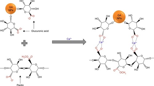
The alga polymeric adhesive is composed of alginate and polyphenol that crosslinked by Ca2+, which have been demonstrated for tissue engineering and many other biomedical applications.Citation38–Citation41 Inspired by this, the bioinspired hydrogels were synthesized from gum arabic, pectin, and divalent calcium ions (). The glucuronic acid at the surface of gum arabic nanoparticles could supply COO− groups. The pectin also contains COO− groups. Crosslinked by the divalent calcium ions, the egg-box-like structures could be formed by the above three components, and finally, the bioinspired hydrogels formed.
Figure 1 Chemical structure of the bioinspired hydrogels.
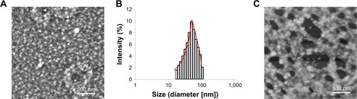
AFM is an important tool used to assess the surface morphology and complex three-dimensional (3D) nano- and microstructure of polysaccharides. The AFM images of the gum arabic in reveal its nanostructure with nanoparticles contained. The average size of the nanoparticles in gum arabic obtained from DLS was around 50 nm (), which was consistent with the AFM result. shows the nanoparticles composed of nanomorphological structure in the bioinspired hydrogels. The nanoparticles in the bioinspired hydrogels could assemble into a network nanostructure and then formed the hydrogels. It has been reported that the nanoparticle could not only enhance the stiffness of the nanocomposite but also increase the cohesive and adhesive forces of the nanocomposite through extra crosslinking and mechanical interlocking interactions.Citation42–Citation44 In other words, the nanoparticles from the gum arabic could be one of the key components to form network nanostructure and then crosslink into hydrogels. To prove this hypothesis, it has been demonstrated that all of the other samples such as individual pectin, divalent cation ions, pectin and divalent cation ions, gum arabic and divalent cation ions, and pectin and gum arabic formed only film structures without apparent network nanostructures. Therefore, they cannot form the hydrogels for wound healing applications. This unique network nanostructure from the bioinspired hydrogels could maintain a physiologically moist microenvironment at the wound site for promoting healing and formation of granulation tissue through the wound healing process.Citation45 The potential adhesive forces developed from the bioinspired hydrogels could also supply the adhesion effects at the wound site, which in turn enhanced the wound contraction and closure in the wound healing process. Moreover, this unique nanostructure could serve as an excellent carrier to accelerate the wound healing process.Citation46–Citation48
Figure 2 Morphological characterization of the bioinspired hydrogels.
Notes: AFM image (A) and DLS analysis (B) of the nanoparticles from gum arabic, the scale bar=500 nm. The nanostructure (network) investigated from the bioinspired hydrogels (C); the scale bar=500 nm.
Abbreviations: AFM, atomic force microscopy; DLS, dynamic light scattering.
Controlled releasing profile and cell proliferation
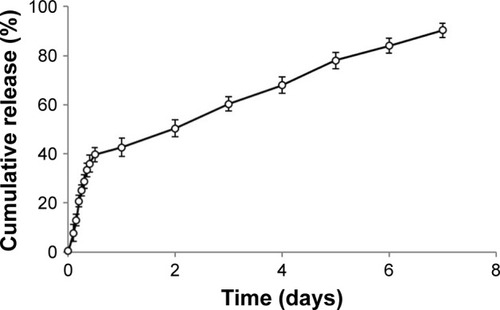
ELISA was utilized to obtain the controlled bFGF release profile from the bioinspired hydrogels (). The bFGF released from the bioinspired hydrogels displayed an initial burst release around 20% at 3 hours and around 40% at 12 hours. After that, a sustained and controlled release profile could be observed for the bFGF release (). It is well known that the bFGF can play as one of the major components in fibroblast cell proliferation in the early stage of wound healing process.Citation49 Therefore, the controlled burst release of bFGF from the non-crosslinking loosen section of the bioinspired hydrogels in this study could enhance the early-stage wound healing process. Moreover, the bFGF has also shown to be useful in chronic wound healing applications as well as the high-quality scar formation in the later stages of wound healing process.Citation50 On the other hand, the bFGF loaded inside the crosslinking section was released slowly could be due to the slow degradation of the egg-boxlike structures formed by the pectin, gum arabic, and Ca2+. So, the controlled and sustained release of bFGF from the bioinspired hydrogels could help the later stages of wound healing processes and have the potential to form scarless wounds finally.
Figure 3 The bFGF release profiles from the bioinspired hydrogels (n=3).
Abbreviation: bFGF, basic fibroblast growth factor.
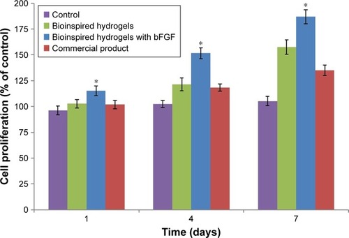
The L929 dermal fibroblast cells were seeded on different samples, including the bioinspired hydrogels with and without bFGF, to test the cell viability and growth through cell proliferation studies (). Compared with the control group without any treatment, the bioinspired hydrogels showed increased cell proliferation rates on days 4 and 7. Moreover, the cell proliferation situation from the bioinspired hydrogels and commercial product was similar on days 1 and 4. For the bioinspired hydrogels with bFGF, the cell growth and viability were significantly higher than all the other samples, including control, bioinspired hydrogels without bFGF, and the commercial product. The experimental results suggested that both bFGF and bioinspired hydrogels could be the important factors that can affect the dermal fibroblast cell proliferation. More importantly, the bioinspired hydrogels with bFGF system showed the best performance in cell proliferation analysis. As the illustration in the introduction,Citation29,Citation34 both Ca2+ and bFGF have the potential to enhance the fibroblast cell proliferation in wound healing processes, which is also compared with the data obtained in this study.
Figure 4 The dermal fibroblast cell proliferation on the bioinspired hydrogels with and without bFGF, negative control (non-treated), and positive control (commercial product; n=3).
Note: Significant differences between samples means are indicated; *P<0.05.
Abbreviation: bFGF, basic fibroblast growth factor.
In vitro wound scratch characterizations
For skin wound healing, cell migration is very important for the wound contraction and the later curing stages. Therefore, studying dermal fibroblast migration affected by different samples may help to target therapies for improved wound healing. The scratch assay method using the L929 dermal fibroblast cell line was used to evaluate the in vitro wound closure effects of the bioinspired hydrogels with bFGF (). At 12 hours, the processes of wound closure just started for the control and commercial product groups in fibroblast scratch assays. However, there is obvious wound closure for the bioinspired hydrogels at 12 hours. More importantly, the extent of wound closure of the bioinspired hydrogels with bFGF together for L929 scratch assays has the highest value. Furthermore, complete wound closure of all the scratches for the bioinspired hydrogels with bFGF together was seen in L929 cultures by 24 hours, compared with all the other samples, including control, bioinspired hydrogels without bFGF, and the commercial product. The bioinspired hydrogels with bFGF enhanced the rate of wound closure significantly at both 12 and 24 hours in L929 fibroblast cell lines. Recent advances using keratinocytes, dermal fibroblasts, and reconstructed epidermis in culture have identified the capacity of local calcium to modulate cell proliferation, maturation, and the creation of epidermal lipid barrier to speed up the wound healing process. These functions that accelerate the wound healing process make the bioinspired hydrogels with bFGF very promising hydrogels for wound healing applications.
Figure 5 The in vitro scratch assays of L929 dermal fibroblast cells treated with control, bioinspired hydrogels with and without bFGF, and commercial product.
Notes: The images (A) and the analyzed results (B) showed the extent of wound closure in scratch assays after 12 and 24 hours for different groups. Error bars indicate SD. Significant differences between samples means are indicated; *P<0.05.
Abbreviation: bFGF, basic fibroblast growth factor.

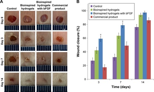
In vivo wound closure studies
To determine the wound closure efficacy of the bioinspired hydrogels with bFGF in vivo, full-thickness skin incisions were created on the back of mice, and then the prepared samples were applied at the wound sites. shows the images of the skin wound taken at different time intervals after treating with control, bioinspired hydrogels with and without bFGF, and the commercial product. There was no sign of inflammation or infection formation in material-covered wounds at different time points. It was observed that the growth of new epidermis was extended to the wound centers in all treated wound lesions, resulting in the reduced area of the wounds. Among the treatment groups, the bioinspired hydrogels with bFGF revealed the most accelerated wound contraction up to 14 days as compared with control, bioinspired hydrogels without bFGF, and the commercial product (). Particularly, the treatment of the bioinspired hydrogels with bFGF led to improved premature healing of the wounds. Three days after transplantation, the wound closure rate of hydrogels with bFGF together reached about 60%, while control, bioinspired hydrogels only, and the commercial product had lower wound closure rates (27%, 42%, and 17%, respectively). However, the bioinspired hydrogels without bFGF still had a valuable wound closure effects compared with the negative and positive controls. Therefore, both of the bioinspired hydrogels and the bFGF could enhance the wound healing in vivo. This result might be due to the material properties of the bioinspired hydrogels, such as nanomorphological structure and Ca2+. These properties could potentially maintain a physiologically moist microenironment and supply an adhesion effect at the wound site, enhancing the wound contraction and closure at the early stage of the wound healing process. Moreover, it is reported that bFGF could promote the fibroblasts proliferation that also could contribute to generating the contractile force at the wound area, and this process mostly occurs at the initial stage of a wound healing process. Both the cell migration that has been demonstrated in the in vitro study and the contractile force generated from the above theory could be the reason of the good performance of the bioinspired hydrogels with bFGF for wound healing applications.
Figure 6 The images (A) and the analyzed results (B) of in vivo wound closure studies for control, bioinspired hydrogels with and without bFGF, and commercial product. Error bars indicate SD.
Note: Significant differences between samples means are indicated; *P<0.05.
Abbreviation: bFGF, basic fibroblast growth factor.
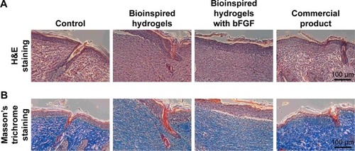
Histological analysis
Histology of wounds covered with the bioinspired hydrogels with bFGF and other samples on the 14th postoperative day is shown in . The surface of the wounds treated with bioinspired hydrogels with bFGF showed a complete formation of new epithelium, which can be observed in stained with H&E. The fibroblasts’ density can also be observed in in which the bioinspired hydrogels with bFGF-treated wound induced the highest fibroblast density. This wound also showed the formation of extracellular proteins with the highest density. The well-proliferated fibroblast and the well-produced collagens in the bioinspired hydrogels with bFGF-treated wound bed contribute to significantly enhance granulation tissue formation. Collagen is a predominant structural protein in skin, which is necessary to effectively reconstruct dermis tissue at the wound sites. After 14 days of treatment, retrieved tissues were processed for Masson’s trichrome staining to identify collagen. A more accumulation of the collagen fibers was observed in the wound tissue covered with the bioinspired hydrogels with bFGF, indicating that collagen deposition by the bioinspired hydrogels with bFGF was more mature than the other samples. Furthermore, the bioinspired hydrogels with bFGF-treated wounds showed more densely packed collagen fibers with a parallel arrangement of the collagen fibers in the extracellular matrix. In contrast, loosely packed collagen fibers with irregular arrangement were observed in other samples.
Figure 7 Histological images of H&E (A) and Masson’s trichrome (B) stained sections after 14 days of wound healing.
Abbreviation: bFGF, basic fibroblast growth factor.
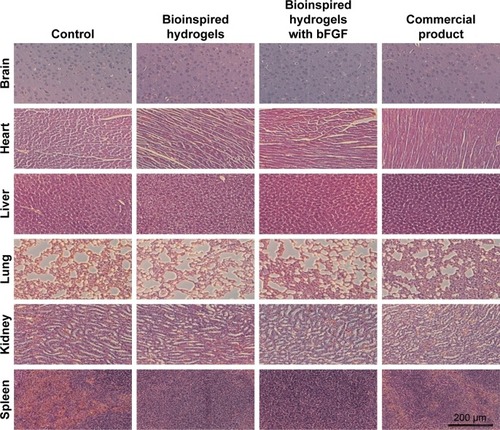
Finally, six major organs, ie, brain, heart, liver, lung, kidney, and spleen, from each experimental animal, were harvested on day 14 and H&E stained for histological evaluation (). The local administrated hydrogels could be accumulated in different organs through the blood circulation system, which could also induce some unexpected toxicities. A thorough examination of the H&E stained organ sections did not reveal any significant signs of toxicity, which further supports the utilization of bioinspired hydrogels with bFGF for wound healing applications.
Figure 8 H&E stained organs revealed no significant signs of toxicity.
Abbreviation: bFGF, basic fibroblast growth factor.
Conclusion
This work demonstrated that bioinspired hydrogels based on the chemical structure and nanomorphology of alga adhesive using gum arabic, pectin, and calcium combined with bFGF showed great promise for wound healing applications. The bioinspired hydrogels with unique nanomorphology, promising biocompatibility, controlled growth factor release, and good cell proliferation make them ideal to promote wound healing. The increased fibroblast cells’ proliferation indicates that the hydrogels with bFGF could potentially enhance the cell migration for wound healing. The experimental data of in vitro and in vivo wound closure show that the bioinspired hydrogels with bFGF could significantly enhance wound re-epithelialization, collagen deposition, and contraction, which accelerated the wound healing process. The histological organ analysis shows that the bioinspired hydrogels with bFGF did not induce noticeable toxicities. Overall, the bioinspired hydrogels with bFGF demonstrate great promise to be utilized for wound healing applications.
Disclosure
The authors report no conflicts of interest in this work.
References
- GuoSDiPietroLAFactors affecting wound healingJ Dent Res201089321922920139336
- MaalejHMoallaDBoissetCRhelogical, dermal wound healing and in vitro antioxidant properties of exopolysaccharide hydrogel from Pseudomonas stutzeri AS22Colloids Surf B Biointerfaces201412381482425454656
- WenYLiuWBagiaCAntibody-functionalized peptidic membranes for neutralization of allogeneic skin antigen-presenting cellsActa Biomater201410114759476725117952
- WuJWuDMutschlerMAChuCCCationic hybrid hydrogels from amino-acid-based poly(ester amide): fabrication, characterization, and biological propertiesAdv Funct Mater2012221838153823
- WuJZhaoXWuDChuCCDevelopment of a biocompatible and biodegradable hybrid hydrogel platform for sustained release of ionic drugsJ Mater Chem B201423866606668
- GongXSelf-assembly technique for biomedical applicationsNano LIFE2015521542002
- AramwitP1-– Introduction to biomaterials for wound healing A2 –ÅgrenMagnus SWound Healing BiomaterialsCambridge, UKWoodhead Publishing2016338
- WenYKolonichHRKruszewskiKMGiannoukakisNGawaltESMengWSRetaining antibodies in tumors with a self-assembling injectable systemMol Pharm20131031035104423419203
- MohamedAXingMNanomaterials and nanotechnology for skin tissue engineeringInt J Burns Trauma201221294122928165
- ZhangZGupteMJJinXMaPXInjectable peptide decorated functional nanofibrous hollow microspheres to direct stem cell differentiation and tissue regenerationAdv Funct Mater201525335036026069467
- LiYZhangWBHsiehIFBreaking symmetry toward nonspherical Janus particles based on polyhedral oligomeric silsesquioxanes: molecular design, “click” synthesis, and hierarchical structureJ Am Chem Soc201113328107121071521682260
- GhavamiNejadARajan UnnithanARamachandra Kurup SasikalaAMussel-inspired electrospun nanofibers functionalized with size-controlled silver nanoparticles for wound dressing applicationACS Appl Mater Interfaces2015722121761218325989513
- GuptaBAgarwalRAlamMS9 – Hydrogels for wound healing applications A2 –RimmerSteveBiomedical HydrogelsCambridge, UKWoodhead Publishing2011184227
- ElsnerJJShefy-PelegAZilbermanMNovel biodegradable composite wound dressings with controlled release of antibiotics: microstructure, mechanical and physical propertiesJ Biomed Mater Res B Appl Biomater201093242543520127990
- LinJLiCZhaoYHuJZhangLMCo-electrospun Nanofibrous membranes of collagen and zein for wound healingACS Appl Mater Interfaces2012421050105722242622
- XieZParasCBWengHDual growth factor releasing multifunctional nanofibers for wound healingActa Biomater20139129351935923917148
- RibeiroMPEspigaASilvaDDevelopment of a new chitosan hydrogel for wound dressingWound Repair Regen200917681782419903303
- BittonRBianco-PeledHNovel biomimetic adhesives based on algae glueMacromol Biosci20088539340018213593
- BittonRBen-YehudaMDavidovichMStructure of algal-born phenolic polymeric adhesivesMacromol Biosci20066973774616967478
- BittonRBerglinMElwingHThe influence of halide-mediated oxidation on algae-born adhesivesMacromol Biosci20077121280128917724788
- SunJYZhaoXIlleperumaWRKHighly stretchable and tough hydrogelsNature201248913313622955625
- GiustoGVercelliCCominoFCaramelloVTursiMGandiniMA new, easy-to-make pectin-honey hydrogel enhances wound healing in ratsBMC Complement Altern Med201717126628511700
- LinHYChenHHChangSHNiTSPectin-chitosan-PVA nanofibrous scaffold made by electrospinning and its potential use as a skin tissue scaffoldJ Biomater Sci Polym Ed201324447048423565688
- MunarinFTanziMCPetriniPAdvances in biomedical applications of pectin gelsInt J Biol Macromol201251468168922776748
- NinanNMuthiahMParkIKElainAThomasSGrohensYPectin/carboxymethyl cellulose/microfibrillated cellulose composite scaffolds for tissue engineeringCarbohydr Polym201398187788523987424
- SamyWMGhoneimAIElgindyNANovel microstructured sildenafil dosage forms as wound healing promotersExpert Opin Drug Deliv201411101525153624940608
- AliBHZiadaABlundenGBiological effects of gum arabic: a review of some recent researchFood Chem Toxicol20094711818672018
- ClarkDTGaziMICoxSWEleyBMTinsleyGFThe effects of Acacia arabica gum on the in vitro growth and protease activities of periodontopathic bacteriaJ Clin Periodontol19932042382438473532
- LansdownABCalcium: a potential central regulator in wound healing in the skinWound Repair Regen200210527128512406163
- LeeSHEliasPMProkschEMenonGKMao-QuiangMFeingoldKRCalcium and potassium are important regulators of barrier homeostasis in murine epidermisJ Clin Invest19928925305381737844
- DlugoszAAYuspaSHProtein kinase C regulates keratinocyte transglutaminase (TGK) gene expression in cultured primary mouse epidermal keratinocytes induced to terminally differentiate by calciumJ Investig Dermatol199410244094147908680
- ArulVKarthaRJayakumarRA therapeutic approach for diabetic wound healing using biotinylated GHK incorporated collagen matricesLife Sci200780427528417049946
- BarrientosSStojadinovicOGolinkoMSBremHTomic-CanicMPerspective article: growth factors and cytokines in wound healingWound Repair Regen200816558560119128254
- EdelmanERMathiowitzELangerRKlagsbrunMControlled and modulated release of basic fibroblast growth factorBiomaterials19911276196261742404
- KreugerJSalmivirtaMSturialeLGiménez-GallegoGLindahlUSequence analysis of heparan sulfate epitopes with graded affinities for fibroblast growth factors 1 and 2J Biol Chem200127633307443075211406624
- CaiSLiuYZheng ShuXPrestwichGDInjectable glycosaminoglycan hydrogels for controlled release of human basic fibroblast growth factorBiomaterials200526306054606715958243
- SaliernoMJGarcíaAJdel CampoAPhoto-activatable surfaces for cell migration assaysAdv Funct Mater2013234859745980
- DimartinoSLirIHaberMAzhariRCharacterization of Biomimetic Adhesives from the Red Alga Gracilaria Conferta for Biomedical ApplicationsBiological and Biomimetic Adhesives: Challenges and OpportunitiesCambridge, UKThe Royal Society of Chemistry2013117131
- de Jesus RaposoMFde MoraisAMde MoraisRMMarine polysaccharides from algae with potential biomedical applicationsMar Drugs20151352967302825988519
- NoeliaFMaria JesusGMDanielaREduardaFHerminiaDMarisaFAlgae polysaccharides’ chemical characterization and their role in the inflammatory processCurr Med Chem201724214917527804878
- TeslerABKimPKolleSHowellCAhanotuOAizenbergJExtremely durable biofouling-resistant metallic surfaces based on electrodeposited nanoporous tungstite films on steelNat Commun20156864926482559
- AhmedMAKandilUFShakerNOHashemAIThe overall effect of reactive rubber nanoparticles and nano clay on the mechanical properties of epoxy resinJ Radiat Res Appl Sci201584549561
- RajaSNOlsonACLimayeAInfluence of three-dimensional nanoparticle branching on the Young’s modulus of nanocomposites: effect of interface orientationProc Natl Acad Sci U S A2015112216533653825971729
- ThoniyotPTanMJKarimAAYoungDJLohXJNanoparticle-hydrogel composites: concept, design, and applications of these promising, multi-functional materialsAdv Sci (Weinh)201521–2140001027980900
- MottaGJCalcium alginate topical wound dressings: a new dimension in the cost-effective treatment for exudating dermal wounds and pressure soresOstomy Wound Manage19892552562610818
- WangYYiSSunLHuangYZhangMCharge-selective fractions of naturally occurring nanoparticles as bioactive nanocarriers for cancer therapyActa Biomater201410104269428424952072
- WangYSunLYiSHuangYLenaghanSCZhangMNaturally occurring nanoparticles from arthrobotrys oligospora as a potential immunostimulatory and antitumor agentAdv Funct Mater2013231721752184
- RiceJJMartinoMMDe LaporteLTortelliFBriquezPSHubbellJAEngineering the regenerative microenvironment with biomaterialsAdv Healthc Mater201321577123184739
- KobayashiFMatsuzakaKInoueTThe effect of basic fibroblast growth factor on regeneration in a surgical wound model of rat sub-mandibular glandsInt J Oral Sci201681162327025261
- AkitaSAkinoKHiranoABasic fibroblast growth factor in scarless wound healingAdv Wound Care (New Rochelle)201322444924527324