
Abstract
Objective
The objective of this study was to verify and confirm the oxidative-mediated hepatotoxicity, inflammatory liver damage, and oxidative stress induced by intraperitoneal administration of gold nanoparticles (GNPs) in vivo; characterize the effect of different natural antioxidants on these hazardous changes; and finally choose the most powerful antioxidant among these different natural antioxidants.
Methods
Ten-nanometer GNPs were dissolved in aqueous solution of 0.01% concentration. A dose of 50 µL of 10 nm GNPs was administered intraperitoneally for 7 days to the rats, whereas the antioxidants were orally administered for the same time period. The antioxidants used in the study were vitamin E (Vit E), α-lipoic acid (ALA), quercetin (Qur), arginine (Arg), and melanin. Forty Wistar-Kyoto male rats were used. Rats were arbitrarily divided into seven groups after acclimatization for 1 week. For serum separation, blood samples were obtained from each animal. Serum liver function markers and tissue oxidative stress and lipid proxidation biomarkers were assessed.
Results
The increase in the levels of gamma-glutamyl transferase, alkaline phosphatase, total protein, alanine aminotransferase, and total bilirubin in the serum of rats and the increase of malondialdehyde in the hepatic tissue and decrease in reduced glutathione when compared with the control in this study confirmed the ability of GNPs to cause hazardous effects.
Conclusion
Treatment of rats with Vit E, ALA, Qur, Arg, and melanin along with GNPs significantly inhibited the inflammatory liver damage, lipid peroxidation, and the oxidative stress induced by GNPs in vivo, but with different responses due to their evaluated normalization values, and it has been confirmed that melanin is the most powerful antioxidant among these different natural antioxidants, ie, it has the most effective potential role against the hepatic inflammatory damage, oxidative stress, and lipid peroxidation.
Introduction
Toxicological studies show that small-sized gold nanoparticles (GNPs) had adverse impacts on human health and other environmental biological species. Numerous other studies have reported that contact to small-sized GNPs causes larger inflammatory and cytotoxic reactions when compared with large-sized GNPs of same weight and concentration.Citation1,Citation4
The exposure to GNPs induced inflammatory, vein intima disruption, lipid change, enlargement of stellate macrophages, increase in absorption of water by hepatocytes, cytoplasmic vacuolization, and death of most or all of the cells.Citation1,Citation3 Moreover, cardiac tissue damage, pneumonia, fibrosis, chronic inflammatory damage all are subject to the volume and length of exposure.Citation5,Citation6
The induced toxicity by GNPs was attributed to alterations in the physical and chemical characteristics of the nanoparticles, mass, form, substance composition, assemblage, great precise surface area, and their ability to dissolve in solvent.Citation7–Citation9 The diseased effects triggered by GNPs lead to swelling of liver, kidney, heart, and lung tissues, including oxidative stress and DNA damage.Citation1,Citation7
Vitamin E (Vit E) may reduce the progression of the hepatic inflammatory damage, which is notably accelerated by oxidative stress, in addition to reducing the cardiac disease linked to chronic kidney failure and urine in the blood. Because of the oxygen-foraging ability of Vit E, it inhibits fatty acid peroxidation and the unsaturated membrane fats.Citation10 It can also inhibit the increase in ROS production and further movement to the nucleus and harm to the DNA. Earlier studies have stated that oxidative destruction caused by various pathological situations caused by the formation of ROS can be inhibited by Vit E supplementation.Citation11,Citation12
α-Lipoic acid (ALA) has been reported to moderately inhibit kidney cysteine reduction and elevate liver cysteine and glutathione (GSH) levels. The production of GSH can be boasted by ALA, which is the major antioxidant inside our cells.Citation13 Post ALA treatment shows reduction in the level of TNF-α when compared to animals injected with zinc oxide nanoparticles (ZnO-NPs). Strong anti-inflammatory actions in vitro and in vivo are demonstrated by ALA by the reduction of inflammatory intermediaries; pro-inflammatory cytokine formation and C-reactive protein (CRP).Citation14
Quercetin (Qur) is an antioxidant, and anti-inflammatory/anti-allergy, which stops the discharge of histamine and other inflammatory proxies in the body.Citation10 Arginine (Arg) has several biological processes and defensive influences and is believed to be temporarily important mostly in inflammatory and oxidative stress conditions.Citation15 Arg can also act as an underlying substance for polyamine production, which has been recognized to be strongly participating in protein production enrichment.Citation13 It has been reported that treatment with either Qur or Arg as protective agents may be useful against DNA, liver, and kidney damage caused by ZnO-NPs.Citation16
Melanin is present in different organs, tissues, and blood of living organisms. Many diseases are accompanied by an increase or decrease in melanin production. It plays essential role in shielding the cell’s nuclei DNA against the harm caused by the ultraviolet radiation. Melanin shows non-oxidative stress and scavenging activities for the free radicals.Citation17–Citation19 In the skin, both pigments function as foragers for free radicals and other oxygen-containing species.Citation13–Citation16 Early studies have shown that melanin combines with chemical species such as oxygen,Citation17,Citation18 hydroxyl radical,Citation20 and hyperoxide.Citation19,Citation20 The ability of melanin to prevent fatty acid peroxidation has established its function as an antioxidant.Citation21
Some researchers have reported that disease-causing mechanisms triggered by in vivo administration of GNPs are dominated by oxidative stress, programmed cell death, and DNA destruction.Citation23,Citation24 Thus, a defensive approach to reduce the generation of inflammatory intermediaries could ameliorate both liver injury and dysfunction.
For the use of GNPs in drug delivery and cancer therapy, knowledge about inhibiting induced toxicity is necessary. The prevention of toxicity triggered by treatment with small-sized GNPs is yet to be known and identified. Thus, this study aimed to confirm and prove the toxicity induced by the administration of GNPs intraperitoneally in vivo, to investigate the protective role of different natural antioxidants such as Vit E, ALA, Qur, Arg, and melanin in the liver inflammatory damage, oxidative stress, and lipid peroxidation, and finally to choose the most powerful antioxidant among these different natural antioxidants.
Materials and methods
GNPs and dosing
Ten-nanometer GNPs (products MKN-Au-010; M K IMPEX Corp; Divn MK Nano, Mississauga, ON, Canada) of spherical shape, determined by the scanning electron microscope, were used in this study; the estimated mean size for GNPs was 9.45±1.33 nm, and GNPs were distributed homogenously in the solution.Citation1–Citation3 The rats were administered GNPs intraperitoneally at a dose of 50 µL for 7 consecutive days, whereas the doses of the different natural antioxidants that were orally administered separately for the same time period were as follows: Vit E, 100 mg/kg body weight (BW)/day; ALA, 200 mg/kg BW/day; Qur, 200 mg/kg/day; Arg, 200 mg/kg/day, and melanin, 100 mg/kg BW/day.
Animals
Forty Wistar-Kyoto male rats weighing 220–240 g (12 weeks old) were obtained from College of Pharmacy Animal house, King Saud University (KSU). Rats were kept in cages under homogenous conditions (temperature 22°C±5°C, humidity 55%±5%, and 12-hour light/dark cycle). The rats had unrestricted right to use of water and fed with usual rat diet. The KSU Animal Use Committee approved the animal experiments, and all experiments were conducted in accordance with the committee’s guidelines approved by KSU Local Animal Care.
Experimental design
All natural antioxidants used in this study were purchased from Sigma-Aldrich Co. (St Louis, MO, USA). Quantification of the purity and content of the prepared melanin was according to the study by ElObeid et al.Citation25 After acclimatization for a period of 1 week, the rats were denied access to food for 24 hours before treatment and randomly separated into seven groups. The biochemical parameters were investigated in the liver of these groups of rats: control group (G1: n=10), GNP group (G2: GNP-intoxicated rats were left untreated; n=6), GNPs + Vit E group (G3: n=6), GNPs + ALA group (G4: n=6), GNPs + Qur group (G5: n=6), GNPs + Arg group (G6: n=6), and GNPs + melanin group (G7: n=6). The rats were administered 50 µL of GNPs intraperitoneally for 7 consecutive days.
Blood sampling and tissue preparation
After final dose administration, the rats were denied of food for 12–14 hours and were euthanized. The blood sample from each animal was obtained using disinfected collection tubes. Serum was separated using a centrifuge at the speed of 3,000 rpm for 10 minutes. Serum was placed in the disinfected tubes and preserved at a temperature of −80°C for several biochemical estimations. The midline incision method was use to collect liver tissues, and then the liver tissues were washed in icy normal saline, blended, ice-covered in liquid nitrogen, and conserved at a temperature of −80°C for biochemical tissue analysis for estimating the reduced GSH and the malondialdehyde (MDA) content.
Serum liver function markers
To evaluate the liver function markers, the levels of gamma-glutamyl transferase (GGT), total bilirubin (TBIL), ALP, aspartate amino transferase (AST), alanine aminotransferase (ALT), and total protein in serum were determined by a biochemical auto analyzer (Type 7170; Hitachi Ltd., Tokyo, Japan).
Oxidative stress biomarkers
Determination of reduced GSH level (µg/mL)
The determination of GSH in hepatocytes was carried out enzymatically according to the modified procedure of Griffith.Citation22
Determination of MDA
MDA level and fatty acid peroxidation index in several organs of rats were analyzed spectrophotometrically as described earlier.Citation23
Statistical analysis
Data are shown as mean normalization (percentage change) for all the samples. To determine the differences between the mean values of the different groups, one-way ANOVA was done. P≤0.05 was used as the limit of statistical significance.
Results
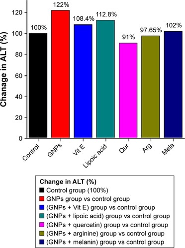
ALT level
demonstrates the activity of liver ALT. ALT was significantly increased to 122% in the G2 rats compared with the control rats (100%). G3, G4, G5, G6, and G7 rats show significant decrease compared with G2, and non-significant change compared to G1. The evaluated normalization values in G3, G4, G5, G6, and G7 rats were 108.4%, 112.8%, 91%, 97.7%, and 102%, respectively. It became evident from that melanin is the most powerful antioxidant among the other different antioxidants used such as Vit E, ALP, Qur, and Arg.
Figure 1 The effect of GNPs and different antioxidant treatments on ALT level in rats.
Abbreviations: ALT, alanine aminotransferase; Arg, arginine; GNPs, gold nanoparticles; Qur, quercetin; Mela, melanin; Vit E, vitamin E.
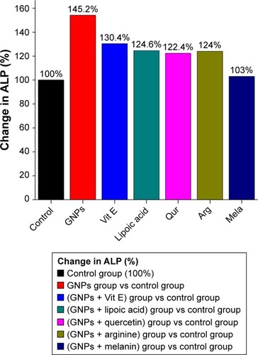
ALP level
shows the activity of liver ALP. ALP significantly increased to normalized value 145.2% in G2 rats compared with the control rats (100%). G3, G4, G5, G6, and G7 rats show significant decrease compared with G2, and non-significant change compared to G1. The evaluated normalization values in G3, G4, G5, G6, and G7 rats were 130.4%, 124.6%, 122.4%, 124%, and 103%, respectively. Also, melanin is proved to be the most powerful antioxidant among the other different natural antioxidants.
Figure 2 The effect of GNPs and different antioxidant treatments on ALP level in rats.
Abbreviations: Arg, arginine; GNPs, gold nanoparticles; Qur, quercetin; mela, melanin; Vit E, vitamin E.
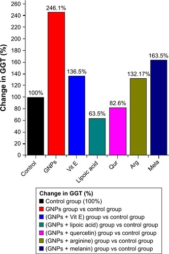
GGT level
As shown in , the liver enzyme GGT significantly elevated G2 to evaluated normalized value 246.12% compared with G1 (100%). G3, G4, G5, G6, and G7 rats show significant decrease compared with G2, and non-significant change compared to G1. The evaluated normalization values in G3, G4, G5, G6, and G7 rats were 136.5%, 63.5%, 82.64%, 132.2%, and 163.5%, respectively. The ALP or Qur with GNPs significantly lowered the marked increase in the level of GGT even below the control value. Moreover, the treatment of GNPs rats with the abovementioned antioxidants directly reveal their protective effect over the disturbance in the GGT level induced by GNPs.
Figure 3 The effect of GNPs and different antioxidants treatment on GGT level in rats.
Abbreviations: Arg, arginine; GGT, gamma-glutamyl transferase; GNPs, gold nanoparticles; Qur, quercetin; Mela, melanin; Vit E, vitamin E.
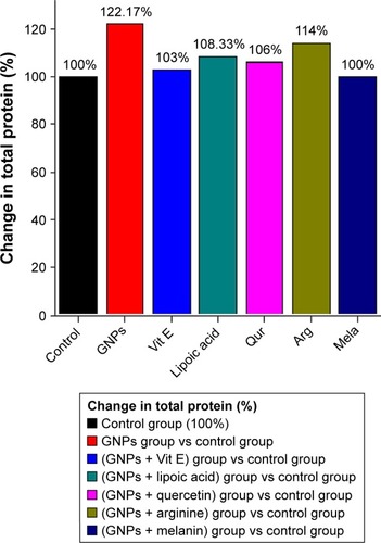
Total protein level
Data presented in show a notable increase in the total protein level in the serum of G2 rats evaluated to have a normalized value of 122.2% compared with G1 rats (100%). G3, G4, G5, G6, and G7 rats showed significant decrease compared with G2, and non-significant change compared to G1. The estimated normalization values in G3, G4, G5, G6, and G7 rats were 103%, 108.3%, 106%, 114%, and 100%, respectively. Also, melanin proved to be the most powerful antioxidant.
Figure 4 The effect of GNPs and different antioxidants treatment on total protein level in rats.
Abbreviations: Arg, arginine; GNPs, gold nanoparticles; Qur, quercetin; Mela, melanin; Vit E, vitamin E.
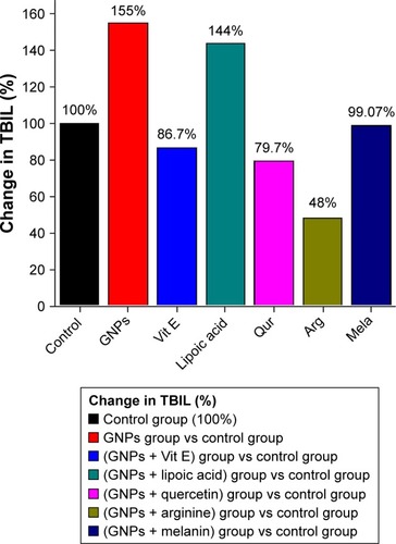
TBIL level
shows a major rise in the total protein level in the serum of G2 rats evaluated to have a normalized value of 158% compared with the control rats (100%). G3, G4, G5, G6, and G7 rats showed significant decrease compared with G2, and non-significant change compared to G1. The estimated normalization values in G3, G4, G5, G6, and G7 rats were 86.7%, 144%, 79.7%, 48%, and 90.1%, respectively, compared to the control rats. Also, melanin proved to be the most powerful antioxidant.
Figure 5 The effect of GNPs and different antioxidants treatment on TBIL level in rats.
Abbreviations: Arg, arginine; GNPs, gold nanoparticles; Qur, quercetin; Mela, melanin; TBIL, total bilirubin; Vit E, vitamin E.
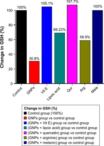
GSH level in rat liver homogenate
shows the GSH levels in liver homogenate of control, GNPs, and antioxidant treatment groups. GSH was significantly reduced in G2 rats evaluated to have a normalized value of 30.8% compared with the control rats (100%). G3, G4, G5, G6, and G7 rats showed significant increase compared with G2, and non-significant change compared with G1. The estimated normalization values in G3, G4, G5, G6, and G7 rats were 105.1%, 68.2%, 107.7%, 58.9%, and 100%, respectively. Also, melanin proved to be the most powerful antioxidant.
Figure 6 Effect of GNPs and different antioxidants treatments on liver GSH level in rats.
Abbreviations: Arg, arginine; GNPs, gold nanoparticles; GSH, glutathione; Qur, quercetin; Mela, melanin; Vit E, vitamin E.
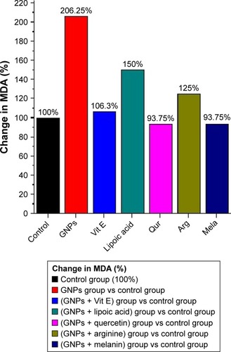
Lipid peroxidation process in rat liver homogenate
shows an obvious significant elevation in MDA activity in G2 rats evaluated to have a normalized value of 206.3% compared with G1 rats (100%). G3, G4, G5, G6, and G7 rats showed significant decrease compared with G2, and non-significant change compared with G1. The estimated normalization values in G3, G4, G5, G6, and G7 rats were 106.3%, 150%, 93.8%, 125%, and 93.8%, respectively. Also, melanin proved to be the most powerful antioxidant.
Figure 7 Effect of GNPs and different antioxidants on liver MDA level in rats.
Abbreviations: Arg, arginine; GNPs, gold nanoparticles; Qur, quercetin; MDA, malondialdehyde; Mela, melanin; Vit E, vitamin E.
Discussion
The different natural antioxidants used in this study were Vit E, ALA, Qur, Arg, and melanin. All these antioxidants have proved their beneficial protective role against the hepatic inflammatory damage, lipid peroxidation, and oxidative stress induced by intraperitoneal administration of GNPs in vivo.
It has been reported that Vit E prevents the progression of liver diseases accelerated by the oxidative stress, protects against the lipid peroxidation, inhibits most of the metal-induced injuries in vitro and in vivo,Citation24 neutralizes the unsaturated membrane lipids and the lipid peroxidation due to its oxygen foraging ability, and intercepts the production of ROS that can damage the DNA of the nucleus. Vit E supplementation shields against oxidative harm triggered by several disease conditions by blocking the generation of ROS.Citation11,Citation12
ALA regenerates vitamin C from its oxidized state, dehydroascorbic acid, and regenerates other antioxidants. It enhances GSH synthesis, the leading antioxidant inside living cells.Citation13 ALA administration decreases the TNF-α level when compared with ZnO-NP-treated animals. ALA exhibits robust anti-inflammatory actions in vitro and in vivo by decreasing inflammatory intermediaries such as pro-inflammatory cytokine formation and CRP.Citation14
In this study, the hepatic tissue GSH levels were significantly reduced, whereas the MDA levels were significantly increased after administration of GNPs. Our data were in consistent with those reported by ElObeid et al.Citation25 GSH effectively wipes up all kinds of poisons and free radicals. Earlier results have elucidated the regulatory influence in the GSH level in animals injected with either Vit E or ALA or post ZnO-NPs administration. MDA the main product of fatty acid peroxidation has been confirmed to function as mutagen and carcinogen and causes DNA damage by the production of deoxyguanosine, deoxyadenosine, and deoxycytidine by triggering the signal transduction pathways leading to cell death or apoptosis.Citation26
The used agent Vit E or ALA plays effective hepatoprotective role against liver dysfunction caused by nanoparticle toxicity. Co-administration of ALA and Vit E to rats injected with ZnO-NPs successfully secures their livers from DNA destruction.Citation27
Findings from this study show that GNPs induce hepatic liver damage as evidenced by the significant increase in the serum liver markers in injected rats compared with rats not injected. These findings indicate that liver is a target organ for small-sized GNP toxicity. Our findings were similar to those of the previous studies.Citation28–Citation30
Our results were in consistent with those reported previously, in which Qur reduces inflammatory damage by inhibiting oxidative stress and cytokine production; it possesses anti-inflammatory effects in vivo,Citation16,Citation31 protects against oxidative damage,Citation17,Citation18 ameliorates oxidative stress in the liver and brain tissues, has antioxidant activity and ROS scavenging properties,Citation20 and inhibits DNA fragmentation and increases DNA wound synthesis.Citation28,Citation29 It has been reported that small-sized NPs intermingle with the DNA in such a manner that may likely be the cause of high harmfulness of these minute particles toward human tumor, since it has been established that breaks in DNA double-strand might give rise to cancer.Citation1–Citation3
In our experiments, the expected pathway involved in the hepatotoxicity induced by GNPs might arise from liver vacuolar degeneration and necrosis arising from the production of ROS, in addition to, an increase in the indicator enzymes such as ALT, ALP, GGT, and also total protein in the serum.
To completely confirm and ensure the hepatoxicity induced by GNPs, additional experiments related to DNA damage, endogenous antioxidants, ROS, and gene cholestasis should be performed. This study also suggests that to understand the mechanism of action of GNPs, any generic oxidative stress-related assay could be carried out with and without NPs, in addition to, the oxidation of protein corona after the incubation of the NPs.
Conclusion
This study highlights hepatic inflammatory damage, lipid peroxidation, and oxidative stress induced by intraperitoneal administration of GNPs in vivo, which were confirmed by the significant increase in serum liver function markers such as ALT, ALP, GGT, and total protein, in addition to significant increase in the hepatic tissue lipid peroxidation biomarker MDA levels in combination with the significant reduction in antioxidant biomarker GSH levels. The treatment with different natural antioxidants successively alleviated the elevation in the serum liver function markers and the tissue oxidative stress biomarker MDA, while it elevated the tissue GSH level. Moreover, the antioxidants Vit E, ALA, Qur, Arg, and melanin markedly reduced the inflammatory response levels, lipid peroxidation, and oxidative stress, but with different normalization values, which might be attributed to the different responses induced by the serum liver markers and the liver tissue biomarkers. It has also confirmed that melanin is the most powerful antioxidant by its evaluated normalization value among these different natural antioxidants, ie, melanin has the most effective potential role against the hepatic inflammatory damage, oxidative stress, and lipid peroxidation.
Acknowledgments
The authors would like to extend their sincere appreciation to the Deanship of Scientific Research at King Saud University for its funding of this research by the research Group Project No RGP-285.
Disclosure
The authors report no conflicts of interest in this work.
References
- AbdelhalimMAJarrarBMHistological alterations in the liver of rats induced by different gold nanoparticle sizes, doses and exposure durationJ Nanobiotechnology201210522276919
- AbdelhalimMAJarrarBMRenal tissue alterations were size-dependent with smaller ones induced more effects and related with time exposure of gold nanoparticlesLipids Health Dis20111016321936889
- AbdelhalimMAJarrarBMGold nanoparticles induced cloudy swelling to hydropic degeneration, cytoplasmic hyaline vacuolation, polymorphism, binucleation, karyopyknosis, karyolysis, karyorrhexis and necrosis in the liverLipids Health Dis20111016621939512
- NelAXiaTMädlerLLiNToxic potential of materials at the nano-levelScience2006311576162262716456071
- AbdelhalimMAExposure to gold nanoparticles produces cardiac tissue damage that depends on the size and duration of exposureLipids Health Dis20111020522073987
- AbdelhalimMAGold nanoparticles administration induces disarray of heart muscle, hemorrhagic, chronic inflammatory cells infiltrated by small lymphocytes, cytoplasmic vacuolization and congested and dilated blood vesselsLipids Health Dis20111023322151883
- LovernSBKlaperRDaphnia magna mortality when exposed to titanium dioxide and fullerene (C60) nanoparticlesEnviron Toxicol Chem20062541132113716629153
- FranklinNMRogersNJApteSCBatleyGEGaddGECaseyPSComparative toxicity of nanoparticulate ZnO, bulk ZnO, and ZnCl2 to a freshwater microalga (Pseudokirchneriella subcapitata): the importance of particle solubilityEnviron Sci Technol200741248484849018200883
- WangZZhouJFanJEffect of rapamycin alone and in combination with sorafenib in an orthotopic model of human hepatocellular carcinomaClin Cancer Res200814165124513018698030
- FryerMJVitamin E as a protective antioxidant in progressive renal failureNephrology200051–217
- QureshiAAReisJCQureshiNPapasianCJMorrisonDCSchaeferDMδ-Tocotrienol and quercetin reduce serum levels of nitric oxide and lipid parameters in female chickensLipids Health Dis2011103921356098
- TahanGAytacEAytekinHVitamin E has a dual effect of anti-inflammatory and antioxidant activities in acetic acid-induced ulcerative colitis in ratsCan J Surg201154533333821933527
- VasdevSFordCAParaiSLongerichLGadagVDietary alpha-lipoic acid supplementation lowers blood pressure in spontaneously hypertensive ratsJ Hypertens200018556757310826559
- ZhangWJWeiHHagenTFreiBAlpha-lipoic acid attenuates LPS-induced inflammatory responses by activating the phosphoinositide 3-kinase/Akt signaling pathwayProc Natl Acad Sci U S A2007104104077408217360480
- HuangCCTsaiSCLinWTPotential ergogenic effects of L-arginine against oxidative and inflammatory stress induced by acute exercise in aging ratsExp Gerontol200843657157718424033
- FaddahLMAbdel BakyNAAl-RasheedNMRole of quercetin and arginine in ameliorating nano zinc oxide-induced nephrotoxicity in ratsBMC Complement Altern Med20121216022551254
- BridelliMGCiatiACrippaPRBinding of chemicals to melanins re-examined: adsorption of some drugs to the surface of melanin particlesBiophys Chem2006119213714516139945
- MeredithPSarnaTThe physical and chemical properties of eumelaninPigment Cell Res200619657259417083485
- PernaGFrassanitoMCPalazzoGFluorescence spectroscopy of synthetic melanin in solutionJ Lumin200912914449
- HerrlingTJungKFuchsJThe role of melanin as protector against free radicals in skin and its role as free radical indicator in hairSpectrochim Acta A Mol Biomol Spectrosc20086951429143517988942
- El-ObeidAEl TahirKEAbdelhalimMAHaseebAProtective action of herbal melanin against carbon tetrachloride induced HepatotoxicityInternational Journal of Chemical Engineering20152116
- GriffithOWDetermination of glutathione and glutathione disulfide using glutathione reductase and 2-vinylpyridineAnal Biochem198010612072127416462
- UtleyHGBernheimFHochsteinPEffect of sulfhydryl reagents on peroxidation in microsomesArch Biochem Biophys196711812932
- ValkoMMorrisHCroninMTMetals, toxicity and oxidative stressCurr Med Chem200512101161120815892631
- ElobeidASKamal-EldinAAbdelhalimMAKHaseebAMPharmacological properties of melanin and its function in healthBasic Clin Pharmacol Toxicol2017120651552228027430
- NiedernhoferLJDanielsJSRouzerCAGreeneREMarnettLJMalondialdehyde, a product of lipid peroxidation, is mutagenic in human cellsJ Biol Chem200327833314263143312775726
- AsukuOAtawodiSEOnyikeEAntioxidant, hepatoprotective, and ameliorative effects of methanolic extract of leaves of Grewia mollis Juss. on carbon tetrachloride-treated albino ratsJ Med Food2012151838821877945
- GurrJRWangASChenCHJanKYUltrafine titanium dioxide particles in the absence of photoactivation can induce oxidative damage to human bronchial epithelial cellsToxicology20052131–2667315970370
- WilmsLCHollmanPCBootsAWKleinjansJCProtection by quer-cetin and quercetin-rich fruit juice against induction of oxidative DNA damage and formation of BPDE-DNA adducts in human lymphocytesMutat Res20055821–215516215781220
- ZhangXJChinkesDLWuZHerndonDNEnternal [corrected] argi-nine supplementation stimulates DNA synthesis in skin donor woundClin Nutr201130339139621277659
- Baky NaARole of quercetin and L-arginine in alleviating zinc oxide nanoparticle hepatotoxicity in ratsChiang Mai J Sci2013404577592