
Abstract
Background
Tendon-derived stem cells (TDSCs) are key factors associated with regeneration and healing in tendinopathy. The aim of this study was to investigate the effects of mechanical stiffness and topographic signals on the differentiation of TDSCs depending on age and pathological conditions.
Materials and methods
We compared TDSCs extracted from normal tendon tissues with TDSCs from tendinopathic Achilles tendon tissues of Sprague Dawley rats in vitro and TDSCs cultured on nanotopographic cues and substrate stiffness to determine how to control the TDSCs. The tendinopathy model was created using a chemical induction method, and the tendon injury model was created via an injury-and-overuse method. Norland Optical Adhesive 86 (NOA86) substrate with 2.48 GPa stiffness with and without 800 nm-wide nanogrooves and a polyurethane substrate with 800 nm-wide nanogrooves were used.
Results
TDSCs from 5-week-old normal tendon showed high expression of type III collagen on the flat NOA86 substrate. In the 15-week normal tendon model, expression of type III collagen was high in TDSCs cultured on the 800 nm NOA86 substrates. However, in the 15-week tendon injury model, expression of type III collagen was similar irrespective of nanotopographic cues or substrate stiffness. The expression of type I collagen was also independent of nanotopographic cues and substrate stiffness in the 15-week normal and tendon injury models. Gene expression of scleraxis was increased in TDSCs cultured on the flat NOA86 substrate in the 5-week normal tendon model (P=0.001). In the 15-week normal tendon model, scleraxis was highly expressed in TDSCs cultured on the 800 nm and flat NOA86 substrate (P=0.043). However, this gene expression was not significantly different between the substrates in the 5-week tendinopathy and 15-week tendon injury models.
Conclusion
Development and maturation of tendon are enhanced when TDSCs from normal tendons were cultured on stiff surface, but not when the TDSCs came from pathologic models. Therapeutic applications of TDSCs need to be flexible based on tendon age and tendinopathy.
Introduction
Tendinopathy and tendon injuries are common ailments whose prevalence have increased over time.Citation1 However, despite numerous investigations in pursuit of regenerative agents that enhance tendon healing, such solutions remain scarce.Citation2–Citation4 Platelet-rich plasma and mesenchymal stem cells, especially adipose-derived stem cells, have been used to enhance tendon healing and regeneration, but their effects are controversial.Citation5–Citation9
Tendon-derived stem cells (TDSCs) are key factors associated with regeneration and tendon healing in tendinopathy and tendon injury. TDSCs are also associated with the pathogenesis of calcific tendinopathy and fatty infiltration by aberrant differentiation.Citation10,Citation11 However, how TDSCs are controlled and induced to engage in the healing and regeneration of tendons in tendinopathy remains unclear. Investigators have attempted to use mechanical stimulation, topographic signals, biochemical factors, electrospun fibers, and combinations of these strategies to regulate stem cell differentiation into tenolineage.Citation11,Citation12
Mechanical stimulation or cyclic stretching is an important factor in differentiating mesenchymal stem cells into a specific lineage, such as chondrocytes or tenocytes.Citation13,Citation14 This mechanical stimulation can be simulated by matrix stiffness or surface topography and influences the fate of stem cells.Citation15
Nanotopographic cues are well-known technology for controlling and manipulating cells into becoming possible therapeutic candidates for treatment of cardiac, renal, and vascular diseases.Citation16–Citation18 Nanotopographic cues affect stem cell adhesion, migration, proliferation, differentiation, and apoptosis by adjusting their structures, surface stiffness, and porosity.Citation19 Tendons are aligned structures, and decreased tendon function caused by tendinopathy and tendon injury is associated with tendon misalignment.Citation20 Therefore, alignment of TDSCs is important in tendon regeneration and healing; well-aligned fibers commit stem cells to tenolineage differentiation.Citation21,Citation22
The aim of this study was to identify the effects of mechanical stiffness and topographic signals on the differentiation of TDSCs depending on age and pathological conditions. In this in vitro study, we compared TDSCs extracted from normal tendon tissues with TDSCs from tendinopathic tendon tissues and cultured them on substrates with and without nanotopographic features and with different surface stiffnesses to determine how to control TDSCs.
Materials and methods
Animal care
Sprague Dawley (SD) rats were obtained from Orient Bio Inc (Seongnam, Republic of Korea). Rats were housed in Samsung Biomedical Research Institute and fed a standard chow diet for the duration of each experiment. Animal care and experimental procedures were approved by the Institutional Animal Care and Use Committee of Samsung Medical Center (Approval No 20130328001 and the guideline name was Samsung Medical Center IACUC guideline).
Tendinopathy model
For the tendinopathy model, a previously demonstrated chemical induction method was utilized.Citation23 Chemically induced tendinopathy was evoked by oral administration of 900 mg/kg ofloxacin (Tarivid®; Jeil Pharm, Seoul, Republic of Korea) to 5-week-old SD rats. To confirm the development of tendinopathy, the left Achilles tendon was extracted for histological examination using H&E staining. Development of tendinopathy was defined by disordered alignment of collagen fibers and increase in vascularity; the right Achilles tendon was extracted for primary culture of TDSCs. For comparison, Achilles tendons were also cultured from normal 5-week-old SD rats.
Tendon injury model
For the tendon injury model, we adopted a model of injury followed by overuse modified from a previously reported method.Citation24 Achilles tendon injuries were surgically induced by cutting the Achilles tendons of 4-week-old SD rats, followed by primary suture repair. After 1 day of recovery, the animals were subjected to a running protocol to induce overuse after injury. The animals ran on treadmills at a rate of 17 m/min for 1 hour, five times per week, for 11 weeks. Then, at 15 weeks of age, the left Achilles tendon of these rats was extracted for histological examination to confirm the tendon injury. Development of tendinopathy was defined by disordered alignment of collagen fibers and increase in vascularity. Concurrently, the right Achilles tendon was extracted for primary culture of TDSCs. For comparison, the Achilles tendons were cultured from normal 15-week-old SD rats. All animals were sacrificed by CO2 inhalation.
Isolation of TDSCs
TDSCs were isolated from the left Achilles tendon as previously described.Citation25,Citation26 The Achilles tendons were carefully removed from the rats to ensure a minimum margin of 0.5 mm between tendon and muscle or enthesis. The tissue was sectioned into 1×1 mm pieces and digested in saline using collagenase (C0130; Sigma-Aldrich Co., St Louis, MO, USA) and dispase (D4693; Sigma-Aldrich Co.). The digest was incubated at 37°C for 2 hours, centrifuged at 2,000 rpm for 15 minutes, and resuspended in DMEM with 20% FBS and 1% penicillin/streptomycin. Cells were cultured until approximately 80% confluence. To isolate TDSCs, single-cell suspensions were cultured in 96-well plates with DMEM with 20% FBS and 1% penicillin/streptomycin.
TDSCs were identified based on colony formation. Identification of TDSCs from selected colonies was verified using positive immunofluorescence staining for OCT4, nucleostemin, SSEA4, and tenomodulin in addition to verification of multipotent differentiation. Multipotency of TDSCs was confirmed based on the differentiation of TDSCs cultured in adipogenic, chondrogenic, and osteo-genic media. Isolated TDSCs cultured in osteogenic medium (A10072-01; Gibco; Thermo Fisher Scientific, Waltham, MA, USA) were evaluated for osteogenic differentiation using Alizarin Red S assay. Those cultured in adipogenic medium (A10070-01; Gibco) were evaluated for adipogenic differentiation using Oil Red O assay. TDSCs to be cultured in chondrogenic medium (A10071-01; Gibco) were first pelleted into a micromass culture by centrifugation and were evaluated using a NovaUltra Safranin O Stain Kit (IW-3011; IHC World, Ellicott City, MD, USA).
Fabrication of nanogrooved polymers for nanotopographic signals and TDSC culture
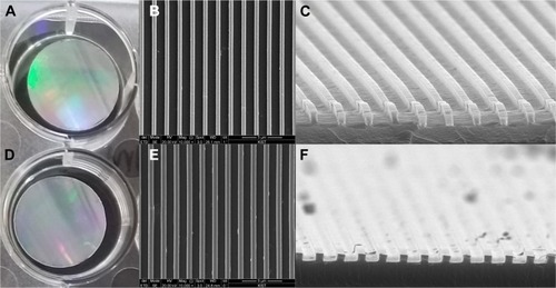
The substrates used in this study were chosen because they have mechanical properties similar to those of native tendons. The Young’s modulus of native tendon ranges from 200 kPa to 8 GPa from embryonic development to adulthood.Citation27–Citation30 To simulate these conditions, a Norland Optical Adhesive 86 (NOA86) substrate, with a Young’s modulus of 2.48 GPa (Minuta Tech, Osan, Republic of Korea), was chosen to provide an environment with mechanical properties comparable to a developing tendon. The NOA86 was coated with a 20 nm layer of 100% silicone and carved using forced capillary lithographyCitation31,Citation32 to create 800 nm-wide nanogrooves that would provide nanotopographic cues to the TDSCs (). For comparison, NOA86 with the same stiffness was prepared without nanogrooves to determine the impact of the nanotopographic cues and a polyurethane (PUA) substrate with 19.8 MPa stiffness was prepared with 800 nm-wide nanogrooves to determine the effect of substrate stiffness.
Figure 1 The NOA86 (A–C) and PUA (D–F) substrates were coated with silicone and carved to create 800 nm-wide nanogrooves for nanotopographic cues.
Note: SEM images show 800 nm grooves on the plates (B and E are 10,000× magnification images and C and F are 50,000× magnification images).
Abbreviations: NOA86, Norland Optical Adhesive 86; PUA, polyurethane; SEM, scanning electron microscopy.
TDSCs were seeded at a density of 20,000 cells per substrate for each substrate type, covered with a 20 nm-diameter coverslip, and cultured in DMEM with 20% FBS and 1% penicillin/streptomycin in an incubator at 37°C and with 5% CO2 for 1 week.
Immunofluorescence staining
The primary antibodies used in this study were rabbit poly-clonal anti-collagen I (1:500, ab292; Abcam, Cambridge, UK) and mouse monoclonal anti-collagen III (1:100, NBP1-05119; Novus Biologicals, Littleton, CO, USA). Secondary antibodies used in this study were donkey polyclonal rabbit IgG antibody (Alexa Fluor® 488) (1:1,000, ab150073; Abcam) and donkey polyclonal mouse IgG antibody (Alexa Fluor® 594) (1:1,000, ab150108; Abcam).
One week after culture, cells were fixed in 4% paraformaldehyde for 15 minutes at room temperature, quenched with 0.25% Triton X-100 in PBS for 10 minutes at room temperature, blocked with 1% BSA in PBS for 30 minutes at room temperature, and incubated with primary antibody overnight at 4°C. The cells were washed with PBS and incubated with a secondary antibody for 2 hours at room temperature in the dark. The cells were rinsed three times in PBS and mounted with fluoroshield mounting medium with DAPI (ab104139; Abcam). Confocal microscopy was performed using a Zeiss Lsm 700 (Carl Zeiss Meditec, Jena, Germany).
Image analysis and alignment
The degree of cell alignment on nanogrooved substrates was determined as previously described.Citation32 Images of cells were obtained after 1 week in culture and processed with ImageJ (The National Institutes of Health). Cell perimeters were outlined, and the angle of the major axis was calculated.
Quantitative real-time PCR (qRT-PCR)
Total RNA was isolated by lysis in TRIzol (15596-018; Thermo Fisher Scientific). cDNA libraries were made from 4 µg of RNA using superscript II reverse transcriptase (18064-014; Invitrogen) in 40 µL of reaction buffer at 42°C for 2 hours. Real-time PCR was performed with 30 ng of cDNA using a QuantiTect SYBR Green PCR Kit (204143; Qiagen NV, Venlo, the Netherlands) in a total volume of 25 µL with specific primers on a Light Cycler apparatus (ABI 7000; Thermo Fisher Scientific). The PCR procedure used was denaturation at 95°C for 15 minutes, 40 cycles at 95°C for 15 seconds, optimal annealing temperature for 30 seconds, and 72°C for 30 seconds. The following primer sequences were used (Genotech Corp., Daejeon, Republic of Korea): scleraxis (forward 5′-AACACGGCCTTCACTGCGCTG-3′ and reverse 5′-CAGTAGCACGTTGCCCAGGTG-3′) and GAPDH (forward 5′-GAGTCCACTGGCGTCTCCAC-3′ and reverse 5′-GGTGCTAAGCAGTTGGTGGT-3′).Citation33,Citation34
The relative expression level of each target gene was calculated using the 2−ΔΔCt method.Citation35 The gene was standardized to GAPDH. Significant changes in gene expression were calculated with respect to stem cell injury, and response to nanotopographic cues and substrate stiffness was evaluated using a Kruskal–Wallis test with post hoc Mann–Whitney U test.
Results
H&E staining of the left Achilles tendon confirmed the pathological findings of tendinopathy and tendon injury models. Upon histological examination, the 5-week-old tendinopathy model showed an irregular pattern of collagen fibers with multiple lipid vacuoles, and the 15-week-old tendon injury model showed a thickened irregular pattern of collagen fibers with abundant polymorphic nuclear cells ().
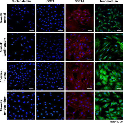
Isolation of TDSCs was validated by identifying cells that positively stained for nucleostemin, OCT4, SSEA4, and tenomodulin (). Each TDSC cell line was successfully differentiated into osteogenic, adipose, and chondrogenic cell lines, demonstrating their multipotent capacity. These results have been described previously.Citation36
Effects of nanotopographic cues and substrate stiffness on the TDSCs
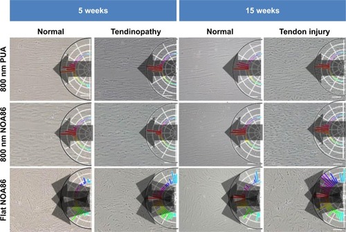
Irrespective of age or pathological status, TDSCs cultured on the 800 nm NOA86 and 800 nm PUA were well aligned along the grooves while cells cultured on the flat NOA86 were not aligned ().
Figure 2 TDSCs were seeded and cultured on the PUA substrate with 19.8 MPa stiffness and 800 nm-wide nanogrooves, NOA86 substrate with 2.4 GPa stiffness and 800 nm-wide nanogrooves, and NOA86 substrate with 2.4 GPa stiffness and flat surface.
Note: Irrespective of age and pathological status, TDSCs cultured on the 800 nm NOA86 and 800 nm PUA substrates were well aligned along the grooves while cells cultured on the flat NOA86 were not.
Abbreviations: NOA86, Norland Optical Adhesive 86; PUA, polyurethane; TDSCs, tendon-derived stem cells.
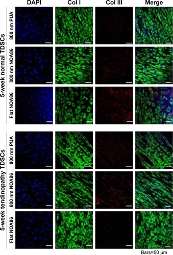
Expression of type I and type III collagen was observed in the TDSCs extracted from 5-week normal and 5-week tendinopathy models cultured on each of the substrates to understand the impact of nanotopographic cues and substrate stiffness on the cells (). In the 5-week normal and 5-week tendinopathic conditions there was no difference in expression of type I collagen among the different substrates. However, expression of type III collagen in the 5-week normal condition was slightly higher on the flat NOA86 than on either of the grooved substrates, while its expression in the 5-week tendinopathic condition was slightly higher on 800 nm NOA86 than on 800 nm PUA or on flat NOA86. Furthermore, higher levels of type III collagen were also found in the 5-week tendinopathic condition than in the normal condition (either with 800 nm NOA86 or with 800 nm PUA).
Figure 3 Expression of Col I and Col III collagen was observed in the TDSCs extracted from 5-week normal and 5-week tendinopathy models cultured on the 800 nm NOA86 (2.4 GPa), flat NOA86, and 800 nm PUA (19.8 MPa) substrates.
Notes: In the 5-week normal and 5-week tendinopathic conditions, there was no difference in expression of type I collagens according to nanotopographic cues and substrate stiffness. Expression of Col III collagen in 5-week normal condition was slightly higher in flat NOA86 than in 800 nm PUA or in 800 nm NOA86, while this expression in 5-week tendinopathic conditions was slightly higher in 800 nm NOA86 than in 800 nm PUA or in flat NOA86.
Abbreviations: Col I, type I collagen; Col III, type III collagen; NOA86, Norland Optical Adhesive 86; PUA, polyurethane; TDSCs, tendon-derived stem cells.
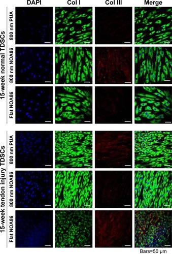
TDSCs in the 15-week tendon injury model showed higher cell densities and higher type I collagen expression than TDSCs in the 15-week normal tendon model irrespective of nanotopographic features or surface stiffness.
In the 15-week normal tendon model, expression of type III collagen was high in TDSCs cultured on the 800 nm NOA86 substrates (). However, expression of type III collagen in the 15-week tendon injury model was independent of nanotopographic cues and substrate stiffness, as was the expression of type I collagen in the 15-week normal and tendon injury models.
Figure 4 Expression of type I and type III collagen was observed in the TDSCs extracted from 15-week normal and 15-week tendon injury models cultured on the 800 nm NOA86 (2.4 GPa), flat NOA86, and 800 nm PUA (19.8 MPa) substrates.
Notes: In the 15-week normal tendon model, expression of type III collagen was high in TDSCs cultured on the 800 nm NOA86 substrates. In the 15-week tendon injury model, expression of type III collagen was similar irrespective of nanotopographic cues and substrate stiffness. The expression of type I collagen was not different between nanotopographic cues and substrate stiffness in the 15-week normal and tendon injury models.
Abbreviations: Col I, type I collagen; Col III, type III collagen; NOA86, Norland Optical Adhesive 86; PUA, polyurethane; TDSCs, tendon-derived stem cells.
Gene expression in the TDSCs based on nanotopographic cues and substrate stiffness
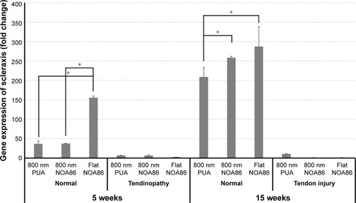
Results from qRT-PCR showed increased gene expression of scleraxis in TDSCs cultured on the flat NOA86 substrate in the 5-week normal tendon model (P=0.001). In the 15-week normal tendon model, scleraxis was highly expressed in TDSCs cultured on the 800 nm and flat NOA86 substrate (P=0.043). However, this gene expression was not significantly different between the substrates in the 5-week tendinopathy and 15-week tendon injury models ().
Figure 5 Gene expression of scleraxis increased in TDSCs cultured on the flat NOA86 substrate in the 5-week normal tendon model (*P<0.01).
Notes: In the 15-week normal tendon model, scleraxis was highly expressed in TDSCs cultured on the 800 nm and flat NOA86 substrate (*P<0.01). However, the gene expression was not significantly different between the substrates in the 5-week tendinopathy and 15-week tendon injury models.
Abbreviations: NOA86, Norland Optical Adhesive 86; PUA, polyurethane; TDSCs, tendon-derived stem cells.
Discussion
In the present study, TDSCs extracted from normal tendons showed similar expression of type I and III collagens irrespective of nanotopographic cues or substrate stiffness. However, scleraxis, a major protein related to development and maturation of tendon, increased under the stiff surface condition.
Expression of type I collagen in TDSCs extracted from the 5-week and 15-week normal and tendinopathic models was observed to be independent of nanotopographic cues and substrate stiffness. However, expression of type III collagen in the 5-week normal condition was slightly higher on flat NOA86 than on either of the grooved substrates, while its expression in the 5-week tendinopathic condition was slightly higher on 800 nm NOA86 than on 800 nm PUA or on flat NOA86. This finding is contradictory to Islam et al’s study that demonstrated increased collagen expression due to increased substrate stiffness by induction of tenogenic differentiation of mesenchymal stem cells.Citation37,Citation38 This contradictory finding might be due to the groove width and depth. The researchers in the previous study used 10 µm groove width and 3 µm depth, whereas we used 800 nm groove width and depth, which was shown to have maximal effects in another prior study.Citation39 We selected 800 nm groove width and depth because this produced optimal regeneration in cardiac stem cells in our previous study.Citation40
The difference of expression between type I and type III collagens according to pathological condition can be explained by type I collagen being the most abundant component of the tendon matrix and exhibiting relatively stable expression, while type III collagen is easily affected by diverse environments.Citation41 Higher levels of type III collagen found in the 5-week tendinopathic condition compared to the normal condition can be explained by increased regenerative capacity in tendinopathic condition.
TDSCs in the 15-week tendon injury model showed higher cell densities than TDSCs in the 15-week normal tendon model irrespective of nanotopographic signals or surface stiffness. We thought that this was due to the increased proliferative properties of stem cells after injury to aid in healing.Citation42 Relatively high expression of type I collagen in the 15-week tendon injury model compared to that in the 15-week normal tendon model might be due to these increased cell densities.
Scleraxis is an important transcription factor that influences the development and maturation of tendon-to-bone attachment.Citation43 Scleraxis was prominently expressed in TDSCs which were extracted from the 5-week normal tendon model and cultured on the flat NOA86 and in TDSCs which were extracted from the 15-week normal tendon model and cultured on the 800 nm and flat NOA86 substrates. These results could be explained by induction of tenogenic differentiation by substrate stiffness, but not by nanotopographic cues.Citation37,Citation38 This finding contradict the results of a previous study, but this might be due to the different depth and width of the grooves used in that study.Citation38 However, in the chemically induced tendinopathy model, scleraxis was not highly expressed regardless of the substrate used. We hypothesize that quinolone caused toxicity in the TDSCs via oxidative stress,Citation44 and this oxidative stress was attenuated by the nanotopographic cues and substrate stiffness. The failure to induce tenogenic differentiation via nanotopographic cues or substrate stiffness in injury-induced tendinopathy might also be due to attenuation of oxidative or mechanical stress.
The results of our study show that therapeutic applications need to be adaptive because the effects of substrate stiffness and nanotopographic cues on the TDSCs were different based on the mechanism of tendinopathy. However, independent of the specific strategy, how to provide the stiffness and nanotopographic cues to the TDSCs in patients with tendinopathy remains a problem. Injection or implantation of TDSCs or mesenchymal stem cells that have been cultured on substrates with adequate nanotopographic cues and stiffness to pathological areas is one possible therapeutic option. However, poor retention and low survival of injected or implanted cells in the pathological areas are major obstacles to this approach.Citation45 Based on our results, further in vivo studies should be conducted to determine how stiffness and nanotopographic cues can be delivered to the TDSCs in the tendinopathy animal model or in patients with tendinopathy.
The present study had several limitations. We used 800 nm width and depth for the nanotopographic cues because these were shown to have optimal effects in the cells in a previous study.Citation39 However, stem cells show different reactions based on the width and depth of grooves,Citation46 and the reactions can differ depending on the source of stem cells. To determine the maximal effects of TDSCs, diverse conditions should be tested. We did not perform this study using TDSCs extracted from human tissues because we could not acquire TDSCs from normal human tendons of different ages due to ethical issues. To extrapolate these results to clinical studies, it will be necessary to investigate characteristics of TDSCs extracted from diverse human pathological tendon tissues.
Conclusion
Our study revealed that scleraxis, a major protein related to development and maturation of tendon, increased when TDSCs from normal tendons were cultured on stiff surface (NOA86), but not when the TDSCs came from pathological models. This suggests that therapeutic applications need to be flexible because the effects of substrate stiffness and nanotopographic cues on the TDSCs were different based on tendon age and tendinopathy.
Author contributions
Sun Jeong Kim, Philip D Tatman, Albert O Gee, Deok-Ho Kim, and Sang Jun Kim contributed to conception and design. Sun Jeong Kim, Da-Hyun Song, Deok-Ho Kim, and Sang Jun Kim contributed to acquisition of data. All authors contributed to interpretation of data, drafting of the article, gave final approval of the manuscript version to be published, and agreed to be accountable for all aspects of the work.
Acknowledgments
This research was supported by a grant of the Korea Health Technology R&D Project through the Korea Health Industry Development Institute (KHIDI), funded by the Ministry of Health and Welfare, Republic of Korea (grant number: HI16C1104) and the National Research Fund (2018R1D1A1B07047084).
Supplementary materials
Figure S1 H&E staining of the left Achilles tendon confirmed the pathological findings of tendinopathy and tendon injury models.
Note: Five-week-old tendinopathy model showed an irregular pattern of collagen fibers with multiple lipid vacuoles (A), and the 15-week-old tendon injury model showed a thickened irregular pattern of collagen fibers with abundant polymorphic nuclear cells (B).
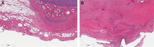
Figure S2 Isolation of TDSCs was validated by identifying cells that positively stained for nucleostemin, OCT4, SSEA4, and tenomodulin. Each TDSC cell line was successfully differentiated into osteogenic, adipose, and chondrogenic cell lines, demonstrating their multipotent capacity.
Abbreviation: TDSCs, tendon-derived stem cells.
Disclosure
The authors report no conflicts of interest in this work.
References
- MaffulliNWongJAlmekindersLCTypes and epidemiology of tendinopathyClin Sports Med200322467569214560540
- RabagoDNouraniBProlotherapy for Osteoarthritis and Tendinopathy: a Descriptive ReviewCurr Rheumatol Rep20171963428484944
- NavaniALiGChrystalJPlatelet Rich Plasma in Musculoskeletal Pathology: A Necessary Rescue or a Lost Cause?Pain Physician2017203E345E35628339434
- ReevesKDSitRWRabagoDPDextrose Prolotherapy: A narrative review of basic science, clinical research, and best treatment recommendationsPhys Med Rehabil Clin N Am201627478382327788902
- FilardoGDi MatteoBKonEMerliGMarcacciMPlatelet-rich plasma in tendon-related disorders: results and indicationsKnee Surg Sports Traumatol Arthrosc20182671984199927665095
- NourissatGOrnettiPBerenbaumFSellamJRichettePChevalierXDoes platelet-rich plasma deserve a role in the treatment of tendinopathy?Joint Bone Spine201582423023425881762
- VeronesiFSalamannaFTschonMMaglioMNicoli AldiniNFiniMMesenchymal stem cells for tendon healing: what is on the horizon?J Tissue Eng Regen Med201711113202321927597421
- AkyolEHindochaSKhanWSUse of stem cells and growth factors in rotator cuff tendon repairCurr Stem Cell Res Ther201510151025012743
- UsuelliFGD’AmbrosiRMaccarioCIndinoCManziLMaffulliNAdipose-derived stem cells in orthopaedic pathologiesBr Med Bull20171241315429253149
- RuiYFLuiPPChanLSChanKMFuSCLiGDoes erroneous differentiation of tendon-derived stem cells contribute to the pathogenesis of calcifying tendinopathy?Chin Med J (Engl)2011124460661021362289
- ChenJLZhangWLiuZYHengBCOuyangHWDaiXSPhysical regulation of stem cells differentiation into teno-lineage: current strategies and future directionCell Tissue Res2015360219520725549759
- HakimiOMurphyRStachewiczUHislopSCarrAJAn electrospun polydioxanone patch for the localisation of biological therapies during tendon repairEur Cell Mater20122434435723090765
- FahyNAliniMStoddartMJMechanical stimulation of mesenchymal stem cells: Implications for cartilage tissue engineeringJ Orthop Res2018361526328763118
- BurkJPlengeABrehmWHellerSPfeifferBKasperCInduction of tenogenic differentiation mediated by extracellular tendon matrix and short-term cyclic stretchingStem Cells Int20162016734237927630718
- LiDZhouJChowdhuryFChengJWangNWangFRole of mechanical factors in fate decisions of stem cellsRegen Med20116222924021391856
- MacGregor-RamiasaMHoppIBachhukaAMurrayPVasilevKSurface nanotopography guides kidney-derived stem cell differentiation into podocytesActa Biomater20175617118028232254
- CarsonDHnilovaMYangXNanotopography-Induced Structural Anisotropy and Sarcomere Development in Human Cardiomyocytes Derived from Induced Pluripotent Stem CellsACS Appl Mater Interfaces2016834219232193226866596
- XingQQianZTahtinenMYapAHYatesKZhaoFAligned Nano-fibrous Cell-Derived Extracellular Matrix for Anisotropic Vascular Graft ConstructionAdv Healthc Mater Epub201729
- ZhangYGordonAQianWChenWEngineering nanoscale stem cell niche: direct stem cell behavior at cell-matrix interfaceAdv Healthc Mater20154131900191426222885
- WangJHGuoQLiBTendon biomechanics and mechanobiology – a minireview of basic concepts and recent advancementsJ Hand Ther201225213314121925835
- ZhangCYuanHLiuHWell-aligned chitosan-based ultra-fine fibers committed teno-lineage differentiation of human induced pluripotent stem cells for Achilles tendon regenerationBiomaterials20155371673025890767
- TehTKTohSLGohJCAligned fibrous scaffolds for enhanced mechanoresponse and tenogenesis of mesenchymal stem cellsTissue Eng Part A20131911–121360137223327653
- KatoMTakadaSKashidaYNomuraMHistological examination on Achilles tendon lesions induced by quinolone antibacterial agents in juvenile ratsToxicol Pathol19952333853927659960
- GlazebrookMAWrightJRJrLangmanMStanishWDLeeJMHistological analysis of achilles tendons in an overuse rat modelJ Orthop Res200826684084618183626
- BiYEhirchiouDKiltsTMIdentification of tendon stem/progenitor cells and the role of the extracellular matrix in their nicheNat Med200713101219122717828274
- RuiYFLuiPPLiGFuSCLeeYWChanKMIsolation and characterization of multipotent rat tendon-derived stem cellsTissue Eng Part A20101651549155820001227
- AnKNSunYLLuoZPFlexibility of type I collagen and mechanical property of connective tissueBiorheology2004413–423924615299256
- KatoYPChristiansenDLHahnRAShiehSJGoldsteinJDSilverFHMechanical properties of collagen fibres: a comparison of reconstituted and rat tail tendon fibresBiomaterials198910138422713432
- MarturanoJEArenaJDSchillerZAGeorgakoudiIKuoCKCharacterization of mechanical and biochemical properties of developing embryonic tendonProc Natl Acad Sci U S A2013110166370637523576745
- SilverFHChristiansenDSnowhillPBChenYLandisWJThe role of mineral in the storage of elastic energy in turkey tendonsBiomacromolecules20001218018511710098
- JiaoATrosperNEYangHSThermoresponsive nanofabricated substratum for the engineering of three-dimensional tissues with layer-by-layer architectural controlACS Nano2014854430443924628277
- KimDHLipkeEAKimPNanoscale cues regulate the structure and function of macroscopic cardiac tissue constructsProc Natl Acad Sci U S A2010107256557020018748
- TanQLuiPPRuiYFWongYMComparison of potentials of stem cells isolated from tendon and bone marrow for musculoskeletal tissue engineeringTissue Eng Part A2012187–884085122011320
- WagenhäuserMUPietschmannMFSieversBCollagen type I and decorin expression in tenocytes depend on the cell isolation methodBMC Musculoskelet Disord20121314022871215
- ChangSChenWYangJAnother formula for calculating the gene change rate in real-time RT-PCRMol Biol Rep20093682165216819109763
- KimSJSongDHKimSJCharacteristics of tendon derived stem cells according to different factors to induce the tendinopathyJ Cell Physiol201823386196620629341108
- IslamAMbimbaTYounesiMAkkusOEffects of substrate stiffness on the tenoinduction of human mesenchymal stem cellsActa Biomater20175824425328602855
- ShiYZhouKZhangWMicrogrooved topographical surface directs tenogenic lineage specific differentiation of mouse tendon derived stem cellsBiomed Mater201712101501328071602
- LiliensiekSJWoodJAYongJAuerbachRNealeyPFMurphyCJModulation of human vascular endothelial cell behaviors by nanotopographic cuesBiomaterials201031205418542620400175
- KimDHKshitizSmithRRNanopatterned cardiac cell patches promote stem cell niche formation and myocardial regenerationIntegr Biol (Camb)2012491019103322890784
- GüngörmüşCKolankayaDGene expression of tendon collagens and tenocyte markers in long-term monolayer and high-density cultures of rat tenocytesConnect Tissue Res201253648549122594477
- RennertRCSorkinMGargRKGurtnerGCStem cell recruitment after injury: lessons for regenerative medicineRegen Med20127683385023164083
- KillianMLThomopoulosSScleraxis is required for the development of a functional tendon enthesisFaseb J201630130131126443819
- PouzaudFBernard-BeauboisKTheveninMWarnetJMHayemGRatPIn vitro discrimination of fluoroquinolones toxicity on tendon cells: involvement of oxidative stressJ Pharmacol Exp Ther2004308139440214569066
- RossiCAPozzobonMDe CoppiPAdvances in musculoskeletal tissue engineering: moving towards therapyOrganogenesis20106316717221197219
- MengsteabPYUtoKSmithASSpatiotemporal control of cardiac anisotropy using dynamic nanotopographic cuesBiomaterials20168611026874887