
Abstract
Background
The investigation of agents promoting recovery of nerve regeneration following neurodegenerative diseases has been the most important issue in neuroscience. Nerve growth factor (NGF) and quercetin as potential flavonoids could possibly have therapeutic applications in the field of degenerative diseases such as Parkinson and Alzheimer.
Materials and methods
The MTT assay was done at 24, 48, and 72 hours to examine the cytotoxicity of superparamagnetic iron oxide nanoparticles (SPIONs) and quercetin. We combined NGF and quercetin with different concentrations of SPIONs as novel compounds to study their effect on neuronal branching morphogenesis of PC12 cells.
Results
Morphological analysis showed a significant growth (P<0.001) in neurite length when PC12 cells were incubated in quercetin solution. We found a significant neurite outgrowth promotion and an increase in the complexity of the neuronal branching trees after exposing PC12 cells to both quercetin and SPIONs. In addition, a higher level of β3-tubulin expression was observed in these cells when treated with both quercetin and SPIONs.
Conclusion
Different photographic analyses indicated that iron oxide nanoparticles function as an important factor in order to improve the efficiency of NGF through improving cell viability, cell attachment, and neurite outgrowth in the shelter of quercetin as an accelerator of these phenomena. The use of the quercetin–SPION complex as a suitable method for improving NGF efficacy and activity opens a novel window for substantial neuronal repair therapeutics.
Introduction
Recovery of nerve function and nerve regeneration have been the most important issues in neuroscience, due to their considerable role in the treatment of injured neurons during a degenerative disease or after an accident.Citation1
Appropriate cell morphology regulation by intrinsic and extrinsic factors is an important requirement for cell to cell communication during the differentiation process. Neuronal cells undergo remarkable changes in cytoskeletal organization and morphology.Citation2 Different factors have been revealed to induce neuritogenesis including different molecular signals such as extracellular matrix (ECM) proteins, growth factors, and mechanical tensile forces.Citation3
Cell cultures of neuronal cell lines or primary neurons are usually used to study compounds controlling plasticity of neuronal processes and differentiation. In particular, PC12 cells (pheochromocytoma cell line) have been extensively used in both neurotoxicological and neurobiological studies as a model of neuronal differentiation.Citation4
The nerve growth factor (NGF) as an important growth factor is critical for the neuritogenesis and maintenance of neurons under in vitro and in vivo conditions. NGF-incubated PC12 cells stop proliferation, outspread neurites, and become electrically impulsive. During differentiation with NGF, the tyrosine kinase receptor A is activated and initiates a number of signaling pathways containing phosphatidylinositol 3-kinase pathway, and the extracellular signal-regulated kinase (ERK) cascade, which obstructs proliferation and encourages neurite growth.Citation4,Citation5
For improving the natural effect of growth factors, nanotechnology has been proposed as a considerable item in therapeutic and diagnostic fields. Especially, magnetic nanoparticles (MNPs) could have different therapeutic applications such as cell-labeling, drug delivery, and medical imaging.Citation6,Citation7
The superparamagnetic iron oxide nanoparticles (SPIONs) with characteristics such as nano size, modified surfaces, monodispersed shape, enhanced magnetization, colloidal stability, bio-distribution, and cellular uptake are considerably focused for their applications in biomedical applications.Citation6–Citation8 Furthermore, the surface of SPIONs could be modified by different organic materials, such as polymers and biomolecules.
Biological molecules in different cells may also be bound to the surface of iron oxide nanoparticles. The interactions between biological molecules and iron oxide nanoparticles are studied to determine the cellular response of the MNPs.Citation7,Citation9,Citation10
It has been shown that metal ions including cobalt, manganese, and iron could modify cell attachment and have an important impact on neuronal differentiation.Citation9,Citation11,Citation12 These ions are related to adhesion molecules (exactly the RGD-dependent ECM). Cell adhesion molecules modulate cell proliferation, differentiation, survival, and migration by interacting with the ECM.Citation13,Citation14
So far, the potential cytotoxicity of MNPs due to the formation of reactive oxygen species (ROS) remains an issue of debate.Citation15 Great efforts have been taken to eliminate the cytotoxic effects of nanoparticles such as coating or combination of iron oxide nanoparticle with different agents.Citation16–Citation18
Recent research has indicated that a variety of natural flavonoids reduce the toxicity of ROS in neural cells by their antioxidant activity.Citation19–Citation21 It is important to note that a selected group of flavonoids (such as quercetin and fisetin) have demonstrated neuroprotective activity and play a critical role in the neurite outgrowth and differentiation of neural cells.Citation20,Citation22
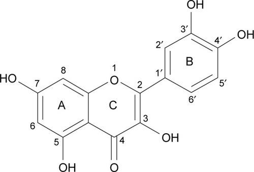
An important natural bioflavonoid is quercetin (3,3′,4′,5′-7-penta-hydroxy flavone, ). This flavonoid is abundantly found in vegetables, edible fruits, and medicinal plants. It has an extensive range of chemotherapeutic applications for many diseases such as anti-viral, anti-cancer, and anti-oxidant.Citation23–Citation25 A number of quercetin actions make it an important neuroprotective agent, including interaction with different proteins and protective effects of neuronal cells from oxidative stress.Citation26
Figure 1 The two dimensional chemical structure of quercetin.
In this study, we suggest a new combination of SPIONs and quercetin to enhance the effect of NGF during neural differentiation of cells. In this combination, the role of iron oxide nanoparticles is more prominent. By increasing the attachment of PC12 cells, iron oxide nanoparticles help to preserve viability of cells and increase the effect of NGF and quercetin. Quercetin could bind to NGF and enhance the effect of NGF.Citation9,Citation27 We used PC12 cells as a model for studying different aspects of developmental cell biology. Based on morphometric assessments, we show that the combination of SPIONs and quercetin promotes neuronal branching morphogenesis of PC12 cells in the presence of NGF.
Materials and methods
Materials
Poly-l-ornithine (PLO), MTT, quercetin (3,3′,4′,5′-7-penta-hydroxy flavone), and PBS were obtained from Sigma-Aldrich Co. (St Louis, MO, USA). Horse serum (HS), T-25, T-75 flasks, 12-well and 96-well plates, and l-glutamine were purchased from Corning Incorporated (Corning, NY, USA). BSA, FBS, penicillin-streptomycin, and RPMI medium were bought from Thermo Fisher Scientific (Waltham, MA, USA). NGF-β (GFM11) obtained from Cell Guidance Systems (St Louis, MO, USA). Iron oxide nanoparticles (Nano-screen MAG-DXS) were purchased from Chemicell GmbH (Berlin, Germany). PC12 cell line (National Center for Biotechnology code: C153) was purchased from Pasteur Institute of Iran (Tehran, Iran).
Cell culture
PC12 cells were cultured in the RPMI medium containing 10% HS, 5% FBS, 1% l-glutamine, and antibiotics. For differentiation, PC12 cells (5,000 cells/cm2) were seeded on PLO/laminin (Sigma-Aldrich Co.)-coated plates and incubated for 24 hours in serum-reduced media (0.5% FBS, 1% HS). Recombinant mouse NGF-β (50 ng/mL) was then added to the medium (every 2 days) to induce the differentiation of PC12 cells. Five treatments were examined: 50 ng/mL (free NGF), 100 ng/mL-NGF, MNPs, NGF-quercetin (10 µM), and NGF + MNPs + quercetin.
Cell viability assay
To examine the cytotoxic effects of quercetin and MNPs on PC12 cells, MTT assay was applied. The cells (30,000/well) were seeded onto the PLO-coated 96-well plates. At 24, 48, and 72 hours after incubation with the MNPs and quercetin at concentrations ranging from 10 to 100 µg/mL for MNPs and 10–50 µg/mL for quercetin, PC12 cells were incubated with 0.5 mg/mL MTT according to the Mosmann method.Citation28 The absorbance at 560 nm was measured using the ELISA reader (Thermo LabSystems Inc., Beverley, MA, USA).
Quantification of nanoparticle intracellular uptake
In order to quantitate the intracellular uptake of nanoparticles by the PC12 cells, following experiments have been performed.
Inductive coupled plasma-atomic emission spectrometer (ICP-AES)
Total MNPs in the cell cytoplasm were measured by evaluating the amount of iron in PC12 cells. PC12 cells were incubated with different concentrations of iron oxide nanoparticles (from 10 to 100 µg/mL). After 24 hours of incubation, the cells were sequestered and lysed with hydrochloric acid. Iron absorption was quantified using an ICP-AES (ICPS-7500; Shimadzu Corp., Kyoto, Japan).Citation29
Flow cytometric analysis
In this study, dextran sulfate-coated MNPs sized 100 nm were used (Nano-screen MAG-DXS). The surface coating of nanoparticles was dextran sulfate with a fluorophore that has an absorbance maximum of 476 nm and an emission maximum of 490 nm. All samples (incubated cells with iron oxide nanoparticles at concentrations ranging from 10 to 100 mg/mL) were analyzed by flow cytometry (FAC-SCalibur; BD Biosciences, San Jose, CA, USA). Green fluorescence was detected using an FL-1 sensor, a 525 nm band-pass filter. Statistics from 10,000 cells per sample were analyzed and three measurements per sample were recorded.
Prussian blue staining
PC12 cells treated with different concentrations of iron oxide nanoparticles were stained with Prussian blue dye for tracing iron in the cell cytoplasm by dipping the cells in a mixture of equal parts of 10% potassium ferrocyanide solution and 20% hydrochloric acid. In this method, trivalent iron in the PC12 cells interacts with the ferrocyanide and results in the formation of ferric ferrocyanide (the blue pigments).Citation30 A light microscope (Inverted Microscope; Nikon, Tokyo, Japan) was used to acquire images of stained cells.
Cresyl violet staining (Nissl staining)
The cresyl violet method was used to show the Nissl substance as an important characteristic of neurons. In the neural differentiation, the maturation grade of neuronal cells is reflected by formational changes in the neuroplasm. The Nissl bodies are rough endoplasmic reticulum (with ribosomes) and are the site of protein synthesis.Citation31 PC12 cells were stained with 0.1% cresyl violet solution for 4–15 minutes and washed three times with PBS to remove excess stain. PC12 cells were photographed by inverted microscopy and scored for the presence of Nissl substance and neurites.
Immunocytochemistry
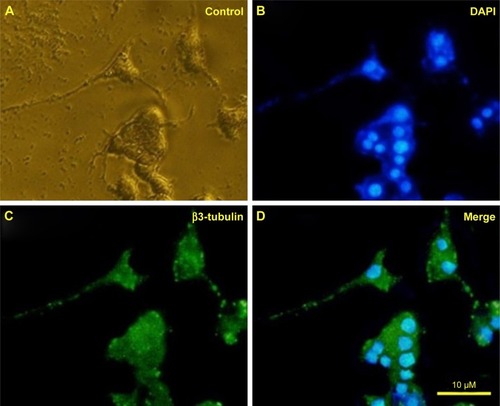
PC12 cells were immunostained against β3-tubulin (in green) as an important neural specific marker. To stain PC12 cells with antibodies against β3-tubulin, cultures were fixed with 4% for 30 minutes and permeabilized with 0.3% Triton X-100 diluted in PBS. PC12 cells were incubated with the primary antibody (β3-tubulin) for 24 hours and then treated with a fluorescein isothiocyanate-conjugated secondary antibody for 1 hour. The nuclei were marked with DAPI for 15 minutes. Light and fluorescent microscopy (Olympus US SZX12 fluorescent stereo microscope; Olympus Corporation, Tokyo, Japan) identified the temporal relationship between neurites morphogenesis and different treatments.
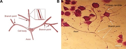
Neuritogenesis in PC12 cells
Neuritogenesis in PC12 cells was studied in a time-dependent method with different treatments either alone or in combination for 5 days. NGF-β (50 ng/µL) was added to the medium of all treatments because this concentration was optimized. Due to lack of serum in the differentiation medium, NGF should be added to the cell culture every 2 days to maintain the viability of the PC12 cells during differentiation process. NGF blocked the apoptotic death of attached cells.Citation9
PC12 cells were incubated with NGF and MNPs with or without quercetin. At different days (from 1 to 5) after incubation, the percentage of neurite outgrowth, number of branching points, and number of neurites originating from soma were quantified (three random images from triplicate tests). Neuritogenesis was measured as described by Kim et al.Citation29 Neurite length was defined as L0 to L4 according to the length of the neurites. L0 was demarcated as cells with no neurites, L1 as cells whose neurite length was shorter than the size of the soma, L2 as cells with neurite length between the original size of the soma and twice the size, and L3 as cells whose neurite length was longer than twice the size of soma.Citation29 Also the number of neurites originating from soma were defined as N1, N2, N3, and N4 (N1, N2, N3, and N4 were defined as one, two, three, and four neurites in the PC12 cells, respectively). Length and number of neurites of 200 cells were measured in three random fields from triplicate experiments.
The effect of different treatments on neuritogenesis
First, the efficiency of free NGF as a differentiating factor was measured. Then, the efficiency of free NGF treatment was compared to other treatments such as quercetin solution and using iron oxide nanoparticles with or without quer-cetin. The PC12 (2×10³) cells were plated on PLO-coated plates and incubated with five different treatments including free NGF, NGF (50 ng/mL) and quercetin (10 µM), quercetin–NGF–MNPs (20 µg/mL), and NGF–MNPs (20 µg/mL). After incubation in different treatments, PC12 cells were washed and fixed with 4% paraformaldehyde for 30 minutes. Percentage of neurite outgrowth, branching points, and differentiation of PC12 cells under different treatments were measured using the light microscope. Light and fluorescent microscopy was used to evaluate both the terminal branch number and the total branch length of neurites.
Statistical analysis
All values of differentiation rate of PC12 cells are presented as mean ± SD. The analysis method was a one-way ANOVA. When the P-value was <0.05 (P<0.05), differences among the treatments were considered statistically significant.
Results
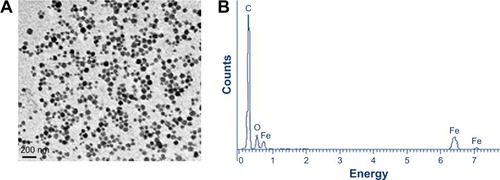
In this study, uniform fluorescent iron oxide (Fe3O4) nanoparticles were used. The surface of the nanoparticles was coated with dextran sulfate with a green fluorophore. The number of particles was ~1.8×10¹⁵/g with a density of 1.25 g/cm3. The size distribution of the dextran sulfate-coated iron oxide nanoparticles was found to be 100 nm (). shows the energy dispersive X-ray pattern of dextran-coated iron oxide nanoparticles (Fe3O4), and their sizes were determined by scanning electron microscopy.
Figure 2 SEM (A) and EDX (B) images of iron oxide nanoparticles used for neurite outgrowth.
Abbreviations: C, carbon; Fe, iron; O, oxygen; SEM, scanning electron microscope; EDX, energy dispersive X-ray analysis.
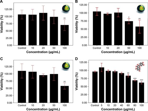
The MTT cell viability assay was employed to examine the cytotoxicity of SPIONs and quercetin. PC12 cells were treated with SPIONs at different concentrations from 10 to 100 µg/mL. shows the effects of different treatments on the viability of PC12 cells. Superparamagnetic nanoparticles in the concentration of 100 µg/mL show significant cytotoxicity in comparison with the control and other groups (P<0.01). Quercetin in the range of 60–100 µg/mL was significantly cytotoxic ().
Figure 3 Effect of different treatments on the viability of PC12 cells. Cell viability was determined by MTT assay. PC12 cells were treated with 10, 20, 50, and 100 µg/mL for 24 (A), 48 (B), and 72 hours (C). (D) PC12 cells were treated with 5, 10, 20, 40, 60, 80, and 100 µg/mL for 24 hours. There were significant differences between the viability of PC12 cells from different treatments (P<0.05). Data are presented as the mean ± SD, n=3, *P<0.05, **P<0.01, and ***P<0.001 vs control group.
Total MNPs in the cell cytoplasm were measured by evaluating the amount of iron in the PC12 cells using ICP-AES (). During incubation with different concentrations of MNPs, iron in the cell cytoplasm increased up to the 35 pg per cell.
Figure 4 Internalization of superparamagnetic iron oxide nanoparticles into the PC12 cells. (A) Uptake of iron oxide nanoparticles by PC12 cells 24 hours after exposure to the iron oxide nanoparticles. Iron oxide nanoparticles were added to the media in a dose-dependent manner ranging from 10 to 100 µg/mL. (B) Fluorescent images of incubated cells with the different concentrations of fluorescent iron oxide nanoparticles. (C) Different histograms using Flowing Software (version 2.5.1) were combined: the red curve represents the maximum fluorescence. In this case, PC12 cells were incubated with 100 µg/mL of iron oxide nanoparticles. (D) Prussian blue method: PC12 cells treated with iron oxide nanoparticles were also stained for tracing the iron in the cell cytoplasm. Arrow represents the position of the iron oxide nanoparticles in PC12 cells (dark blue dots).
Abbreviation: MNPs, magnetic nanoparticles.
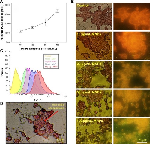
The fluorescent microscopic study of the PC12 cells treated with different concentration of iron oxide nanoparticles exhibited a bright green color emission as shown in . These data show that the majority of superpara-magnetic nanoparticles were internalized into the cells.
Also as it is shown in , treatment of PC12 cells with different concentration of iron oxide nanoparticles (10– 50 µg/mL) led to an increase in fluorescence up to 89%±0.7% intensity (yellow, violet, and blue curves). Surprisingly, during the use of maximum concentration of MNPs, the fluorescence intensity of about 97% was recorded (red curve).
PC12 cells incubated with iron oxide nanoparticles were also stained for tracing the iron in the PC12 cell cytoplasm (). The red arrow represents the position of the MNPs in PC12 cells (dark blue dots).
Based on the internalization and MTT assay results and considering published articles, a dose of 20 µg/mL for nanoparticle and a dose of 10 µM for quercetin were chosen. Differentiation was studied by observing the morphology of PC12 cells. First, the efficacy of free NGF as a differentiating factor was experienced. As shown in , when PC12 cells were treated with free NGF, neurite outgrowth and neurite arborization were observed. While the use of NGF with quercetin led to an increase in neurite outgrowth between 1 and 5 days after differentiation induction (exactly neurite length). As shown in , the Nissl substance was represented dark blue due to the staining of ribosomal RNA. The distribution of the neurite length of PC12 cells 5 days after the induction of differentiation is shown in . Neurite length was significantly affected by the quercetin treatment. The percentage of the PC12 cells with longer neurite (L3 and L4) increased when quercetin was added to the cells (). To examine the effect of MNPs on neuronal differentiation, PC12 cells were incubated with a combination of NGF and iron oxide nanoparticles. The results showed that the number of neurites originating from the soma and branching points of the cells increased when PC12 cells were incubated with iron oxide nanoparticles ().
Figure 5 Typical images of differentiated PC12 cells at 1, 3, and 5 days after treatment with NGF (50 ng/mL) and quercetin (10 µM) and NGF + quercetin. Quercetin induces PC12 cell differentiation (magnification: 200×).
Abbreviation: NGF, nerve growth factor.
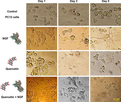
Figure 6 Neurite outgrowth of PC12 cells. (A) Cresyl violet staining: the Nissl substance (rough endoplasmic reticulum) appeared dark blue due to the staining of ribosomal RNA. Magnification ×400. (B) Distribution of the neurite length of PC12 cells 5 days after the induction of differentiation (L0 refers to the cells without neurites; L1 refers to the cells with neurites whose length is shorter than the size of the cell body; L2 refers to the cells with neurites whose length is between the original and twice the size of the cell body; L3 refers to the cells with neurites whose length is longer than twice the size of the cell body). (C) Neurite length of PC12 cells under different treatments. Cells were scored positive if one or more neurites with length >1 cell body diameter was observed. The results presented are the mean ± SD of 10 independent experiments. *P<0.05, **P<0.01.
Abbreviations: NGF, nerve growth factor; Q, quercetin; MNP, magnetic nanoparticle.
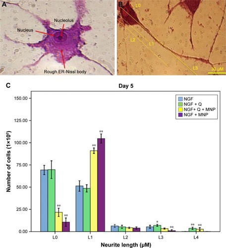
Figure 7 Compartment between different treatment effects on neurite morphology. PC12 cells were treated with NGF (A), NGF + MNPs (B), NGF + quercetin (C), and NGF + quercetin + MNPs (D). For each treatment, 100 cells in each of three separate fields were counted.
Abbreviations: MNPs, magnetic nanoparticles; NGF, nerve growth factor.
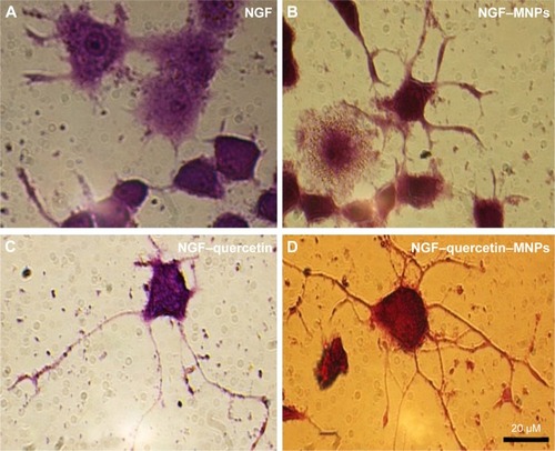
PC12 cells incubated with NGF and MNPs developed more branching points, more neurites per cell, and maximum percentage of cells expressing neurites in comparison with other groups (). Surprisingly, the treatment of PC12 cells with iron oxide nanoparticles together with NGF and quercetin demonstrated considerable enhancement in the neurite lengths and branching points in comparison with those treated with free NGF ( and ). Neural-specific marker (β3-tubulin) was studied immunohistochemically using specific monoclonal antibodies in PC12 cells incubated with NGF and SPIONs ().
Figure 8 Effect of different treatments (NGF–MNPs–quercetin) on morphological parameters of neuronal differentiation, comparing between the four treatments: free NGF, non-conjugated MNPs with free NGF, NGF–MNPs + quercetin, and NGF + quercetin (A, B). (C) The number of neurites originating from the soma. Scale bar set at 30 µM. (D) The number of branching points. ANOVA test, *P<0.05, **P<0.01.
Figure 9 Immunofluorescence images of differentiated PC12 cells 5 days after treatment with iron oxide nanoparticles. (A) Control, (B) cells stained with DAPI, (C) cells stained with β3-tubulin, and (D) the merge of (B) and (C). Green and blue fluorescence represent β3-tubulin and nucleus, respectively. Nuclei marked with DAPI.
Abbreviations: MNPs, magnetic nanoparticles; NGF, nerve growth factor; Q, quercetin.
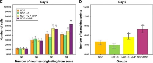
Several studies have shown that 1 day after incubation of PC12 cells on the PLO-coated plate, neuritogen-esis and neurite arborization have begun as an important neuronal differentiation stage (). shows neuritogenesis in PC12 cells cultured for 3 days in NGF + MNP treatment. Neurite outgrowth in PC12 cells treated with NGF + quercetin was considerable, although the neuronal processes have fewer branches than comparable processes in MNP treatment (). Measurement of branching points, neurites per cell, and percentage of cells expressing neurites in cells cultured in different treatments show that PC12 cells grown in MNP treatment have significantly more branches than those treated with NGF or quercetin ( and ). The branching frequency on the NGF + MNPs + quercetin was significantly higher than the branching frequency of neurons in NGF + MNP treatment ( and ).
Ethics statement
No animal or human subjects were used in this study.
Discussion
PC12 cells are an important model system by which to study how growth factors and external signals initiate neuronal differentiation. In this study, we have described an important nano-based combination of iron oxide nanoparticles and quercetin to enhance the effect of NGF during neural differentiation of cells. The data from the MTT assay demonstrated that quercetin and dextran sulfate-coated SPIONs possess low cytotoxicity at low concentration. Increasing the concentration of iron oxide nanoparticles leads to cellular dysfunction and the development of neurological diseases.Citation17,Citation32 Pisanic et al reported that Fe at 1.5 mM concentration is very toxic to PC12 cells.Citation33 Based on the data presented in this study, when the concentration of iron oxide nanoparticles was <50 µg/mL, there was no significant difference in the viability of PC12 cells. Under our experimental conditions, iron oxide nanoparticle treatment increased the efficacy of NGF compared to those that were incubated with free NGF. Flow cytometry technique, Prussian blue staining, and ICP-AES allowed us to observe and quantify the intracellular MNP content in PC12 cells.
A considerable issue in this study is the significant increase of neurite length and branching point when MNPs and quercetin were applied together. Kim et al demonstrated that MNPs taken up by PC12 cells improved neurite outgrowth, especially the percent of neurite-bearing cells.Citation29 This behavior is observed in a dose-dependent manner. Sagara et al found that a special group of bio-flavonoids could trigger neuritogenesis by an ERK-dependent process.Citation26
Cell adhesion regulates different growth factor signaling in different cell lines. Cell anchorage to the ECM is rec-ognized to have considerable effects on cell proliferation and differentiation.Citation34 The growth factors such as NGF are activated by their receptor tyrosine kinase, but only under the suitable condition of the cell anchorage.Citation9,Citation13,Citation35
How can iron oxide nanoparticles affect differentiation? Several authors have reported a number of changes in genes related to cell morphology and behavior including receptors of growth factors, ion channels, and the cytoskeleton.Citation5,Citation29,Citation36
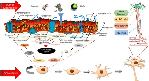
Berry et al demonstrated extensive changes in gene expression of human fibroblasts when treated with MNPs.Citation37 These genes were associated with the receptors of growth factors, ion channels, collagen, laminin, cytoskeleton, and different proteins related to cell movement and cell interactions ().Citation38 Kim et alCitation29 proposed a hypothesis to presume the state of Fe ions. In this hypothesis, Fe ions were released from internalized MNPs due to acidic pH inside the internal organelles and promoted neuritogenesis.
Figure 10 Schematic representation of different treatments (NGF plus iron oxide nanoparticle and quercetin) effects on PC12 cell signaling during differentiation. Growth factors such as NGF are optimally activated by their ligands but only under the appropriate condition of cell attachment. MNPs and quercetin as the neuroprotective agents induced neurite outgrowth by an ERK-dependent process.
Abbreviations: ERK, extracellular signal-regulated kinase; NGF, nerve growth factor; MNP, magnetic nanoparticle; ECM, extracellular matrix.
How can quercetin affect differentiation? The data presented in our study demonstrated that neurite length was significantly affected by the quercetin treatment. This flavonoid is capable of inducing PC12 cell differentiation and can surprisingly enhance the efficacy of NGF function.Citation27
Tangsaengvit et al demonstrated that quercetin at a very low concentration of 1 nm improved the viability of P19-derived neurons, considerably increased neuritogenesis.Citation39 Ishige et al demonstrated that certain flavonoids can protect different cell lines from oxidative stress. Some of these fla-vonoids (such as quercetin and fisetin) are also capable of inducing PC12 cell differentiation.Citation19
The bioflavonoids fisetin and quercetin were reported to have a stimulatory effect on alkaline phosphatase activity through the ERK signaling pathway.Citation40,Citation41 Quercetin has an additional hydroxyl group at C-5 than fisetin. Therefore, based on quercetin antioxidant properties, this bioflavonoid has the ability to improve differentiation through modulation of signal cascades.Citation19,Citation26
Sagara et al reported that four-oxo group and 2,3-double bond in the C ring of quercetin play a significant role in quercetin neuroprotective effects.Citation42,Citation43 They indicated that Ras-ERK cascade is essential for quercetin-induced PC12 cell differentiation. Also, the results of Sagara et al showed that three different mitogen/extracellular signal-related kinase inhibitors block both flavonoid-induced ERK activation and differentiation of PC12 cells.Citation26
In this study, we examined neurite branching morphogenesis in PC12 cells under different treatments. We present in vitro results that demonstrate neuritogenesis and neurite arborization in the presence of extracellular factors. We established that iron oxide nanoparticles can enhance neurite branching and produce more complex branching trees and that quercetin can increase the length of neurites. Combination of MNPs with quercetin leads to considerable enhancement in the neurite lengths and branching points.
How do neurite branches form during the development of PC12 cells? Actin rearrangements are extremely controlled by actin-associated proteins, which have essential roles in neuritogenesis and neurite arborization (). A highly dynamic area is found at the tips of growing neurites, where extreme rearrangements of actin filaments and microtubules occur during neurite elongation.Citation44 Recent studies have shown the importance of enabled/vasodilator-stimulated phosphoprotein proteins, formins, actin-related protein 2/3, and cordon-bleu in the neurite morphogenesis.Citation45,Citation46
In the present study, we have described a new effective combination of quercetin and iron oxide nanoparticles with neuroprotective effects that are capable of promoting neuritogenesis at a high level. This combination has great potential for increasing neuronal differentiation and may function as a novel therapeutics in the field of neuronal regeneration and repair.
Conclusion
While it is well recognized that different cells can respond to various external signals during neuritogenesis and neurite arborization, only limited studies have examined the effects of SPIONs and quercetin on neural differentiation parameters. The results of this study suggest that SPIONs and quercetin have considerable effects on neuritogenesis, specifically the neurite arborization. Findings of the present study showed that the combination of iron oxide nanopar-ticles with a flavonoid can be an appropriate candidate for improving the efficiency of NGF during differentiation.
Data sharing statement
The data sets used and/or analyzed during the present study are available from the corresponding author upon reasonable request.
Author contributions
All authors contributed toward data analysis, drafting and revising the paper, gave final approval of the version to be published and agree to be accountable for all aspects of the work.
Acknowledgments
We thank the vice chancellor for research and technology of the University of Isfahan. The authors are grateful to the University of Isfahan for providing support to undertake this work. This work was supported by the grant from the University of Isfahan.
Disclosure
The authors report no conflicts of interest in this work.
References
- SchmidtCELeachJBNeural tissue engineering: strategies for repair and regenerationAnnu Rev Biomed Eng20035129334714527315
- KalilKDentEWBranch management: mechanisms of axon branching in the developing vertebrate CNSNat Rev Neurosci201415171824356070
- StollGMüllerHWNerve injury, axonal degeneration and neural regeneration: basic insightsBrain Pathol19999231332510219748
- GreeneLATischlerASEstablishment of a noradrenergic clonal line of rat adrenal pheochromocytoma cells which respond to nerve growth factorProc Natl Acad Sci Unit States Am197673724242428
- TsujiMInanamiOKuwabaraMInduction of neurite outgrowth in PC12 cells by alpha-phenyl-N-tert-butylnitron through activation of protein kinase C and the Ras-extracellular signal-regulated kinase pathwayJ Biol Chem200127635327793278511438521
- GuptaAKGuptaMSynthesis and surface engineering of iron oxide nanoparticles for biomedical applicationsBiomaterials200526183995402115626447
- ItoAShinkaiMHondaHKobayashiTMedical application of functionalized magnetic nanoparticlesJ Biosci Bioeng2005100111116233845
- LitovskySMadjidMZarrabiACasscellsSWWillersonJTNaghaviMSuperparamagnetic iron oxide-based method for quantifying recruitment of monocytes to mouse atherosclerotic lesions in vivo: enhancement by tissue necrosis factor-alpha, interleukin-1beta, and interferon-gammaCirculation2003107111545154912654614
- HongJ-HNohK-MYooY-EIron promotes the survival and neurite extension of serum-starved PC12 cells in the presence of NGF by enhancing cell attachmentMol Cells2003151101912661755
- Enteshari NajafabadiRKazemipourNEsmaeiliABeheshtiSNazifiSUsing superparamagnetic iron oxide nanoparticles to enhance bioavailability of quercetin in the intact rat brainBMC Pharmacol Toxicol20181915930253803
- Kotake-NaraETakizawaSQuanJWangHSaidaKCobalt chloride induces neurite outgrowth in rat pheochromocytoma PC-12 cells through regulation of endothelin-2/vasoactive intestinal contractorJ Neurosci Res200581456357115948191
- LeinPGallagherPJAmodeoJHowieHRothJAManganese induces neurite outgrowth in PC12 cells via upregulation of alpha(v) integrinsBrain Res2000885222023011102576
- RothJAHorbinskiCHigginsDLeinPGarrickMDMechanisms of manganese-induced rat pheochromocytoma (PC12) cell death and cell differentiationNeurotoxicology200223214715712224755
- HäfeliUORiffleJSHarris-ShekhawatLCell uptake and in vitro toxicity of magnetic nanoparticles suitable for drug deliveryMol Pharm2009651417142819445482
- YarjanliZGhaediKEsmaeiliARahgozarSZarrabiAIron oxide nanoparticles may damage to the neural tissue through iron accumulation, oxidative stress, and protein aggregationBMC Neurosci20171815128651647
- SinghNJenkinsGJSAsadiRDoakSHPotential toxicity of superparamagnetic iron oxide nanoparticles (SPION)Nano Reviews2010115358
- OberdörsterGStoneVDonaldsonKToxicology of nanoparticles: a historical perspectiveNanotoxicology200711225
- StrohAZimmerCGutzeitCIron oxide particles for molecular magnetic resonance imaging cause transient oxidative stress in rat macrophagesFree Radic Biol Med200436897698415059638
- IshigeKSchubertDSagaraYFlavonoids protect neuronal cells from oxidative stress by three distinct mechanismsFree Radic Biol Med200130443344611182299
- ChoiD-YLeeY-JHongJTLeeH-JAntioxidant properties of natural polyphenols and their therapeutic potentials for Alzheimer’s diseaseBrain Res Bull2012872–314415322155297
- LauFCShukitt-HaleBJosephJAThe beneficial effects of fruit polyphenols on brain agingNeurobiol Aging200526112813216194581
- CaoGSoficEPriorRLAntioxidant and prooxidant behavior of flavonoids: structure-activity relationshipsFree Radic Biol Med19972257497609119242
- KumariAYadavSKPakadeYBSinghBYadavSCDevelopment of biodegradable nanoparticles for delivery of quercetinColloids Surf B2010802184192
- WuT-HYenF-LLinL-TTsaiT-RLinC-CChamT-MPreparation, physicochemical characterization, and antioxidant effects of quercetin nanoparticlesInt J Pharm20083461–216016817689897
- MurakamiAAshidaHTeraoJMultitargeted cancer prevention by quercetinCancer Lett2008269231532518467024
- SagaraYVanhnasyJMaherPInduction of PC12 cell differentiation by flavonoids is dependent upon extracellular signal-regulated kinase activationJ Neurochem20049051144115515312169
- ChanGKLHuWWHZhengZXQuercetin potentiates the NGF-induced effects in cultured PC12 cells: identification by HerboChips showing a binding with NGFEvid Based Complementary Altern Med20182018119
- MosmannTRapid colorimetric assay for cellular growth and survival: application to proliferation and cytotoxicity assaysJ Immunol Methods1983651–255636606682
- KimJALeeNKimBHEnhancement of neurite outgrowth in PC12 cells by iron oxide nanoparticlesBiomaterials201132112871287721288566
- AlexiouCJurgonsRSeligerCBrunkeOIroHOdenbachSDelivery of superparamagnetic nanoparticles for local chemotherapy after intraarterial infusion and magnetic drug targetingAnticancer Res2007274A2019202217649815
- SmithSW“Reticular” and “Areticular” nissl bodies in sympathetic neurons of a lizardJ Cell Biol1959617784
- NelAXiaTMädlerLLiNToxic potential of materials at the nano-levelScience2006311576162262716456071
- PisanicTRBlackwellJDShubayevVIFiñonesRRJinSNano-toxicity of iron oxide nanoparticle internalization in growing neuronsBiomaterials200728162572258117320946
- KeeganKHalegouaSSignal transduction pathways in neuronal differentiationCurr Opin Neurobiol19933114198453284
- YooY-EHongJ-HHurKCOhE-SChungJ-MIron enhances NGF-induced neurite outgrowth in PC12 cellsMol Cells200417234034615179052
- PangLSawadaTDeckerSJSaltielARInhibition of MAP kinase kinase blocks the differentiation of PC-12 cells induced by nerve growth factorJ Biol Chem19952702313585135887775407
- BerryCCCharlesSWellsSDalbyMJCurtisASGThe influence of transferrin stabilised magnetic nanoparticles on human dermal fibroblasts in cultureInt J Pharm2004269121122514698593
- PolakPShefiONanometric agents in the service of neuroscience: manipulation of neuronal growth and activity using nanoparticlesNanomedicine20151161467147925819887
- TangsaengvitNKitphatiWTadtongSBunyapraphatsaraNNukoolkarnVNeurite outgrowth and neuroprotective effects of querce-tin from Caesalpinia mimosoides lamk on cultured P19-derived neuronsEvidence Based Complementary Altern Med20132013217
- MaherPAkaishiTAbeKFlavonoid fisetin promotes ERK-dependent long-term potentiation and enhances memoryProc Natl Acad Sci200610344165681657317050681
- ProuilletCMazièreJ-CMazièreCWattelABrazierMKamelSStimulatory effect of naturally occurring flavonols quercetin and kaempferol on alkaline phosphatase activity in MG-63 human osteoblasts through ERK and estrogen receptor pathwayBiochem Pharmacol20046771307131315013846
- PuFMishimaKIrieKNeuroprotective effects of quercetin and rutin on spatial memory impairment in an 8-arm radial maze task and neuronal death induced by repeated cerebral ischemia in ratsJ Pharmacol Sci2007104432933417666865
- OraveczKKalkaDJeneyFCantzMZs-NagyIHydroxyl free radicals induce cell differentiation in SK-N-MC neuroblastoma cellsTissue and Cell2002341333811989968
- DentEWGuptonSLGertlerFBThe growth cone cytoskeleton in axon outgrowth and guidanceCold Spring Harb Perspect Biol201133a00180021106647
- BearJEGertlerFBEna/VASP: towards resolving a pointed controversy at the barbed endJ Cell Sci2009122121947195319494122
- KorobovaFSvitkinaTArp2/3 complex is important for filopodia formation, growth cone motility, and neuritogenesis in neuronal cellsMol Biol Cell20081941561157418256280