This article refers to:
Mosselhy DA, He W, Hynönen U, et al. Int J Nanomedicine. 2018;13:7939–7957.
On page 7945, parts were labeled incorrectly. The correct figure is as follows:
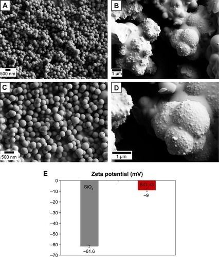
Figure 1 SEM images of the materials.
Notes: (A, C) Smooth spherical pristine SiO2 NPs and (B, D) granular rough aggregated network of SiO2-G nanohybrids at the magnifications of 10,000× (A, B) and 20,000× (C, D). (E) The zeta potential of the pristine SiO2 NPs and SiO2-G nanohybrids in deionized water. The error bars represent the standard errors of the means.
Abbreviations: SEM, scanning electron microscope; SiO2 NPs, silica nanoparticles; SiO2-G, silica–gentamicin.