
Abstract
Background
Magnetic nanoparticles (MNPs) have been successfully tested for several purposes in medical applications. However, knowledge concerning the effects of nanostructures on elderly organisms is remarkably scarce.
Purpose
To fill part of this gap, this work aimed to investigate biocompatibility and bio-distribution aspects of magnetic nanoparticles coated with citrate (NpCit) in both elderly and young healthy mice.
Methods
NpCit (2.4 mg iron) was administered intraperitoneally, and its toxicity was evaluated for 28 days through clinical, biochemical, hematological, and histopathological examinations. In addition, its biodistribution was evaluated by spectrometric (inductively coupled plasma optical emission spectrometry) and histological methods.
Results
NpCit presented age-dependent effects, inducing very slight and temporary biochemical and hematological changes in young animals. These changes were even weaker than the effects of the aging process, especially those related to the hematological data, tumor necrosis factor alpha, and nitric oxide levels. On the other hand, NpCit showed a distinct set of results in the elderly group, sometimes reinforcing (decrease of lymphocytes and increase of monocytes) and sometimes opposing (erythrocyte parameters and cytokine levels) the aging changes. Leukocyte changes were still observed on the 28th day after treatment in the elderly group. Slight evidence of a decrease in liver and immune functions was detected in elderly mice treated or not treated with NpCit. It was noted that tissue damage or clinical changes related to aging or to the NpCit treatment were not observed. As detected for aging, the pattern of iron biodistribution was significantly different after NpCit administration: extra iron was detected until the 28th day, but in different organs of elderly (liver and kidneys) and young (spleen, liver, and lungs) mice.
Conclusion
Taken together, the data show NpCit to be a stable and reasonably biocompatible sample, especially for young mice, and thus appropriate for biomedical applications. The data showed important differences after NpCit treatment related to the animals’ age, and this emphasizes the need for further studies in older animals to appropriately extend the benefits of nanotechnology to the elderly population.
Introduction
Advances in nanotechnology strongly affect biomedical applications, enabling new approaches to be developed for diagnosis and therapy.Citation1 Several nanostructured materials are potentially useful in this area, and among these, platforms based on magnetic nanoparticles (MNPs) may be highlighted.Citation2–Citation4 MNPs based on iron oxides, such as magnetite (Fe3O4) and maghemite (γ-Fe2O3), have been widely investigated for in vivo applications because of their potential biocompatibility. To achieve the indispensable colloidal and chemical stability,Citation5 MNPs are dispersed in a carrier solvent and coated with a stabilizing molecular layerCitation6 provided by several different molecules, such as dextran,Citation7 dimercaptosuccinic acid,Citation8 polyaspartic acid,Citation9 or citrate.Citation10
Several MNP-based systems have succeeded in pre-clinical and clinical tests/purposes, such as delivery and controlled release of drugs to specific sites, enhancement of contrast in magnetic resonance imaging, and magnetic hyperthermia for tumor treatment.Citation11–Citation13 However, despite the increasing number of studies and publications related to the biomedical use of MNPs, little is known about their use in elderly organisms.
Although aging is not a disease, it is the main risk factor for chronic diseases.Citation14 Age-related physiological, immunological, and genetic changes, associated with the concomitant presence of different diseases and possible drug interactions, make the treatment of elderly patients particularly challenging.Citation15,Citation16 In this context, new therapeutic strategies using MNPs may represent powerful alternatives in the treatment of age-associated diseases, like neoplasms. However, the lack of studies makes it difficult to assess nanoparticle treatments in elderly organisms.
Li et alCitation17 emphasized that understanding the effects of nanostructure exposure on the elderly is critical, and therapies developed that are based on nanoparticle use should involve suitable animal models with special attention to nanotoxicity. In fact, the possible toxicity of MNPs is a priority in nanomedicine.Citation18 Kaur et alCitation19 highlighted that although the use of MNPs for therapeutic purposes, such as hyperthermia, has been announced as an important biomedical advancement, concerns about their biocompatibility need to be addressed individually for each formulation and administration, especially in elderly organisms. Besides that, some studies show under representation of elderly patients in clinical trials and, consequently, a lack of standardized treatments and clear management guidelines for these patients.Citation20 In this context, the present work aimed to evaluate the effects of age on biocompatibility and biodistribution aspects of maghemite nanoparticles coated with citrate (NpCit) administered to elderly and young mice.
Materials and methods
Synthesis and characterization of magnetic nanoparticles coated with citrate
The NpCit sample was synthesized and kindly supplied by Dr Emilia CO Lima (UFG, Goiania, GO, Brazil). Maghemite (γ-Fe2O3) nanoparticles were obtained via the oxidation of magnetite nanoparticles synthesized by the alkaline hydrolysis of ions Fe(II) and Fe(III).Citation21 γ-Fe2O3 nanoparticles were functionalized with citrate ions as described previously.Citation22 The functionalized nanoparticles were dispersed in water, yielding stable colloidal suspension at neutral pH. The total iron concentration in the colloidal suspension and the Fe(III)/Fe(II) ratio were determined by atomic absorption and colorimetric analysis, respectively. Diffraction pattern was analyzed using a Shimadzu XRD-600 diffractometer. The nanoparticle shape and size dispersity were determined by transmission electron microscopy (TEM; Jeol JEM-1011). The stability of NpCit colloidal suspension was verified through several characteristics, namely the polydispersity index (PDI), zeta potential, pH, and hydrodynamic size, using a Zetasizer NanoZS (Malvern Instruments). The measurements were performed in triplicate, at 25°C, with a fixed detection angle of 173°.
Animals, NpCit treatment, and experimental design
Elderly (E) non-isogenic Swiss mice aged 14–16 months (weighing 40±5 g) and young (Y) mice aged 03–04 months (weighing 30±5 g) were used. All the procedures of housing and handling of animals were carried out according to the international practices for animal use and care, after being approved by the Animal Ethics Committee of the Institute of Biological Sciences at the University of Brasilia/Brazil (CEUA, reference number 17672/2016).
A single injection of NpCit containing 2.4 mg iron (150 µL) was administered intraperitoneally into the animals.Citation23 PBS was used in elderly (EC) and young (YC) control groups (n=09).
Biological material was collected at 1, 7, and 28 days after the NpCit injection. Elderly treated groups (n=07) were accordingly named E1, E7, and E28, while young treated groups (n=05) were named Y1, Y7, and Y28. The biodistribution and biocompatibility aspects were evaluated. Comparisons of data obtained from the treated groups and the corresponding controls, as well as from the elderly and the corresponding young groups, were performed.
Biocompatibility tests
Biocompatibility evaluations of NpCit sample were performed through clinical observations (loss of weight, diarrhea, alopecia, inappetence, motor disorders, and salivary gland secretions).Citation24 Changes in biochemical and hemogram indexes, production levels of inflammatory cytokines tumor necrosis factor alpha (TNF-α) and nitric oxide (NO), and histological analyses were examined.
All animals (elderly and young) were monitored from 2 weeks before the beginning of the experiments, and twice a week during the experimental period. The animals were weighed at the time of NpCit or PBS administration, immediately before euthanasia, and weekly in the case of groups euthanized 28 days after treatment (E28/Y28).
Biochemical analyses were performed specifically to assess hepatic, renal, tissue damage, and nutritional status due to administration of NpCit. After obtaining a total of 700 µL of blood by cardiac puncture, the serum was separated by centrifugation and used for evaluations of alanine amino-transferase (ALT), aspartate aminotransferase (AST), lactate dehydrogenase (LDH), creatinine K, urea, and albumin, using a ChemWell-T automatic biochemical analyzer (Labtest, Brazil).
Blood samples (300 µL) were used for evaluating the possible effects of NpCit on the inflammatory, allergic, and anemia processes through cell count and cell type identification (leukogram, platelet, and erythrogram), using a multiple automated hematology analyzer for veterinary use, the Sysmex pocH-100iV Diff (Curitiba/Paraná, Brazil).
Effects of NpCit injection on the induction of inflammatory process were also investigated through quantification of levels of TNF-α in the serum of mice by the ELISA, according to the manufacturer’s instructions (eBioscience and R&D System). Plates were read at 450 nm using a SpectraMax M3 spectrophotometer (Molecular Devices, Sunnyvale, CA, USA). In addition, measurement of NO production was detected by using Griess reagent. After incubating the plate with this reagent at room temperature, the OD was determined in a SpectraMax M3 (Molecular Devices) at a test wavelength of 540 nm.
For analyses of morphology and acute toxicity of NpCit, the brain, spleen, liver, lungs, and kidneys were removed from elderly and young animals. Fragments of these organs were fixed in 4% buffered paraformaldehyde, dehydrated in alcohol solutions, diaphanized in xylol, and embedded in paraffin. Tissue sections (3–5 µm) were stained with H&E and analyzed under a Zeiss Axiophot light microscope.
NpCit distribution analyses
Biodistribution aspects of NpCit nanoparticles were evaluated in two different ways: quantification of iron in the organs and histological analysis.Citation25 After euthanasia, part of the organs (liver, spleen, lung, kidneys, and brain) and the blood were processed for determination of the amount of iron through inductively coupled plasma optical emission spectrometry (ICP-OES) using an Optima 8000 ICP-OES Spectrometer.Citation26 For histological analysis, slides were submitted to Perls’ staining methods with nuclear flash red. In 3–5 µm sections, the presence of iron clusters in the tissues was detected with a Zeiss Axiophot light microscope.
Statistical analyses
Statistical analyses were performed using SPSS software version 18 and Prisma version 6.01. The normality of the continuous variables was evaluated by the Shapiro–Wilk test. Differences between the analyzed groups were investigated through ANOVA (for data following normal distribution) or Kruskal–Wallis (in the case of non-normal distribution) test. For significant results of ANOVA, pairwise comparisons were performed by Student’s t-test or Mann–Whitney U test according to the distribution of normality.
Results
Nanoparticle characterization
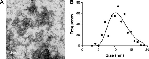
The characterization data of the NpCit sample are summarized in . The visual observation showed the NpCit sample to be in red-brown color, characteristic of maghemite, and absence of detectable nanoparticle precipitation, even in the presence of a magnetic field, despite the high concentration of nanoparticles (18×1018 particles/mL). Transmission electron micrographs showed NpCit nanoparticles with spherical-like shapes (). The random counting of 441 MNPs in TEM revealed a mean diameter of 10.8±2.7 nm, as obtained by the log normal curve (). Measurements, especially zeta potential (−40 mV) and PDI (0.290), indicated the high colloidal stability of the sample at pH =7.Citation21
Table 1 Characterization of the maghemite (γ-Fe2O3) nanoparticles functionalized with citrate ions (NpCit) and the magnetic colloidal suspension
Figure 1 (A) TEM micrograph (scale bar: 200 nm) and (B) histogram of MNP size distribution.
Abbreviations: TEM, transmission electron microscopy; MNP, magnetic nanoparticle.
Biocompatibility evaluation
The intraperitoneal treatment of mice with NpCit sample (2.4 mg iron) did not induce obvious clinical or behavioral changes such as hair loss, diarrhea, inappetence, salivary gland secretions, and motor dysfunction throughout the entire experimental period in all elderly and young animals investigated. Further, NpCit did not induce significant variation in the animal weight in elderly and young treated groups when compared to their corresponding controls.
The biochemical analyses () showed through comparisons with non-treated controls that elderly animals (EC) presented lesser amounts of ALT and albumin than young animals (YC). However, the administration of NpCit induced different effects in elderly and young animals. In the elderly group, a temporary significant decrease in the urea and an increase in the albumin levels were observed at day 1 after NpCit treatment (E1) in comparison to the control (EC). Levels of the enzymes AST, creatinine K, ALT, and LDH were not significantly different in all the treated elderly groups (E1, E7, and E28) compared to EC, at any point of time.
Table 2 Biochemical analysis of the elderly and young mice after treatment with NpCit (2.4 mg iron)
In the young animals, NpCit administration caused significant decreases in urea, albumin, and LDH levels at day 7 after NpCit injection (Y7) compared to the YC group. In this group, NpCit did not cause alterations in the ALT, AST, and creatinine K levels in all treated young groups (Y1, Y7, and Y28) compared to the control (YC).
The leukogram analysis () showed, through comparisons between the elderly groups and their corresponding young groups, that the elderly animal groups EC, E1, E7, and E28 presented significant decreases in the rate of lymphocytes (small cell rate [lymphocytes], W-SCR) and in the absolute counting of lymphocytes (absolute small cell count [lymphocytes], W-SCC) when compared to YC, Y1, Y7, and Y28, respectively. In addition, the same elderly groups showed significant increases in the mean cell rates of basophils, eosinophils, and monocytes (W-MCR), when compared to the young groups.
Table 3 Effects of NpCit (2.4 mg iron) treatment on the leukogram and platelet parameters of elderly and young mice
In the elderly groups, the NpCit treatment induced significant decreases in the rate of lymphocytes (W-SCR) observed in the day 1 group (E1) and in the day 28 group (E28) compared to the EC group. In contrast, significant increases in the mean cell rate (basophils, eosinophils, and monocytes) were also observed in groups E1 and E28, compared to EC.
In the young groups (Y1, Y7, and Y28), different from the elderly groups, NpCit did not induce any significant alterations in the leukocyte parameters as compared to the control young group (YC).
Regarding platelets, significant differences were not found either in comparison between the control elderly and young groups, or in comparison of NpCittreated elderly animals with the control elderly mice (). The only significant alterations in platelet count were observed in the young group at day 7 (Y7) in comparison with both the control young animals (YC) and the treated elderly animals, also at day 7 (E7).
The erythrogram analysis () revealed that the aging process caused significant alterations in most of the parameters investigated. In fact, the elderly groups in comparison to their corresponding young groups showed significant decreases in the total erythrocyte counts (RBC) for E1, E7, and E28 groups compared to their corresponding young groups Y1, Y7, and Y28. Besides, significant decreases in the hemoglobin (HGB) concentration, mean corpuscular volume, and hematocrit values were observed for EC, E1, E7, and E28 compared to YC, Y1, Y7, and Y28 groups, respectively. On the other hand, mean corpuscular hemoglobin was significantly higher in EC group than in YC group, while the red cell distribution width was higher in EC, E1, and E28 than in YC, Y1, and Y28 groups, respectively ().
Table 4 Erythrogram analysis of the elderly and young mice observed at different time points after NpCit (2.4 mg iron) administration
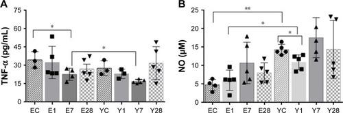
Concerning the pro-inflammatory cytokines, NpCit administration in elderly mice triggered a decrease in TNF-α levels at day 7 (E7) compared to the untreated mice (EC). There was also a significant decrease of TNF-α levels in young mice submitted to NpCit administration at day 7 (Y7) compared to the corresponding elderly mice (E7), as shown in . Moreover, there was a decrease in NO levels in the serum of untreated control elderly mice (EC) and treated young mice at day 1 (Y1), when both groups were compared to control young mice (YC) ().
Figure 2 Effects of NpCit (2.4 mg iron) injection on the TNF-α and NO levels in serum of elderly and young mice.
Notes: (A) TNF-α levels were detected by ELISA and (B) NO levels were detected by Griess assay. Asterisks indicate significant (*P<0.05) and highly significant (**P<0.01) differences.
Abbreviations: NpCit, nanoparticles coated with citrate; TNF-α, tumor necrosis factor alpha; NO, nitric oxide; EC, elderly control; E1, E7, and E28, elderly groups investigated at day 01, day 07, and day 28 after NpCit treatment, respectively; YC, young control; Y1, Y7, and Y28, young treated groups investigated at day 01, day 07, and day 28, respectively.
The histological analysis of tissues after H&E staining showed no abnormalities in all tissues analyzed for both young and elderly animals, treated or not treated with NpCit sample (data not shown). Briefly, the brains had no pathologies in the white and gray matter; in the spleens, both red and white pulps were easily distinguishable and there were no large numbers of white cells; the observed livers presented hepatocytes in polyhedral shape, radiating from a central vein; in the lungs, there were no signs of fibrosis; and in the kidneys, cells from glomerulus, proximal, and distal convoluted tubules presented normal patterns.
NpCit biodistribution
The NpCit biodistribution assays in the organs and blood performed by ICP-OES are shown in . Comparisons between EC and YC animals showed that although the amount of iron in the blood was similar in both groups (), the spleen, liver, kidney, lung, and brain presented a significantly higher concentration of iron in the elderly group () than in the young group.
Figure 3 Biodistribution of iron as obtained by inductively coupled plasma optical emission spectrometry (ICP-OES).
Notes: Concentration of iron in the blood (µg/mL) (A) and in the organs (µg/g) in decreasing order from spleen to brain (B–F). Asterisks indicate significant (*P<0.05) and highly significant (**P<0.01) differences.
Abbreviations: EC, elderly control; E1, E7, and E28, elderly groups investigated at day 01, day 07, and day 28 after NpCit treatment, respectively; YC, young control; Y1, Y7, and Y28, young treated groups investigated at day 01, day 07, and day 28, respectively.
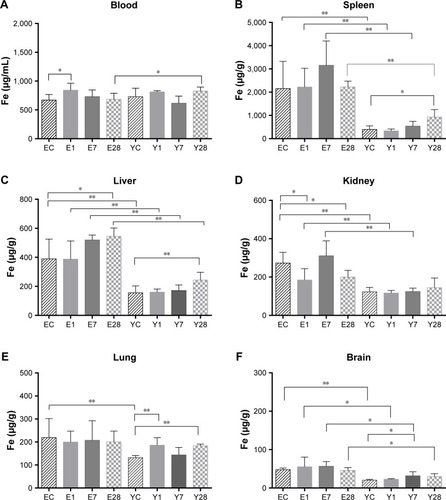
In fact, the distribution of iron in both animal groups was also different; while the EC group presented the iron in descending order, spleen > blood > liver > kidney > lung > brain, in the YC mice, the order was blood > spleen > liver > lung > kidney > brain.
Concerning the effects induced by NpCit in the elderly groups, there were significant changes in comparison with the control untreated mice: an increase of iron levels in the blood and decrease in the kidneys at day 1 and an increase in the liver at day 28. The significant effects of NpCit on the young treated groups when compared to the YC group were different from the elderly groups and characterized by iron level increases in the lungs at day 1, in the brain at day 7, and in the spleen, liver, and lungs at day 28.
Due to the initial differences in the amount of iron between elderly and young groups, the comparison of elderly treated animals (E1, E7, and E28) with the corresponding young treated groups (Y1, Y7, and Y28) led to significant differences, particularly in the spleen, liver, and brain, as shown in .
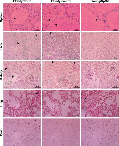
Through microscopic observation after Perls’ staining (), it was possible to observe iron aggregates in all the analyzed organs, except in the brain. In general, the concentration was higher in the elderly animals than in the young animals, in both NpCit treated and control groups. The highest iron concentration was observed in the spleen of elderly mice of the day 7 group. In this group, the iron concentration was higher than in the EC group or the corresponding young group (Y7). In the young groups, highest concentrations in the spleen were observed at day 28.
Figure 4 Effects of NpCit on iron distribution of elderly (left column) and young (right column) animals. Organ sections of elderly control animals without treatment are presented in the central column. Histological sections of spleen, liver, kidneys, lungs, and brain were submitted to Perls’ blue staining. Arrows indicate blue positive aggregates which are more evident in the spleen. Micrographs are illustrative and were taken at day 7 and day 28 of treatment, respectively, from elderly and young animals. Scale bar =50 µm.
Abbreviation: NpCit, nanoparticles coated with citrate.
To facilitate the comparison between the effects related to aging and/or the NpCit treatment, a table containing a summary of the main effects of aging and/or NpCit treatment in mice is presented ().
Discussion
This work was performed to compare the effects of citrate-coated MNPs on healthy elderly and young mice, focusing on biocompatibility and biodistribution aspects. Citrate is a biocompatible small molecule that has been widely accepted as the capping agent to stabilize water-dispersible MNPs for biomedical applications, and it is even commercially available. This is attributed to the fact that citrate is capable of adsorption onto the surface of MNPs (especially iron oxides) by coordinating via one or two carboxylate groups, leaving at least one carboxylate group exposed to the bulk medium. This makes the nanoparticle surface hydrophilic, negatively charged in a wide range of pH (including the physiological one), and cytocompatible, preventing particle agglomeration and providing functional groups to be used for further surface modification. Moreover, citrate detachment from the surface of MNPs under physiological conditions is negligible, and the physical–chemical characteristics of citrate-capped MNPs can be preserved over years.Citation21,Citation27 Indeed, previous in vivo investigations have shown the biocompatibility and non-mutagenic effects of citrate coatings,Citation28–Citation30 in spite of adverse effects described in in vitro tests.Citation31
However, citrate-coated MNPs can be colloidally unstable in a biological system when dispersed in a medium with high ionic strength, such as PBS buffer. Sharma et alCitation32 reported coagulation of citrate-stabilized iron oxide particles in PBS, a high increase of the hydrodynamic diameter (>15-fold), and formation of precipitates. To avoid this undesirable effect, in our work, dilution/dispersion of citrate-capped MNPs was performed in water (ie, low ionic strength medium). Therefore, zeta potential (-40 mV), hydrodynamic size (~78 nm), and PDI (0.290) indicated the high colloidal stability of diluted NpCit samples at pH =7.
Szigeti et alCitation27 showed that citric acid as a surface-capping agent to control size and biocompatibility and to avoid agglomeration of the synthesized particles is able to chelate Ca2+ ions. However, physiological Ca2+ concentration does not affect in vivo colloidal properties. The authors affirm that at the physiological pH of 7.4, the zeta potential of citrate-coated MNPs and the steric stabilization effect of citrate coating further enhance the colloidal stability. Thus, even if there are other ions in addition to Ca2+ and other complexing molecules, the citrate-coated samples present considerable biological stability and thus suitability for use in in vivo tests.Citation27,Citation33,Citation34
Concerning the in vivo biocompatibility tests, differences related to the aging process and some significant changes induced by the NpCit sample were observed. Liver and renal functions represent important predictors of toxicity and aging. Dysfunctions in the hepatobiliary system represent more than 50% of toxicity events occasioned by chemical agents.Citation35 These are traditionally assessed by measuring the serum levels of AST and ALT.Citation36,Citation37 AST is widely distributed in the organismCitation38 and was not altered by mice aging or after NpCit treatment, suggesting no damage in the cytoplasmic and mitochondrial membranes.Citation36 In accordance with this, the administration of NpCit did not induce changes in the ALT enzyme levels of young or elderly animals, despite the presence of higher levels of iron in the liver. However, ALT which is usually produced in high concentrations in the liver presented a twofold reduction in the EC animals, thus indicating possible liver cell lesions in this groupCitation38–Citation41 with a subsequent decrease in the mitochondrial number,Citation14 an aging characteristic not detected in this study.
Kidney function was evaluated by serum creatinine and urea levels.Citation37 The absence of creatinine level changes that are related to aging or NpCit treatment is indicative of preserved kidney function.Citation42,Citation43 Indeed, no increase was observed in serum urea, which may derive from a decreased glomerular filtrationCitation37 related to aging or nephrotoxicity. On the other hand, a decrease in serum urea may be associated with liver impairment, since this organ is responsible for the production of urea during protein catabolism.Citation42 NpCit led to a reduction of urea levels in both elderly and young animals, but this effect was temporary and not observed at the 28th day after treatment, even if the iron concentration was still higher than usual at this time.
Other biochemical changes were investigated through albumin and LDH levels. Albumin is an abundant serum protein produced in the liver.Citation44 In this study, the albumin level in elderly animals appeared lower than in young animals, another sign of possible kidney or liver dysfunction consistent with the aging process. However, no significant differences in the LDH enzyme levels were observed between elderly and young groups, nor were they seen after NpCit treatment, indicating once again that there was no damage to the body’s tissues or any production of excessive free radicals related to the NpCit treatment or even to aging.
The hemogram analysis showed the predominance of myeloid cells in the elderly group and higher lymphoid cells in the young group, considered a hallmark difference between elderly and young mice.Citation45 Lymphocytes are crucial for the immune response,Citation42 and the decrease in lymphocyte-dependent immunity could increase oxidative stress and foment diseases related to aging.Citation46 Remarkably, these differences were even more pronounced and durable after NpCit treatment of elderly mice. At the same time, NpCit did not affect leukocyte populations in young groups, evidencing one important aspect of nanoparticle applications to be taken into account in older organisms. On the other hand, myeloid cells are responsible for the phagocytosis of foreign particles, and their increase in the elderly group could explain why the increase of iron in the blood was observed only on the first day after the NpCit injection. Interestingly, erythrogram differences were essentially only related to the aging process.
The pro-inflammatory cytokine TNF-α may be associated with the triggering mechanisms of typical age-related diseases and inflammatory processes.Citation47 Accordingly, a tendency to having a higher level of TNF-α was observed in the EC group. Nevertheless, significant TNF-α decreases were temporarily induced 7 days after NpCit treatment. The other cytokine investigated, NO, was also modulated after the NpCit injection. NO is involved in many physiological and pathological processes and can have opposite actions such as damage or protection of cells during inflammatory response.Citation48 In this study, NO levels in the elderly mice were lower than in the young mice, despite a higher number of phagocytes observed in the elderly animals, whether they were treated or not with NpCit.Citation48 Low levels of NO production are typically involved in liver damage, and they can enhance the risk of several diseases associated with older individuals, which was not evidenced by biochemical data. In addition, although NpCit did not affect NO production in the elderly mice, it induced a temporary significant reduction of NO levels in young mice 1 day after the treatment. In a previous study, the NP-induced neurotoxicity was correlated with the increase of NO production,Citation34 indicating that different NP samples may have diverse actions in organisms.
The findings that the effects induced by NpCit in the young group were slight and no longer observed after the seventh day after treatment suggest the biocompatibility of the sample. This supposition is strongly supported by both the absence of histological alterations in all the organs investigated and absence of clinical changes usually associated with aging or toxicity.Citation24 It is reasonable to extend this conclusion to the elderly mice, but since they still presented some leukocyte alterations at day 28 after NpCit treatment, longer investigations should be carried out before finally reaching conclusions about the sample’s biocompatibility and safeness in biomedical applications for the elderly.
The presence of excessive iron was observed until the 28th day of NpCit treatment in both the elderly group (liver and kidneys) and the young group (spleen, liver, and lungs) as revealed by ICP-OES and histology studies. GrottoCitation49 has pointed out that an elevated amount of iron in tissues can damage lipid structures, proteins, and DNA, causing severe tissue damage or mutations. However, as mentioned earlier, signs of structural damage, including necrosis and even inflammation, were never observed throughout the whole study period, despite obvious high concentration of MNPs in some organs. Accordingly, young mice exposed to a bolus dose of dextran-coated magnetite NPs did not show morphological or ultramicroscopic alterations, even when the MNPs were retained for long term (6 months) in the liver and spleen.Citation50
It is well established that excessive iron, such as iron coming from metabolized NPs, is gradually deposited in various organs,Citation51,Citation52 especially in the spleen and liver,Citation32,Citation53,Citation54 corroborating the findings of the present study in both elderly and young animals. However, some differences related to age were found. Although the amount of iron detected in the blood of EC animals was slightly lower than that of YC groups, all the organs (spleen, liver, kidney, lung, and brain) of older mice presented a significantly higher concentration of iron than those of young animals, highlighting an important difference in iron biodistribution related to age.
Furthermore, the elderly and young animals presented a completely different iron biodistribution profile after NpCit injection. The significantly higher iron concentration in the liver at the 28th day after the injection, relative to their respective controls, was the only similar result between the two animal groups. The difference in iron distribution is another important aspect, because several diseases are associated with alterations in iron metabolismCitation55 and should be considered when investigating the biodegradation of iron oxide nanoparticles designed as a diagnostic or therapeutic agent for those diseases, especially in older animals.Citation34
In agreement with our results, some literature data have shown that older animals are more susceptible to the adverse effects of nanoparticles than young animals.Citation17 Inhalation of SiO2 nanoparticles under identical conditions caused more pulmonary and cardiovascular damage in elderly rats than in young animals.Citation56 Aluminum or copper nanoparticles induced higher neurotoxicity in older adult rats than in young adult rats, suggesting that the elderly animals are more vulnerable to brain damage induced by nanoparticles.Citation34 Furthermore, aged mice present more intestinal permeability and consequently more NP absorption, and a higher liver deposition and hence more liver injuries, which can culminate in increased oxidative stress and inflammation levels after NP administration.Citation57 In spite of the relevance of these data, an extensive search in the literature () concerning the administration of nanomaterials in older organisms and its effects reveals a huge gap in the area. Investigations have been performed in classical aging models such as Caenorhabditis elegansCitation58,Citation59 and Drosophila melanogaster,Citation60 and ratsCitation61 which showed opposite effects of NPs, and in animals deficient for antioxidant enzymes,Citation62 due to their similarities with aged organisms. Certainly, more information concerning nanotoxicity and biodistribution may come from the data obtained from aged patients treated with nanomaterials.Citation63–Citation65 In a recent review of patients with lung cancer treated with nanoparticles, Herrera et alCitation66 included data related to the safety and tolerability of nanomaterials, while highlighting the underrepresentation of elderly patients in the clinical trials.
Table 5 Nanoparticle investigations in aged organisms
Despite all the age-dependent effects observed in the present study, it is important to remember that the investigated elderly animals did not present any histological alterations or the clinical characteristics of old mice, thus evidencing they were in good health. Certainly, studies focusing on the safety of iron-based MNPs for animals bearing chronic diseases are mandatory for a better understanding of the effects of nanostructured materials on older organisms.
Conclusion
The NpCit sample induced age-dependent effects on mice. In the young animals, it caused very slight and temporary biochemical and hematological changes, such as changes in albumin and urea levels and HGB parameters. The changes associated with the natural aging process were more numerous and profound in the elderly than those induced by NpCit in the young animals. These aging changes made the elderly mice susceptible to NpCit effects that were either synergistic (leukocytes) or opposite (erythrocytes, albumin, TNF-α, and NO levels) to the aging changes and sometimes more durable (leukocytes). Iron distribution was also age dependent and showed different patterns until the 28th day of treatment. Remarkably, no significant changes were seen in the clinical studies or in the histological features after the treatment, indicating the biocompatibility of NpCit and its potential use in biomedical applications. However, the differences in data concerning the biodistribution and biocompatibility of nanoparticles between elderly and young animals emphasize the need for more studies performed for a longer time window to appropriately extend the benefits of nanotechnology to the elderly population.
Acknowledgments
We acknowledge Dr Emilia CO Lima from the Federal University of Goiás, Goiânia, GO, Brazil, for providing the NpCit sample, the Coordination for Further Training of Graduate Staff (CAPES), the Brazilian National Council for Technological and Scientific Development (CNPq), the Foundation to Support Research in the Federal District (FAPDF), the Network of Nanobiotechnology CON-NANO (CAPES), the National Institute of Science and Technology in Nanobiotechnology (INCT-Nanobiotecnologia), the Center for Nanoscience and Nanobiotechnology of the University of Brasília (CNANO-UnB), and the Dean of Research and Post-Graduation of the University of Brasília (DPP-UnB) for financial support.
Supplementary material
Table S1 Summary of effects of aging and/or NpCit treatment in mice
Disclosure
The authors report no conflicts of interest in this work.
References
- MohammadiZZackMSSeidiKBaratiMAkbarzadehAZarghamiNThe effect of chrysin loaded PLGA-PEG on metalloproteinase gene expression in mouse 4T1 tumor modelDrug Res (Stuttg)201767421121610.1055/s-0042-12213628166590
- HaleyBFrenkelENanoparticles for drug delivery in cancer treatmentUrol Oncol Semin Orig Investig200826576410.1016/j.urolonc.2007.03.015
- SteichenSDCaldorera-MooreMPeppasNAA review of current nanoparticle and targeting moieties for the delivery of cancer therapeuticsEur J Pharm Sci201348341642710.1016/j.ejps.2012.12.00623262059
- KossatzSGrandkeJCouleaudPEfficient treatment of breast cancer xenografts with multifunctionalized iron oxide nanoparticles combining magnetic hyperthermia and anti-cancer drug deliveryBreast Cancer Res201517111710.1186/s13058-014-0509-425567532
- CassimSMGiustiniAJBakerIDevelopment of novel magnetic nanoparticles for hyperthermia cancer therapyProc SPIE Int Soc Opt Eng2011790179011524619487
- BuskeNSonntagHGötzeTMagnetic fluids – their preparation, stabilization and applications in colloid scienceColloids and Surfaces19841219520210.1016/0166-6622(84)80099-2
- LacavaLMLacavaZGMAzevedoRBUse of magnetic resonance to study biodistribution of dextran-coated magnetic fluid intravenously administered in miceJ Magn Magn Mater200225236736910.1016/S0304-8853(02)00654-6
- ChavesSBLacavaLMLacavaZGMLight microscopy and magnetic resonance characterization of a DMSA-coated magnetic fluid in miceIEEE Trans Magn20023853231323310.1109/TMAG.2002.802495
- SadeghianiNBarbosaLSGuedesMHAMagnetic resonance of polyaspartic acid-coated magnetite nanoparticles administered in miceIEEE Trans Magn200541104108411010.1109/TMAG.2005.855334
- LacavaZGMAzevedoRBMartinsEVBiological effects of magnetic fluids: toxicity studiesJ Magn Magn Mater199920143143410.1016/S0304-8853(99)00002-5
- IvkovRMagnetic nanoparticle hyperthermia: A new frontier in biology and medicine?Int J Hyperthermia20136736870370510.3109/02656736.2013.857434
- CandidoNMCalmonMFTabogaSRHigh efficacy in hyperthermia-associated with polyphosphate magnetic nanoparticles for oral cancer treatmentJ Nanomed Nanotechnol20140503111
- WuWJiangCZRoyVALDesigned synthesis and surface engineering strategies of magnetic iron oxide nanoparticles for biomedical applicationsNanoscale2016847194211947410.1039/c6nr07542h27812592
- SheedfarFBiaseSDKoonenDLiver diseases and aging: friends or foes?Aging Cell201312695095410.1111/acel.1212823815295
- HastyPVijgJGenomic priorities in agingScience200229655711250125110.1126/science.107180811951000
- JenkinsEODealAMAndersCKAge-specific changes in intrinsic breast cancer subtypes: a focus on older womenOncologist201419101076108310.1634/theoncologist.2014-018425142841
- LiYZhangYYanBNanotoxicity overview: Nano-threat to susceptible populationsInt J Mol Sci20141533671369710.3390/ijms1503367124590128
- CaleroMChiappiMLazaro-CarrilloACharacterization of interaction of magnetic nanoparticles with breast cancer cellsJ Nano-biotechnology201513111510.1186/s12951-014-0062-4
- KaurPAliruMLChadhaASHyperthermia using nanoparticles – promises and pitfalls punitInt J Hyperthermia2016321768810.3109/02656736.2015.112088926757879
- OrucevicABreast cancer in elderly caucasian women – an institution-based study of correlation between breast cancer prognostic markers, tnm stage, and overall survivalCancers (Basel)2015731472148310.3390/cancers703084626264027
- ShresthaSJiangPSousaMHCitrate-capped iron oxide nanoparticles impair the osteogenic differentiation potential of rat mesenchymal stem cellsJ Mater Chem B20164224525610.1039/C5TB02007G
- MoraisPCSantosRLPimentaACMPreparation and characterization of ultra-stable biocompatible magnetic fluids using citrate-coated cobalt ferrite nanoparticlesThin Solid Films2006515126627010.1016/j.tsf.2005.12.079
- SilvaACOliveiraTRMamaniJBMagnetohyperthermia for treatment of gliomas: experimental and clinical studiesEinstein20108336136710.1590/S1679-45082010RW175726760156
- WhiteheadJCHildebrandBASunMA clinical frailty index in aging mice: comparisons with frailty index data in humansJ Gerontol A Biol Sci Med Sci201469662163210.1093/gerona/glt13624051346
- KückelhausSTedescoACOliveiraDMOptical emission spectroscopy as a tool for the biodistribution investigation of cobalt-ferrite nanoparticles in miceJ Appl Phys200597102003200610.1063/1.1852311
- SousaMHGeraldoJDepeyrotJChemical analysis of size-tailored magnetic colloids using slurry nebulization in ICP-OESMicrochem J201197218218710.1016/j.microc.2010.09.002
- SzigetiKHegedusNRaczKThallium labeled citrate-coated prussian blue nanoparticles as potential imaging agentContrast Media Mol Imaging2018411010.1155/2018/2023604
- KückelhausSGarciaVAPLacavaLMBiological Investigation of a citrate-coated cobalt-ferrite-based magnetic fluidJ Appl Phys200393106707670810.1063/1.1558665
- BonadioRSArcanjoACLimaECDDNA methylation alterations induced by transient exposure of MCF-7 cells to maghemite nanoparticlesNanomedicine201712232637264910.2217/nnm-2017-024129111877
- NevesWPSousaCRSMiranda-VilelaALComparative efficacy of a biocompatible citrate-functionalized magnetic fluid mediating radiofrequency hhyperthermia and magnetohyperthermia to treat ectopic ehrlichsolid-tumor-bearing elderly miceJ Cancer Sci Ther20179392393
- PernodetNFangXSunYAdverse effects of citrate/gold nanoparticles on human dermal fibroblastsSmall20062676677310.1002/smll.20050049217193121
- SharmaACornejoCMihalicJPhysical characterization and in vivo organ distribution of coated iron oxide nanoparticleSci Rep201881491610.1038/s41598-018-23317-229559734
- FengQLiuYHuangJChenKHuangJXiaoKUptake, distribution, clearance, and toxicity of iron oxide nanoparticles with different sizes and coatingsSci Rep20188111310.1038/s41598-017-17765-529311619
- SharmaAMuresanuDFPatnaikRSize- and age-dependent neurotoxicity of engineered metal nanoparticles in ratsMol Neurobiol201348238639610.1007/s12035-013-8500-023821031
- LeeWMAcute liver failure in the United StatesSemin Liver Dis200323321722610.1055/s-2003-4264114523675
- SutherlandRJBiochemical evaluation of the hepatobiliary system in dogs and catsVet Clin North Am Small Anim Pract19891958999272678713
- QuimbyFWLuongHRClinical chemistry of the laboratory mouseFoxJGBartholdSWDavissonMTNewcomerCEQuimbyFWSmithALThe Mouse in Biomedical Research: Normative Biology, Husbandry, and ModelsCaliforniaElsevier2007171216
- DewarHARowellNRSmithAJSerum glutamic oxalacetic transaminase in acute myocardial infarctionBr Med J1958251051121112513584872
- BruceRToddJKLeduneLSerum transaminase: its clinical use in diagnosis and prognosisBr Med J1958251051125112813584873
- AlmiersjoOBengnmarkSEngevikLSerum enzyme changes after hepatic dearterialization in manAnn Surg196816791710.1097/00000658-196801000-000024169542
- ChalifouxALagacéAEnzymes sériques pour le diagnostic de la nécrose hépatique aigue expérimentaleCan J Comp Med19693331781864242768
- ThrallMABakerDCCampbellTWHematologia e Bioquímica Clínica VeterináriaRocaSão Paulo2007259260
- Pulchinelli JuniorAJuniorAJCGimenesACClinical laboratory findings in the elderlyJ Bras Patol Med Lab201248316917410.1590/S1676-24442012000300004
- FarrugiaAAlbumin usage in clinical medicine: tradition or therapeutic?Transfus Med Rev2010241536310.1016/j.tmrv.2009.09.00519962575
- LiJGárciaCCRiedtTMurine hematopoietic stem cell reconstitution potential is maintained by osteopontin during agingSci Rep2018811929311619
- AmesBNShigenagaMKHagenTMOxidants, antioxidants, and the degenerative diseases of agingProc Natl Acad Sci U S A19939017791579228367443
- EwersIRizzoLVFilhoJKImunologia e envelhecimentoEinstein20086Supl 11320
- HabibSAliABiochemistry of nitric oxideIndian J Clin Biochem201126131710.1007/s12291-011-0108-422211007
- GrottoHZWFisiologia e metabolismo do ferroRev Bras Hematol Hemoter201032281710.1590/S1516-84842010005000050
- EstevanatoLCintraDBaldiniNPreliminary biocompatibility investigation of magnetic albumin nanosphere designed as a potential versatile drug delivery systemInt J Nanomedicine201161709171710.2147/IJN.S2132321980234
- NallathambyPDMortensenNPPalkoHANew surface radiola-beling schemes of super paramagnetic iron oxide nanoparticles (SPIONs) for biodistribution studiesNanoscale20157156545655510.1039/C4NR06441K25790032
- HanXYaoPChengCPreparation and in vivo biodistribution of ultra-small superparamagnetic iron oxide nanoparticles with high magnetic targeting responseJ Nanosci Nanotechnol201818287988610.1166/jnn.2018.1411029448510
- CançadoRDSobrecarga e quelação de ferro na anemia falciformeRev Bras Hematol Hemoter200729331632610.1590/S1516-84842007000300025
- RodriguesDFreitasMCostaVMQuantitative histochemistry for macrophage biodistribution on mice liver and spleen after the administration of a pharmacological-relevant dose of polyacrylic acid-coated iron oxide nanoparticlesNanotoxicology201711225626610.1080/17435390.2017.129186528166432
- CrielaardBJLammersTRivellaSTargeting iron metabolism in drug discovery and deliveryNat Rev Drug Discov201716640042310.1038/nrd.2016.24828154410
- ChenZMengHXingGAge-related differences in pulmonary and cardiovascular responses to SiO2 nanoparticle inhalation: nanotoxicity has susceptible populationEnviron Sci Technol200842238985899219192829
- WeiYLiYJiaJAggravated hepatotoxicity occurs in aged mice but not in young mice after oral exposure to zinc oxide nanoparticlesNanoImpact20163411110.1016/j.impact.2016.09.003
- KimJTakahashiMShimizuTEffects of potent antioxidant, platinum nanoparticle, on the lifespan of Caenorhabditis elegansMech Ageing Dev2008129632233118400258
- ScharfAPiechulekAvon MikeczAEffect of nanoparticles on the biochemical and behavioral aging phenotype of the nematode caenorhabditis elegansACS Nano2013712106951070310.1021/nn403443r24256469
- ZhangYWangZLiXDietary Iron Oxide Nanoparticles Delay Aging and Ameliorate Neurodegeneration in DrosophilaAdv Mater20162871387139310.1002/adma.20150389326643597
- GatéLDisdierCCosnierFBiopersistence and translocation to extrapulmonary organs of titanium dioxide nanoparticles after subacute inhalation exposure to aerosol in adult and elderly ratsToxicol20172656169
- ShibuyaSOzawaYWatanabeKPalladium and platinum nanoparticles attenuate aging-like skin atrophy via antioxidant activity in micePLoS One20149101910.1371/journal.pone.0109288
- ZhengQYaoYNanKWeekly intravenous nanoparticle albumin-bound paclitaxel for elderly patients with stage IV non-small-cell lung cancer: a series of 20 casesJ Biomed Res201226315916410.7555/JBR.26.2011010623554745
- OkumaYHosomiYTakahashiSA phase II study of nanoparticle albumin-bound paclitaxel plus carboplatin as the first-line therapy in elderly patients with previously untreated advanced non small cell lung cancerCancer Chemother Pharmacol201678238338810.1007/s00280-016-3092-927339149
- ValerioMRAnconaCMarcheseAImpressive objective response to nab-paclitaxel plus trastuzumab as fifth line therapy in an elderly HER-2 positive breast cancer patientJ Cancer Ther20170811933994
- HerreraDAAshaiNPerez-SolerRNanoparticle albumin bound-paclitaxel for treatment of advanced non-small cell lung cancer: an evaluation of the clinical evidenceExpert Opin Pharmacother20192019510210.1080/14656566.2018.154629030439289