Abstract
The integration of nanotechnology with biology has produced major advances in molecular diagnostics, therapeutics, and bioengineering. Recent advances have led to the development of functionalized nanoparticles (NPs) that are covalently linked to biological molecules such as antibodies, peptides, proteins, and nucleic acids. These functionalized NPs allow for development of novel diagnostic tools and methods, particularly for pathogens, as rapid and sensitive diagnostics are essential for defining the emergence of infection, determining the period that preventive measures should be applied, for evaluating drug and vaccine efficacy, and for controlling epidemics. In this study, we show that functionalized NPs conjugated to monoclonal antibodies can be used to rapidly and specifically detect respiratory syncytial virus in vitro and in vivo. These results suggest that functionalized NPs can provide direct, rapid, and sensitive detection of viruses and thereby bridge the gap between current cumbersome virus detection assays and the burgeoning need for more rapid and sensitive detection of viral agents.
Introduction
Semiconductor quantum dots are excellent fluorescent nanoprobes for biological and biomedical applications because of their unique size-dependent optical and electronic properties (CitationPenn et al 2003; CitationAlivisatos et al 2005; CitationHotz 2005). These nanoparticles (NPs) possess unique optical properties in comparison with traditional organic dyes including size- and composition-tunable emission, narrow emission spectra, and excellent photostability (CitationHotz 2005). Recent improvements in surface chemistry have augmented the ability to make functionalized NPs to create bioconjugated NPs that allow for specific targeting or signal enhancement (CitationFarokhzad et al 2006; CitationFujiwara et al 2006; CitationGoodman et al 2006; CitationIpe et al 2006; CitationSuk et al 2006). Bioconjugated NPs, coupled with spectroscopy or other spectral imaging tools, eg, flow cytometry and histological techniques, provides a novel and powerful platform for mapping the molecular and disease profiles associated with infection by different pathogens (CitationZhao et al 2004; CitationDriskell et al 2005; CitationYao et al 2006; CitationMcVeigh 2006). Another advantage of bioconjugated NPs over conventional pathogen detection methods is that the NPs may be conjugated with many biomolecules thereby enhancing the signal and the binding avidity by mitigating a multivalency effect. This is particularly important for targeting NPs and for increasing the delivery or efficacy of bioconjugated molecules. However, only limited reports are available which address the utility of NPs for detection or diagnosis virus infection (CitationAgrawal et al 2005, Citation2006; CitationBentzen et al 2005; CitationDriskell et al 2005; CitationLi, Cu, et al 2005; CitationLi, Liu, et al 2005; CitationWabuyele and Vo-Dinh 2005; CitationWan et al 2005; CitationFuentes et al 2006; CitationLiu, Cao, et al 2006; CitationSouza et al 2006).
Respiratory syncytial virus (RSV) is a single-stranded negative sense RNA virus in the Paramyxoviridae family that is the primary cause of morbidity and life-threatening lower respiratory tract disease in infants and young children worldwide, as well as an important pathogen of the elderly and immune compromised (CitationBader and McKinsey 2005; CitationEbbert and Limper 2005; CitationMejias et al 2005) The disease burden associated with RSV infection is considerable as RSV is a leading cause of hospitalization for infants and young children worldwide having infection rates approaching 70%–80% in the first year of life with many patients requiring hospitalization (CitationMcCarthy and Hall 2003; CitationLeung et al 2005). Unfortunately, despite five decades of research, no safe and effective RSV vaccine is available and few effective prophylactic or therapeutic treatments are available. Thus, there is a critical need for the development of novel tools and platforms to augment virus diagnosis and foundational studies required for development of disease intervention strategies.
The surface topography of viruses lends to detection by antibodies, a feature that has been successfully used for bio-conjuagted NP detection of RSV (CitationAgrawal et al 2005, Citation2006; CitationBentzen et al 2005). RSV has two major surface proteins, ie, G and F proteins, that are primarily responsible for virus attachment and fusion, respectively (CitationTripp 2005). Both G and F proteins are recognized by the immune system as neutralizing antigens; thus a majority of antibodies are directed toward these proteins (CitationTripp 2005). The RSV F protein is more conserved than the G protein among RSV strains (CitationTripp 2005), thus antibodies reactive to the F protein are ideal target developing bioconjugated nanoparticles.
Taking advantage of the composition-tunable emission of NPs and their multivalent specificity when conjugated to monoclonal antibodies, we tested the ability of these bioconjugated NPs to detect RSV infection in vitro and in vivo using in a single-step format. We show that bioconjugated NPs can be used to rapidly and sensitively detect RSV infection in both cell lines and mice, and that the detection can be done in a single step format. This single-step format dramatically improves virus detection time, potentially saves costs, and reduces background staining that is currently associated with conventional virus detection methods.
Experimental procedures
Bioconjugated nanoparticles
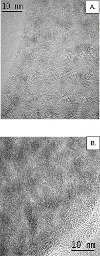
To develop bioconjugated nanoparticles to detect RSV infection, semiconductor cadmium telluride (CdTe) quantum dots (QDs) were synthesized as previously described (CitationWang et al 2002; CitationLiu, Chen, et al 2006). QDs have been reported to be an excellent quantum particle for use in biological systems based on very high photoluminescence quantum efficiencies, and the ability to cover the whole visible spectral range depending on particle size (CitationLiu, Chen, et al 2006). Thus, two different sized CdTe Qds with emission peaks at 585 nm and 540 nm were prepared () and used in the preparation of bioconjugated nanoparticles (NPs) for detection of RSV infection. These particles were prepared for bioconjugation as previously described (CitationWang et al 2002), and conjugated to monoclonal antibodies reactive to RSV F protein, clone 131-2A. Briefly, thioglycolate (TGA)-coated CdTe particles were mixed 1:1 with RSV anti-F protein monoclonal antibody (4 mg/ml) and the solution was stabilized with 10X phosphate-buffered saline (PBS), pH 7.2 to a final 1 X PBS concentration. EDC/NHS (1-Ethyl-3-(3-dimethylaminopropyl) carbodiimide HCl/N-Hydroxysulfosuccinimide) was added to bring to QD solution to a final concentration of 50 mM/5mM, respectively. The reaction was incubated for 2 h at room temperature, then overnight at 4 °C. Precipitated bioconjugated NP aggregates were separated by centrifugation to remove unwanted aggregates from single, bioconjugated nanoparticles. The final concentration of the purified NP-antibody conjugate stock solution was 0.625 mM.
Figure 1 HRTEM images of green and orange CdTe nanoparticles showing an average size of 3 nm (A) and 5 nm (B), respectively.
Abbreviations: CdTe, cadmium telluride; HRTEM, high resolution transmission electron microscopy.
Vero cell and virus propagation
Vero cells (African green monkey kidney cells) were grown in Dulbecco’s Modified Eagle’s Media (DMEM) containing 10% fetal bovine sera (DMEM-10%). For some experiments involving NPs, Vero cells were propagated on Lab-Tek chamber slides to 80%–90% confluency using DMEM-10%.
Respiratory syncytial virus strain A2 (RSV) was propagated in Vero cells as previously described (CitationTripp et al 1999). Briefly, semi-confluent Vero cells were prepared and washed with PBS. RSV was diluted in DMEM and the cells infected at a multiplicity of infection (MOI) of 1. The virus was allowed to adsorb for 2 h at 37 °C after which DMEM-10% was added and the cells incubated at 37 °C for 4 days. At day 4 post-infection (pi), the virus was recovered by removing the cell culture supernatant, freeze-thawing the infected cells, and centrifuging the cell lysate to remove debris and recover the virus from the cell lysate supernatant.
Detection of RSV infection in vitro
The bioconjugated nanoparticles conjugated to anti-RSV F protein monoclonal antibody (RSV-NPs), ie, both 585 nm and 540 nm QDs, were tested for their ability to detect RSV infection of Vero cells. RSV-infected Vero cells were propagated on Lab-Tek chamber slides to 80%–90% confluency and subsequently infected with RSV at a MOI = 1 as previously described (CitationTripp et al 1999). At day 4 pi, the media from the cells was removed, the cells were washed gently with PBS, fixed with acetone:methanol (60:40), and air dried. RSV-NPs (diluted 1:10,000) were added to the cells and incubated for 1 hour at 37 °C, followed by three washes with PBS/0.05% Tween 20. Cells were visualized by fluorescent microscopy (40X) on an Olympus CKX41 inverted microscope (Olympus, Center Valley, PA), and photographed with an Olympus Q-Color3 digital camera. Images were analyzed using the Olympus Q-Capture software.
The most widely used assay to quantitate virus titers in both the laboratory and clinical setting is the virus plaque assay. Virus plaques on the cell lawn are enumerated by immunostaining to specifically detect virus plaques due to confounding background staining that is often associated with the counterstain procedure. RSV-NPs were evaluated for detection of RSV infection using a modification of an immunostaining plaque assay as previously described (CitationTripp et al 1999). Briefly, the modification involved substituting RSV-NPs in place of the primary (anti-RSV monoclonal antibody: clone 131-2A), secondary (anti-mouse whole molecule immunoglobulin G (IgG)-alkaline phosphatase conjugated), and detection substrate, eg, Vector black alkaline phosphatase, to generate a single-step nanoparticle-based assay. In this modified assay, Vero cells were plated onto 24-well flat-bottom plates 24-h prior to RSV infection. Subsequently, the cell supernatants were removed and RSV diluted in DMEM was added to the cells at a MOI = 1. The virus was allowed to adsorb for 1 h at 37 °C after which the cells were overlayed with 2% methylcellulose in DMEM-10% as previously described (CitationTripp et al 1999), and cells incubated for 6 days at 37 °C, 5% CO2. At day 6 pi, the overlays were removed, and cells were fixed with acetone: methanol (60:40), and allowed to air dry. Cells were blocked for 30 minutes with Powerblock (10% casein in PBS) at room temperature. RSV-NPs, either 540 nm or 585 nm bioconjugated QDs, were diluted 1:1,000 from 0.625 mM stock solution in PBS. For positive control wells, anti-RSV monoclonal antibody (clone 131-2A) was diluted 1:500 in PBS containing Powerblock, added to the appropriate wells, and incubated 1 hour for RSV-NPs, or 2 hours for the primary monoclonal antibody at 37 °C. Cells were washed 3 times with PBS/0.05% Tween. For standard immunostaining plaque assay, secondary antibody (anti-mouse IgG-alkaline phosphatase conjugated) was diluted 1:500 in Powerblock, added to cells, incubated for 1 hour at 37 °C, washed twice with PBS, and developed using a Vector black alkaline phosphatase substrate kit according to the manufacturer’s protocol. For RSV-NPs, the plates were directly scanned for virus plaques using an Amersham Biosciences Typhoon 9210 scanner with a fluorescence excitation (532 nm) and emission (526 SP) filter. The virus plaques were enumerated and compared to plaque numbers obtained by standard counting using an Olympus SZ dissecting scope.
Detection of RSV infection in vivo
To determine the ability of RSV-NPs to detect RSV infection in lung tissue, the natural site of RSV infection and replication (CitationTripp 2005), 6–8 week old female BALB/c mice were intranasally infected with 106 plaque-forming units (pfu) of RSV as previously described (CitationTripp et al 1999). At day 4 pi, RSV-infected or naïve BALB/c mice were intravenously administered by tail vein 0.05 ml of RSV-NPs, either 540 nm or 585 nm bioconjugated QDs, diluted 1:1000 in PBS from a 0.625 mM stock solution. At day 5 pi, the lungs from RSV-NP-treated or PBS carrier-treated mice were removed and prepared for thin sectioning and immunohistochemistry (IHC) analysis as previously described (CitationLi, Sarmento, et al 2005). Briefly, lung tissue was frozen as tissue blocks and sectioned (20 μm) on a cryostat. The frozen sections were placed on slides which were immersed in acetone at 4 °C for 15 min, air dried for 30s, and then stored in PBS until immunostaining was performed. Briefly, slides were stained with anti-RSV monoclonal antibody (clone 131-2A) diluted 1:500 in PBS and incubated for 2 h at 37 °C. The slides were washed 2 times with PBS, and a secondary antibody (FITC conjugated anti-mouse IgG) diluted 1:500 in PBS was added to slides and incubated for 1 h at 37 °C, and washed twice with PBS. The lung sections from RSV-NP-treated mice and untreated mice that were immunostained as noted above were directly visualized using a fluorescent microscope.
Results and discussion
Current antibody-based methods used for virus detection are cumbersome, have limited sensitivity due to substantial background staining, and are costly as they often require multiple antibodies and enzyme substrate for detection. In an effort to ameliorate these issues, and reduce the time for virus detection, we synthesized semiconductor CdTe QDs with emission at either 540 nm or 585 nm to be conjugated to monoclonal antibodies to detect RSV-infected cells. These bioconjugated NPs (RSV-NPs) that are specific to RSV F protein were tested for their ability to detect RSV infection of cells both in vitro and in vivo. As expected, no differences in virus detection were observed using either 540 or 585 nm NPs.
As shown in the high resolution transmission electron microscopy (HRTEM) images in , the green emission CdTe NPs had an average size of approximately 3 nm (A) and the orange CdTe NPs of 5 nm (B). Given the reported difficulties conjugating proteins to CdTe NPs (CitationTan et al 2004; CitationGao et al 2005; CitationMcVeigh 2006), the CdTe NPs were produced with a thioglycolate coating to allow direct anti-RSV F protein monoclonal antibody conjugation via an EDC/Sulfo-NHS conjugation reaction. These bioconju-gated NPs (RSV-NPs) were stable for >1 year at 4 °C, and did not precipitate in the PBS diluent at room temperature or at 4 °C.
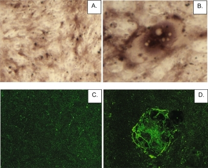
To determine the ability of the RSV-NPs to detect RSV infection, in vitro studies were performed using RSV-infected Vero cells. In these studies, we compared RSV detection using conventional antibody staining methods, ie, primary and secondary antibody staining plus enzyme substrate, to that of a single-step detection method using RSV-NPs. Conventional antibody staining methods produced substantial background staining in mock-infected Vero cells (), however did detect RSV plaque formation in the Vero cell lawn (). In contrast, RSV-NPs used in a single-step method produced little background (), and readily detected RSV plaques (). These results show that RSV-NPs can be used to detect RSV in a single-step fashion, and that this method can be used to greatly reduce the amount of time and reagent needed for RSV detection compared to conventional immunostaining procedures (CitationTripp et al 1999).
Figure 2 RSV-NP detection of RSV-infected Vero cells. Vero cells were mock-infected (A, C) or RSV-infected at a MOI = 1 (B, D) and fixed with acetone: methanol (60:40). Cells were immunostained with by conventional methods (A, B) or by RSV-NPs (C, D), and analyzed using an immunofluorescence microscopy (40X; A, C or 100X; B, D).
Abbreviations: MOI, multiplicity of infection; RSV-NP, respiratory syncytial virus-nanoparticles.
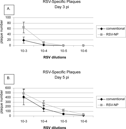
Extending the in vitro detection findings (), we determined if RSV-NPs could be used to quantitate RSV titers in a single step fashion, thereby shortening the detection time needed to quantitate virus titers by plaque assay (). In these studies, we compared a conventional immunostaining plaque assay which requires 5 or 6 days of in vitro culture post-RSV infection (CitationTripp et al 1999) to rapid single-step detection by RSV-NPs. For comparison of the different plaque assay methods, virus titers were assessed at days 2, 3, 5, or 6 pi. The results showed that at days 5 or 6 pi, conventional immunostaining () and the single-step RSV-NP detection method () were similarly sensitive for detecting RSV plaques in the Vero cell lawn. However, RSV-NPs could detect RSV plaques earlier (days 2 or 3 pi) compared with conventional immunostaining which was comparatively ineffective (), indicating that the single-step RSV-NPs procedure was more sensitive at detecting limiting levels of RSV. This result is not unexpected given the multivalent properties of RSV-NPs.
Figure 3 RSV-NP virus plaque assay. RSV was serially diluted ten-fold and the dilutions used to infect Vero cells for determination of virus titers. The viral titer determined from a conventional immunostaining plaque assay (A). were compared with RSV-NPs detection (B). Both 540 nm and 585 nm QDs were evaluated as RSV-NPs (B). RSV plaques were enumerated using an Amersham Biosciences Typhoon 9210 scanner.
Abbreviations: RSV-NP, respiratory syncytial virus-nanoparticles; QDs, quantum dots.
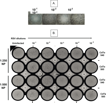
Figure 4 Dynamic range and sensitivity of detection of RSV-NP plaque assay compared with a conventional plaque assay. The viral titer of RSV-infected Vero cells was determined at day 3 pi (A) and day 5 pi (B) using conventional immunostaining plaque assay or single-step detection using 1:250 dilution of RSV-NPs derived from 585 nm QDs. RSV plaques were enumerated using an Amersham Biosciences Typhoon 9210 scanner.
Abbreviations: RSV-NP, respiratory syncytial virus-nanoparticles; QDs, quantum dots.
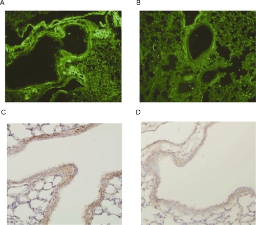
To evaluate the efficacy of RSV-NPs to detect RSV infection in vivo, BALB/c were either intranasally infected with 106 pfu RSV or mock-treated, and at day 4 pi, RSV-infected or mock-treated mice were intravenously administered 0.05 ml of RSV-NPs from the stock solution (0.625 mM) by tail vein. At day 5 pi, the lungs from RSV-NP-treated or mock-treated mice were removed, frozen, and prepared for thin sectioning and IHC analysis using conventional methods, i.e. primary and secondary antibody staining, or directly visualized by fluorescence microscopy in the case of RSV-NP treatment. Treatment with RSV-NPs provided clear and rapid detection of RSV-infected lung tissue () with very limited background staining as assessed in lung tissue from mock-treated mice similarly administered RSV-NPs (). Importantly, the magnitude and location of RSV-NPs staining along the epithelial cells of the alveoli in the lungs was similar to that of conventional IHC staining (), and the background staining for RSV-NPs was lower in naïve mice compared to conventional IHC staining (). The selective staining by RSV-NPs of only RSV-infected lung epithelial cells suggests antibody-mediated specific targeting to sites of infection. These results suggest that RSV-NPs may be useful to determine the progression of RSV infection in the lungs, and as a result, aid our understanding of sites of infection, and help to define disease intervention strategies that employ bioconjugated NP targeting strategies.
Figure 5 RSV-NPs detection of RSV-infected lung tissue. Lung tissue sections from RSV-infected (A, C) or naïve, mock-treated BALB/c mice (B, D) were stained by IHC using RSV-NPs (A, B), or by conventional IHC (C, D), and analyzed using an immunofluorescence microscopy.
Abbreviations: IHC, immunohistochemistry; RSV-NP, respiratory syncytial virus-nanoparticles.
Conclusions
The results from this study show that RSV-NPs can be used to detect RSV infection both in vitro and in vivo and suggest that beyond diagnostics, bioconjugated NPs may be useful for multiplexed detection of viral and/or host cell antigens, as well as for targeting sites of virus infection with antiviral agents that may be linked to the NPs. Indeed, bioconjugated NPs have been used to identify the presence and progression of RSV infection in a human epithelial cell line, ie, HEp-2 cells (CitationBentzen et al 2005), as well as used in a variety of intracellular tracking and related studies (CitationAlivisatos et al 2005; CitationBruchez 2005; CitationGao et al 2005; CitationHotz 2005; CitationShenoy et al 2006; CitationMcVeigh 2006). Importantly, we show here that RSV-NPs can be applied as tools to enhance virus detection by increasing sensitivity of virus detection while decreasing the detection time. Application of RSV-NPs for virus detection in animal models can provide a path to assist our ability to dissect important parameters of RSV disease pathogenesis such as determining sites of virus infection and virus load in the lungs of infected animals. This understanding is critical to move the field forward in antiviral disease intervention strategies.
Acknowledgements
Ralph Tripp would like to thank the Georgia Research Alliance for supporting aspects of this study, and Dr. Zhen Fu for help with IHC studies. Wei Chen would like to thanks the US Army Medical Research Acquisition Activity (W81XWH-05-C-0101), the UTA Startup fund, and the LERR fund.
References
- AgrawalATrippRAAndersonLJ2005Real-time detection of virus particles and viral protein expression with two-color nanoparticle probesJ Virol798625815956604
- AgrawalAZhangCByasseeT2006Counting single native biomolecules and intact viruses with color-coded nanoparticlesAnal Chem7810617016478096
- AlivisatosAPGuWLarabellCl2005Quantum dots as cellular probesAnnu Rev Biomed Eng7557616004566
- BaderMSMcKinseyDS2005Viral infections in the elderly. The challenges of managing herpes zoster, influenza, and RSVPostgrad Med11845851416329530
- BentzenEHouseFUtleyTJ2005Progression of respiratory syncytial virus infection monitored by fluorescent quantum dot probesNano Lett5591515826092
- BruchezMP2005Turning all the lights on: quantum dots in cellular assaysCurr Opin Chem Biol9533716125995
- DriskellJDKwartaKMLipertRJ2005Low-level detection of viral pathogens by a surface-enhanced Raman scattering based immunoassayAnal Chem7761475416194072
- EbbertJOLimperAH2005Respiratory syncytial virus pneumonitis in immunocompromised adults: clinical features and outcomeRespiration72263915942295
- FarokhzadOCChengJTeplyBA2006Targeted nanoparticle-aptamer bioconjugates for cancer chemotherapy in vivoProc Natl Acad Sci U S A10363152016606824
- FuentesMMateoCRodriguezA2006Detecting minimal traces of DNA using DNA covalently attached to superparamagnetic nanoparticles and direct PCR-ELISABiosens Bioelectron2115748016129594
- FujiwaraKWataraiHItohH2006Measurement of antibody binding to protein immobilized on gold nanoparticles by localized surface plasmon spectroscopyAnal Bioanal Chem3884754
- GaoXYangLPetrosJA2005In vivo molecular and cellular imaging with quantum dotsCurr Opin Biotechnol16637215722017
- GoodmanCMChariNSHanG2006DNA-binding by functionalized gold nanoparticles: mechanism and structural requirementsChem Biol Drug Des6729730416629827
- HotzCZ2005Applications of quantum dots in biology: an overviewMethods Mol Biol30311715923671
- IpeBIYoosafKThomasKG2006Functionalized gold nanoparticles as phosphorescent nanomaterials and sensorsJ Am Chem Soc12819071316464092
- LeungAKKellnerJDDaviesHD2005Respiratory syncytial virus bronchiolitisJ Natl Med Assoc9717081316396064
- LiXQSarmentoLFuZF2005Degeneration of neuronal processes after infection with pathogenic, but not attenuated, rabies virusesJ Virol7910063816014967
- LiYCuYTLuoD2005Multiplexed detection of pathogen DNA with DNA-based fluorescence nanobarcodesNat Biotechnol23885915951805
- LiYTLiuHSLinHT2005Gold nanoparticles for microfluidics-based biosensing of PCR products by hybridization-induced fluorescence quenchingElectrophoresis2647435016283695
- LiuHHCaoXYangY2006Array-based nano-amplification technique was applied in detection of hepatitis E virusJ Biochem Mol Biol392475216756752
- LiuYChenWJolyAG2006Comparison of water-soluble CdTe nanoparticles synthesized in air and in nitrogenJ Phys Chem B11016992700016927992
- McCarthyCAHallCB2003Respiratory syncytial virus: concerns and controlPediatr Rev24301912949266
- McVeighER2006Emerging imaging techniquesCirc Res988798616614313
- MejiasAChavez-BuenoSJafriHS2005Respiratory syncytial virus infections: old challenges and new opportunitiesPediatr Infect Dis J24S18996discussion S196–7. 16378045
- PennSGHeLNatanMJ2003Nanoparticles for bioanalysisCurr Opin Chem Biol76091514580566
- ShenoyDFuWLiJ2006Surface functionalization of gold nanoparticles using hetero-bifunctional poly (ethylene glycol) spacer for intracellular tracking and deliveryInt J Nanomedicine151816467923
- SouzaGRChristiansonDRStaquiciniFI2006Networks of gold nanoparticles and bacteriophage as biological sensors and cell-targeting agentsProc Natl Acad Sci USA10312152016434473
- SukJSSuhJChoyK2006Gene delivery to differentiated neuro-typic cells with RGD and HIV Tat peptide functionalized polymeric nanoparticlesBiomaterials2751435016769110
- TanWWangKHeX2004Bionanotechnology based on silica nanoparticlesMed Res Rev246213815224383
- TrippRA2005Pneumovirus and Metapneumovirus: respiratory syncytial virus and human metapneumovirusMahyBTopley and Wilson’s Microbiology and Microbial InfectionsLondonHodder Arnold2783806
- TrippRAMooreDJonesL1999Respiratory syncytial virus G and/or SH protein alters Th1 cytokines, natural killer cells, and neutrophils responding to pulmonary infection in BALB/c miceJ Virol73709910710438795
- WabuyeleMBVo-DinhT2005Detection of human immunodeficiency virus type 1 DNA sequence using plasmonics nanoprobesAnal Chem7778101516316192
- WanZWangYLiSS2005Development of array-based technology for detection of HAV using gold-DNA probesJ Biochem Mol Biol3839940616053706
- WangSMamedovaNKotovNA2002Antigen/Antibody imunocomplex from CdTe nanoparticle bioconjugatesNano Letters281722
- YaoGWangLWuY2006FloDots: luminescent nanoparticlesAnal Bioanal Chem3855182416715275
- ZhaoXHilliardLRMecherySJ2004A rapid bioassay for single bacterial cell quantitation using bioconjugated nanoparticlesProc Natl Acad Sci USA101150273215477593