 ?Mathematical formulae have been encoded as MathML and are displayed in this HTML version using MathJax in order to improve their display. Uncheck the box to turn MathJax off. This feature requires Javascript. Click on a formula to zoom.
Abstract
Calcium phosphate nanoparticles (nanoCaP) conjugated with cis-diamminedichloro-platinum (CDDP, cisplatin) were prepared through the electrostatic binding of an aquated species of cisplatin to the nanoCaP in a chloride-free solution. The agglomeration of the nanoCaP that typically occurs during synthesis of CaP was controlled through the addition of DARVAN® 811 immediately after precipitation and before drug conjugation. In vitro drug release studies were completed and showed a sustained release of CDDP from the nanoconjugates over time. The cytotoxicity of the nanoCaP/CDDP was compared to that of the free drug in an in vitro cell proliferation assay using the CDDP resistant A2780cis human ovarian cancer cell line. The CDDP released from the nanoconjugates was equally effective as the free drug against the A2780cis cell line. Direct addition cytotoxicity studies revealed that the sterically-stabilized, negatively-charged drug nanoconjugates are unable to overcome drug resistance and had an increased IC50 value as compared to the free drug.
Introduction
Attachment of a pharmaceutical to a particulate drug delivery system is a tactic that has long been employed to sustain drug delivery. Particulate drug carrier systems for tumor specific targeting of anti-cancer drugs include liposomes (CitationGregoriadis 1995; CitationAllen and Moase 1996; CitationVicent and Duncan 2006), polymer microspheres (CitationFung and Saltzman 1997; CitationLiu et al 2003; CitationLin et al 2005; CitationFoger et al 2006) and recently nanoparticles (CitationBarbe et al 2004; CitationAmbruosi et al 2006; CitationDong and Feng 2006; CitationFarokhzad et al 2006). Although liposomal systems have made the most headway in the clinic, they are under further optimization to be safer to normal tissues and long-circulating in blood, yet able to efficiently accumulate and transfer drug in a sustained manner to targeted sites (CitationHong et al 1999). For biodegradable polymer-based drug delivery systems, there are concerns that polymer acidic byproducts or degrading polymer fragments can adversely affect the drug they are delivering or the tissues they interact with. Often an undesirable late stage, uncontrolled and massive drug release is observed with polymer-based drug delivery systems (CitationSpenlehauer et al 1989). Bioceramics, such as calcium phosphates (CaP), represent another class of materials suitable for use as a carrier for drugs, non-viral gene delivery, antigens, enzymes, and proteins. CaP can be produced at a low cost and is simple to manufacture. Hydroxyapatite (HA) is a type of calcium phosphate that has a similar chemical structure to bone mineral, and hence has excellent biocompatibility, bioactivity, and high affinity to proteins (CitationGorbunoff 1984a, Citation1984b; CitationSpenlehauer et al 1989), DNA (CitationWelzel et al 2004; CitationBisht et al 2005), chemotherapy drugs (CitationBarroug et al 2004), and antigens (CitationHe et al 2000).
Localized pharmaceutical treatments utilizing CaP as a drug carrier have been achieved by injections (CitationHe et al 2000) or surgical placement of disks, pellets or particulates (CitationYamamura et al 1994; CitationItokazu et al 1998; CitationRogers-Foy et al 1999; CitationBenghuzzi 2000; CitationMizushima et al 2006). The localized drug release from these CaP-based controlled release systems minimized the high concentration of drugs typically required in the bloodstream and other organs to achieve therapeutic outcomes. The CaP also provided a means to minimize unnecessary systemic toxicity and reduce the need for repeated dosing often required of most drugs. Due to the low solubility of the HA type of CaP in physiological conditions, HA remains for long periods after in vivo subcutaneous placement. The large sintered disks and large particle sizes of HA utilized in the previously researched formulations would remain in vivo long after drug release. This led to our interest in investigating nano-sized CaP particles that could speed carrier resorption, allow greater tumor or tissue perfusion, and perhaps overcome drug resistance through intracellular drug/particle uptake observed with particulate drug delivery formulations (CitationMinko et al 1998). Particle enhanced endocytosis may endow the particulate drug delivery systems with an ability to bypass p-glycoprotein efflux pump and lead to sequestration of anticancer drugs in acidic intracellular compartments, yielding high cytotoxicity (CitationLee et al 2005).
In order to use nanoCaP crystals as an efficient drug carrier for localized chemotherapy treatment, it is important that the chemotherapy drug can be loaded and released in a controlled manner. Moreover, it is also important that the drug released is chemically active and effective over a long period of time. Therefore we investigated these variables using Cisplatin (CDDP), a commonly used chemotherapy drug with high antitumor activity (CitationLong and Repta 1981; CitationBarroug et al 2004) as the model drug. Our approach can be generalized to other drugs and biomolecules due to the versatility of the CaP crystal surface to bind both positively and negatively charged molecules through simple adsorption. In the present study, we describe the preparation of nanoCaP/CDDP conjugates with controlled size and purity. The in vitro drug release profile of the nanoconjugates is investigated. The cytotoxicity of the released drug and directly added nanoCaP/CDDP is compared to that of the free drug in an in vitro cell proliferation assay using the CDDP resistant A2780cis human ovarian cancer cell line.
Experimental procedures
Nano-calcium phosphate particle synthesis
NanoCaP was synthesized by co-precipitation from the addition of equal volumes of a 30 mM Ca(NO3)2 solution and a 30 mM K2HPO4 solution which are both filtered through 0.1 μm filtration device (Millipore, Boston, USA) separately, followed by immediate addition of 1.67(v/v)% of 0.2 μm filtered DARVAN®811 (sodium polymethacrylate, MW = 3,300, R.T. Vanderbilt Company, Inc. Norwalk, CT, USA) as a dispersing agent. All reagents are ACS grade and purchased from Sigma Chemical Co., (St. Louis, MO), unless noted otherwise. After 1 hr stirring, a pellet of nanoCaP was collected by centrifugation at 12,000 rpm (20,076 g) for 30 min. Before binding, the nanoCaP pellet was redispersed in ultrapure H2O as a wash step, and then collected by centrifugation at 12,000 rpm for 30 min.
NanoCaP/CDDP conjugate synthesis
CDDP (Sigma Chemical Co., St. Louis, MO) was bound to nanoCaP by following a slight modification of the electrostatic conjugate preparation procedure we developed previously (CitationBarroug and Glimcher 2002). Following the recommendation of Dr. S. Lippard, (Chemistry Dept., Massachusetts Institute of Technology, Cambridge, MA, USA), the aquated form of CDDP was prepared and used instead of CDDP to more efficiently form the conjugates. Aquated CDDP was prepared by reacting 90 mM AgNO3 solution with CDDP solution (about 1000 μg/mL) at a 2:1 molar ratio. The reaction mixture was placed on a thermal rocker (Lab-Line®, model 4637) for 12–24 hrs and kept protected from light. The silver chloride precipitate was removed by several centrifugation steps at 3000 rpm (1000 g) for 20 min. The remaining supernatant was filtered through a 0.2 μm filter. The final concentration of aquated CDDP was determined by Pt analysis using an Atomic Absorption Spectrophotometer (AAS) (Model 5100, Perkin Elmer, Shelton, CT, USA).
The nanoconjugates were formed by adding 0.625 mL of 20 mM potassium phosphate buffer (KPB, pH = 6) to 31.55 mg of a wet nanoCaP pellet (which corresponds to 5 mg of dry CaP as determined by oven drying), and sonicating for 10 sec. Aquated CDDP (0.625 mL with initial binding CDDP concentration C0) was added, and the sample was put in a thermorocker at 37 °C, speed 5 (LAB-LINE® thermorocker, Model 4637, Barnstead Thermolyne, IL, USA) for 4 hrs. The conjugates thus formed were centrifuged at 12,000 rpm (20,076 g) for 30 min. The supernatant, which contained unbound CDDP, was decanted and measured for final binding supernatant CDDP concentration (Cf) by AAS. The pellet was washed with 0.25 mL 10 mM KPB buffer and centrifuged at 12,000 rpm(20,076 g), 30 min. The supernatant from this KPB wash was decanted and measured by AAS to determine KPB wash supernatant CDDP concentration (CKPB). This pellet was rinsed with 0.21 mL of 0.9% NaCl solution for 30 min. on the thermorocker (37 °C, speed 5) after brief sonication. The sample was centrifuged again at 12,000 rpm for 30 min. to collect the nanoCaP/CDDP conjugates. The supernatant was decanted and measured for saline wash supernatant CDDP concentration (Cw). The drug loading was calculated by the following equation:
Three batches of nanoconjugates were synthesized aseptically for the different studies. Volumes of the precipitation solutions were varied proportionally depending on the yield of conjugates required. The drug loading of the nanoconjugates was controlled by changing the initial aquated CDDP concentration (C0). The drug loading of the nanoconjugates used for the direct addition cytotoxicity study was 112 μg/mg obtained by using 1052 μg/mL aquated CDDP. The drug loading efficiency was 0.85. The drug loading of nanoconjugates used for in-vitro drug release study was 88 μg/mg by using 900 μg/mL aquated CDDP and the drug loading efficiency was 0.78. The drug loading of the conjugates used for cytotoxicity test was 35 μg/mg by using 552 μg/mL aquated CDDP and drug loading efficiency was 0.5.
Physical and chemical characterization of nanoCaP and nanoCaP/CDDP conjugates
Samples were prepared for transmission electron microscopy (TEM) by dispersing nanoCaP/CDDP conjugates in ultrapure H2O at about 1 mg/mL concentration with an Ultrasonic 1000L Cell Disruptor (Ultrasonic Power Corporation, IL, USA) for 1 minute. One drop of this liquid was immediately transferred by a micropipette to a 3 mm diameter Formvar coated copper TEM grid and slowly evaporated to dryness. The samples on the TEM grid were analyzed using a 100cx JEOL TEM at 80 kV in brightfield (BF) modes.
The chemical structure of nanoCaP was determined by FTIR as follows. Infrared absorption spectra were obtained from nanoCaP in a KBr pellet using a Bruker Tensor 27 Fourier transform infrared (FTIR) spectrometer with a resolution of 0.1cm−1. X-ray diffraction analysis was used to determine the crystal structure of nanoCaP. The samples were scanned with Cu-Kα x-ray radiation from a Philips XRD 2500 at 40 KV and 20 mA, using a step size of 0.02° and a step time of 1.2 s over a 2θ range of 10–70. The particle size of the nanoCaP and nanoCaP/CDDP conjugates were measured on samples dispersed in ultrapure H2O at about 1 mg/mL concentration by Ultrasonic 1000L Cell Disruptor. The particle size and Z-potential of nanoparticles was measured on 90 Plus particle sizer coupled with Z-potential analyzer (Brookhaven Instruments, NY, USA).
In vitro drug release studies
Release studies were conducted by dispersing 40 mg of nanoCaP/CDDP conjugates (88 μg/mg loading), by mixing and brief vortexing, in 0.8 mL PBS and rocking at 37 °C, 20 cycle/min. Supernatants were collected at 1hr, 6 hr, 1, 3, 7, 12, and 16 days, after centrifugation at 9,000 rpm (7,000 g) for 10 minutes. The released drug in the unfiltered supernatant was measured by AAS. Full replacements of release media were made at each time point.
In vitro cytotoxicity testing
For in vitro cytotoxicity activity studies, the CDDP-resistant cell line was used: A2780cis human ovarian carcinoma cell line (Sigma, 93112517) and cultured according to supplier’s descriptions. Briefly, cells were cultured in RPMI1640 medium, supplemented with 2 mM Glutamine and 10% Fetal Bovine Serum (FBS) in a humid atmosphere at 37 °C and 5% CO2. Cells were supplemented with 1μm CDDP to the culture media every 2–3 passages, post-attachment. The CellTiter96® AQueous One (Promega Corporation, Madison, WI, USA) colorimetric proliferation assay was used to determine the IC50 value (50% inhibitory concentration) evaluated from 12 two-fold dilutions of CDDP in 0.9% saline (free drug), nanoconjugates, nanoCaP and free drug, or CDDP released from the nanoconjugates. The highest concentrations of test samples were prepared in the drug master plate prior to dilution as follows: CDDP was dissolved in 0.9% saline at 1000 μg/mL and diluted in PBS to prepare a free drug solution of 200 μg CDDP/mL. CDDP released from the nanoconjugates was obtained from the supernatant of 40 mg of nanoconjugates (loaded at 35 μg CDDP/mg nanoCaP) incubated in 0.8 mL PBS for 3d on a rocker at 37 °C, 20 cycles/min. Three days were necessary to achieve a CDDP concentration high enough to obtain an IC50 value. Five milligrams of nanoCaP/CDDP, synthesized aseptically with a drug loading of 112 μg CDDP/mg CaP, was dispersed in 0.8 mL PBS (CDDP 700 μg/mL if totally released) for the highest nanoconjugate concentration directly added. To confirm that the particulate nanoconjugates were diluted evenly across the wells, measurements of the total Pt concentration in all the wells of the drug master plate were made by AAS after dissolving the nanoconjugate solutions in dilute HCl. Five milligrams of nanoCaP was dispersed in 0.8 mL free drug solution for the nanoCaP not conjugated to free drug sample, directly added to cells.
Preliminary investigations of the growth rate of A2780cis were conducted to determine the proper cell seeding number that would remain in the linear range of the assay throughout the study. The cytotoxicity assay was conducted as follows: twenty-four hours after seeding 2000 A2780cis cells in 50 μl of media on 96 well plates, 50 μl PBS, or PBS with drug, carrier or nanoconjugates was added to the wells. Five replicates were tested for each sample. Following two days of continuous exposure, 20 μl of CellTiter96® AQueous One (Promega) colorimetric proliferation reagent was added to each well, and then the plates were incubated for 4 more hours before being read on a Spectramax Plus384 spectrophotometer (Molecular Biosciences, Sunnyvale, CA, USA) at an absorbance value of 490nm. Absorbance values were converted to IC50 values using the four parameter logistic equation:
| Y | = | observed absorbance |
| Amax | = | absorbance of control cells |
| Amin | = | absorbance of cells in presence of highest agent concentration |
| x | = | drug concentration (μg/ml) |
| n | = | slope of curve |
Samples were analyzed for statistically significant differences using the Student’s T-test (p <0.05).
Results
Physical and chemical characterization of the nanoCaP and the nanoCaP/CDDP conjugates
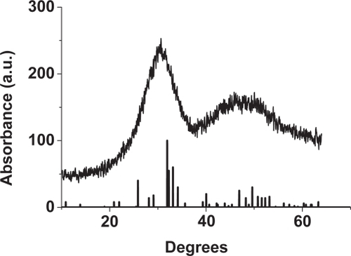
A combination of several methods was used to fully characterize the nanoconjugate shape, size, chemistry and structure. TEM images showed that the nanoconjugates are spherical and well dispersed (). The mean particle size of the nanoCaP precipitated with DARVAN 811 before conjugation with CDDP was 129 ± 33 nm (50% below 125.4 nm, 90% below 181.3 nm), and the zeta-potential = −45.59 mV. The size and zeta-potential slightly decreased after adsorbance of CDDP: 106.5 ± 35.4 nm (50% below 101.1 nm, 90% below 163.3 nm), zeta-potential = −27.9 mV (). Solutions of nanoconjugates remain stably dispersed for periods of up to at least two weeks. The FTIR spectra of the nanoCaP and the nanoconjugates have similarities to HA (CitationElliott 1994; CitationZhang and Colwell 2004; CitationLiu et al 2005), and not other calcium phosphate phases, except that several peaks associated with the DARVAN 811 are present in the nanoparticles (). However, there is a lack of resolution of the P-O absorption bands, indicating that the sample may contain amorphous calcium phosphate (CitationLegeros et al 2005). The R-COO- stretch in the DARVAN 811 is changed from 1573 cm−1 to 1559 cm−1 which is possibly due to intermolecular bridge R-COO-Ca complex formation with the CaP (CitationZhang et al 2006). The X-ray diffraction spectra () of the nanoCaP contains broad peaks characteristic of HA. The broad peaks of the nanoCaP relative to the HA standard peaks indicates that the crystals are nanometer in size, poorly crystalline or perhaps amorphous. The sample does not show any evidence of contamination from other crystalline calcium phosphate phases.
Figure 1 (a) TEM image of nanoCaP/CDDP conjugates. (b) Particle size analysis of nanoCaP/CDDP conjugates redispersed in H2O at 1 mg/mL concentration. NanoCaP/CDDP particle size = 106.5 ± 35.4 nm (mean size ± standard error, 50% below 101.1 nm, 90% below 163.3 nm), Z-potential = −27.9 mV. The control NanoCaP: Particle size = 129 ± 33 nm, 50% below 125.4 nm, 90% below 181.3 nm. Z-potential = −45.59 mV.
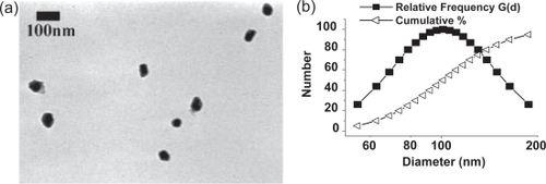
Figure 2 FTIR spectrum of (a) DARVAN 811 (b) Commercial hydroxyapatite standard and (c) NanoCaP formed in the presence of DARVAN 811 (d) NanoCaP/CDDP conjugates.
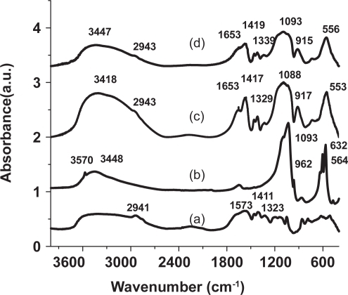
Figure 3 XRD spectrum of nanoCaP particles compared to a hydroxyapatite standard (JCPDS, #09-0432).
In vitro drug release from nanoCaP/CDDP conjugates
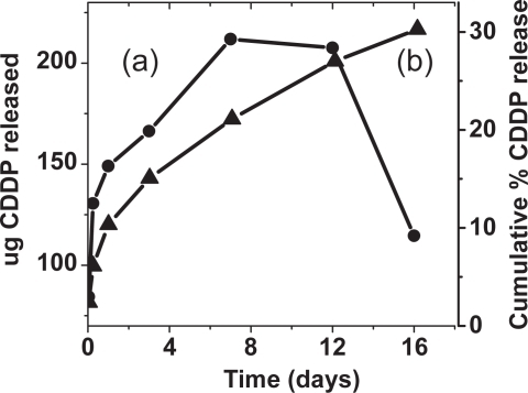
The amount of CDDP released from the nanoconjugates into PBS, pH = 7.4 during gentle rocking at 37 °C at various time points is shown in . The results are also expressed as a percentage of the total amount bound (). There is a burst release of drug in the first day, followed by a slower, but continuous, release of drug over the time tested. After 16-days in PBS with eight solution changes, 30% of the bound drug released.
Figure 4 Drug release profile of nanoCaP/CDDP conjugates (88 ug/mg loading). (a) Amount of CDDP released over time in PBS, pH = 7.4 (b) Cumulative release over time of CDDP in PBS, pH = 7.4.
Cytotoxicity of nanoCaP/CDDP conjugates
The effect of the CDDP conjugated to nanoCaP on the proliferation of A2780cis cancer cells was evaluated indirectly and directly by (a) addition of the CDDP released from the nanoconjugates during incubation in PBS for three days, and (b) direct addition of the nanoconjugates to the cells in culture. The IC50 value obtained for the nanoconjugate-released CDDP was not significantly different from the free drug (p > 0.05) (), indicating the conjugation procedure and the release process do not adversely affect CDDP. The IC50 value obtained after direct addition of the nanoconjugates is also shown in . Determination of the IC50 value for directly added nanoconjugates was complicated by the fact that the nanoCaP and the nanconjugates themselves have an absorbance maximum at 490 nm, the same as the formazan product produced by the viable cells in the assay. Therefore, it was necessary to deduct the interference of the nanoCaP using the readings from wells prepared using the same conditions as above (same seeding cell number, same nanoconjugate or nanoCaP concentration and volume, same culture time) without the addition of CellTiter96® reagent, as shown in . The IC50 values obtained this way indicate that the addition of the carrier alone (nanoCaP) to a free drug solution slightly, but significantly, increases the IC50 value relative to the free drug alone. This provides indirect evidence that the nanoCaP itself is not cytotoxic at the concentration tested. The IC50 value of the nanoconjugates was found to be significantly higher than the free drug (17.6 ± 2.7 vs 3.2 ± 0.2) indicating that a portion of the CDDP attached to the nanoconjugates is protected from direct interaction with the cells during the two-day test period.
Figure 5 Comparison of IC50 values obtained for the cytotoxicity testing of CDDP released after incubation of the nanoconjugates in PBS: (a) Free drug control, (b) CDDP released from the nanoCaP/CDDP conjugates. (c)–(e) are from the direct addition study: (c) Free drug control, (d) NanoCaP + Free drug. (e) Nano-CaP/CDDP conjugates particles directly added to the cells. (*) denotes significant difference (p < 0.05, Student’s T-test) from free drug control.
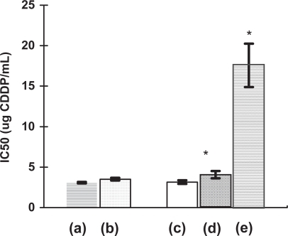
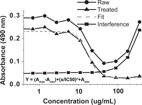
Figure 6 Demonstration of IC50 value determination of nanoCaP/CDDP conjugates on A2780Cis cancer cell lines. NanoCaP and nanoCaP/CDDP conjugate particles have interference around 490 nm at higher concentrations (see interference plot above). This interference was determined at the same test conditions but without adding Celltiter96 solution. It is subtracted from the raw data to give treated data (treated = raw–interference). The 4-parameter sigmoidal fit of this treated data is used to calculate the IC50 value.
Discussion
In our previous experiments with CDDP release from different types CaP, we observed that the less crystalline the CaP, the slower the release of drug (CitationBarroug et al 2004). This effect was correlated to particle surface area: particles with higher surface areas bind more drug and release it more slowly and less completely than particles with lower surface areas. Therefore, varying the crystallinity is one means of controlling drug release in inorganic particle-based drug delivery systems. Given that the nanoCaP of this study appears to be even less crystalline than the CaP tested in our previous work, it is not surprising that the initial burst release and the cumulative drug release from the nanoconjugates in the present study was even lower than that observed previously for CaP. The reduction of a burst release and enhanced sustained release possible with the nanoconjugates is desirable for in vivo applications. While there is low cumulative release in neutral PBS, the nanoconjugates are completely soluble in acidic solutions. This property may make the nanoCaP/CDDP drug delivery system particularly suited for in vivo intratumoral drug delivery applications in which the acidic pH of tumor tissue will lead eventually to complete drug release.
The reduced cytotoxicity of the nanoconjugates relative to free drug seen in the direct addition studies also confirms that a large portion of CDDP attached to the CaP is not released over two days in neutral pH cell culture medium. From the in vitro release studies (), approximately 12% of the total CDDP bound would be expected to be released by the end of the two day incubation with cells. The 12% is probably an overestimate since the release study was conducted with solution agitation and multiple total solution replacements, while the cell culture media was not disturbed or replaced. Assuming 10% release, of the 350 μg/ml available for release from the conjugates, only 35 ug/ml of free drug would have been available at the highest concentration to the cells compared to the 100 ug/ml in the free drug wells. If the concentrations of the nanoconjugates were adjusted to this theoretical value and the IC50 values recalculated, the IC50 would be 1.7 μg/mL, which is less than free drug (3.2 μg/mL). Therefore it appears that more of the CDDP than expected from cell-free in vitro release studies is being released when in contact with cells. This would be possible if some endocytosis of the nanoconjugates occurred as has been reported for particle based drug delivery systems (CitationMinko et al 1998).
Previous studies with CaP have shown that unsintered microcrystals of HA are readily taken up by cancer cells (CitationAoki 1994), therefore we had anticipated extensive particle-assisted drug transport. If this had been the case, then the nanoconjugates would have overcome the drug resistance of the A2780cis cells (lower IC50 than free drug), which they did not. There is literature indicating that negatively charged particles are less likely than neutral particles to be taken up by cells (CitationBonhomme et al 1992), and since the nanoCaP/CDDP is negatively charged, cellular uptake may have been reduced because of the negative surface charge. Endocytotic uptake can be categorized into nonspecific fluid phase endocytosis, adsorptive endocytosis and specific receptor-mediated endocytosis (CitationYi et al 2005). Adsorptive endocytosis requires nanoparticle adsorption on the cell membrane to obtain cytotoxicity and has been observed for doxorubicin-loaded polyalkylcyanoacrylate nanoparticles (CitationVauthier et al 2003). CaP crystals with unmodified surfaces have been shown to adsorb to cells in culture (CitationMandel and Riese 1991), therefore particle uptake of our nanoCaP was expected. However, in the present study we modified the surface of the nanoCaP with DARVAN 811 to prevent the adhesion of nanoparticles to each other through steric stabilization. This modification appears to have also prevented or greatly reduced cell membrane adhesion required for CaP particle-assisted drug transport. Overcoming this may require applying an additional surface modification such as a tumor cell targeting ligand (eg, folic acid, VEGF) (CitationMarcucci and Lefoulon 2004; CitationMinko et al 2004).
Conclusion
Stably dispersed nanoparticles of CaP were synthesized by the addition of DARVAN 811 immediately after precipitation. Aquated CDDP was simply and efficiently adsorbed to the surface of CaP nanoparticles through electrostatic interactions. The high surface area of the CaP nanoparticles led to a reduction of the CDDP burst release and greater sustained release relative to more crystalline, micron-sized agglomerated particles of CaP previously tested. In vitro cytotoxicity testing showed that the CDDP released from the nanoconjugates retained complete activity during conjugation and release and had comparable cytotoxicity to free drug. The nanoCaP alone was not cytotoxic. CDDP release from CaP nanoconjugates in neutral pH was slow and complete release was limited (30%), therefore the direct addition studies showed reduced cytotoxicity of the nanoconjugated CDDP relative to free drug. Particle assisted drug transport was not a highly active mechanism in this formulation, possibly due to the negative surface charge or steric stabilization by the DARVAN 811. However, we postulate that in acidic environments such as tumor tissues, the CaP nanoconjugates can slowly dissolve and completely release the adsorbed drug. The favorable properties of nanoCaP/CDDP conjugates warrant their further investigation in intratumoral anti-cancer drug delivery applications.
Acknowledgements
The authors gratefully acknowledge financial support from The Susan G. Komen Breast Cancer Foundation (Basic, Clinical and Translational Breast Cancer Research Grant BCTR0504344).
References
- AllenTMMoaseEH1996Therapeutic opportunities for targeted liposomal drug deliveryAdvanced Drug Delivery Reviews2111733
- AmbruosiAKhalanskyASYamamotoH2006Biodistribution of polysorbate 80-coated doxorubicin-loaded [14C]-poly(butyl cyanoacrylate) nanoparticles after intravenous administration to glioblastoma-bearing ratsJournal Of Drug Targeting149710516608736
- AokiH1994Medical applications of hydroxyapatiteTokyoTakayama Press System Center Co., Inc1069
- BarbeCBartlettJKongLG2004Silica particles: a novel drug-delivery systemAdvanced Materials16195966
- BarrougAGlimcherMJ2002Hydroxyapatite crystals as a local delivery system for cisplatin: adsorption and release of cisplatin in vitroJournal of Orthopaedic Research202748011918306
- BarrougAKuhnLTGerstenfeldLC2004Interactions of cisplatin with calcium phosphate nanoparticles: in vitro controlled adsorption and releaseJournal of Orthopaedic Research22703815183424
- BenghuzziH2000Long-term sustained delivery of 3′-azido-2′,3′-dideoxythymidine in vivo by means of HA and TCP delivery devicesBiomedical Sciences Instrumentation36343810834256
- BishtSBhaktaGMitraS2005pDNA loaded calcium phosphate nanoparticles: highly efficient non-viral vector for gene deliveryInternational Journal of Pharmaceutics2881576815607268
- BonhommeLMathieuMCAmdidoucheD1992Intratumor treatment of C3H mouse mammary carcinoma with 5-fluorouracil adsorbed on activated charcoal particlesAnti-Cancer Drugs326161525407
- DongYFengS-S2006Nanoparticles of poly(D,L-lactide)/methoxy poly(ethylene glycol)-poly(D,L-lactide) blends for controlled release of paclitaxelJournal Of Biomedical Materials Research. Part A78121916596586
- ElliottJ1994Structure and chemistry of the apatites and other calcium orthophosphatesElsevier389
- FarokhzadOCKarpJMLangerR2006Nanoparticle-aptamer bioconjugates for cancer targetingExpert Opinion On Drug Delivery33112416640493
- FogerFNoonpakdeeWLoretzB2006Inhibition of malarial topoisomerase II in Plasmodium falciparum by antisense nanoparticlesInternational Journal of Pharmaceutics3191394616713146
- FungLKSaltzmanWM1997Polymeric implants for cancer chemotherapyAdvanced Drug Delivery Reviews262093010837544
- GorbunoffMJ1984aThe interaction of proteins with hydroxyapatite : I. Role of protein charge and structureAnalytical Biochemistry136425326721142
- GorbunoffMJ1984bThe interaction of proteins with hydroxyapatite : II. Role of acidic and basic groupsAnalytical Biochemistry13643396721143
- GregoriadisG1995Engineering liposomes for drug delivery: progress and problemsTrends in Biotechnology13527378595139
- HeQMitchellARJohnsonSL2000Calcium phosphate nanoparticle adjuvantClinical and Diagnostic Laboratory Immunology789990311063495
- HongRLHuangCJTsengYL1999Direct comparison of liposomal doxorubicin with or without polyethylene glycol coating in C-26 tumor-bearing mice: Is surface coating with polyethylene glycol beneficial?Clinical Cancer Research536455210589782
- ItokazuMSugiyamaTOhnoT1998Development of porous apatite ceramic for local delivery of chemotherapeutic agentsJournal of Biomedical Materials Research3953689492212
- LeeESNaKBaeYH2005Doxorubicin loaded pH-sensitive polymeric micelles for reversal of resistant MCF-7 tumorJournal of Controlled Release1034051815763623
- LegerosRZMijaresDLeGerosJP2005Amorphous calcium phosphate (ACP): formation and stabilityKey Engineering Materials2846710
- LinRShi NgLWangC-H2005In vitro study of anticancer drug doxorubicin in PLGA-based microparticlesBiomaterials2644768515701377
- LiuTYChenSYLiuDM2005On the study of BSA-loaded calcium-deficient hydroxyapatite nano-carriers for controlled drug deliveryJournal of Controlled Release1071122115982777
- LiuZBallingerJRRauthAM2003Delivery of an anticancer drug and a chemosensitizer to murine breast sarcoma by intratumoral injection of sulfopropyl dextran microspheresThe Journal of Pharmacy and Pharmacology5510637312956895
- LongDReptaA1981Cisplatin: chemistry, distribution and biotransformationBipharm Drug Disposition2116
- MandelNRieseR1991Crystal-cell interactions: crystal binding to rat renal papillary tip collecting duct cells in cultureAmerican Journal of Kidney Diseases: The Official Journal of the National Kidney Foundation1740261848963
- MarcucciFLefoulonF2004Active targeting with particulate drug carriers in tumor therapy: fundamentals and recent progressDrug Discovery Today92192814980540
- MinkoTDharapSSPakunluRI2004Molecular targeting of drug delivery systems to cancerCurrent Drug Targets538940615134222
- MinkoTKopeckovaPPozharovV1998HPMA copolymer bound adriamycin overcomes MDR1 gene encoded resistance in a human ovarian carcinoma cell lineJournal of Controlled Release: Official Journal of the Controlled Release Society54223339724909
- MizushimaYIkomaTTanakaJ2006Injectable porous hydroxyapatite microparticles as a new carrier for protein and lipophilic drugsJournal of Controlled Release: Official Journal of the Controlled Release Society110260516313993
- Rogers-FoyJMPowersDLBrosnanDA1999Hydroxyapatite composites designed for antibiotic drug delivery and bone reconstruction: a caprine modelJournal of Investigative Surgery122637510599002
- SpenlehauerGVertMBenoitJP1989In vitro and In vivo degradation of poly(D,L lactide/glycolide) type microspheres made by solvent evaporation methodBiomaterials10557632605288
- VauthierCDubernetCChauvierreC2003Drug delivery to resistant tumors: the potential of poly(alkyl cyanoacrylate) nanoparticlesJournal of Controlled Release931516014636721
- VicentMJDuncanR2006Polymer conjugates: nanosized medicines for treating cancerTrends in Biotechnology24394716307811
- WelzelTRadtkeIMeyer-ZaikaW2004Transfection of cells with custom-made calcium phosphate nanoparticles coated with DNAJournal of Materials Chemistry14221317
- YamamuraKIwataHOsadaT1994Anticancer effects of adriamycin-loaded hydroxyapatite implants determined in a swarm rat chondrosarcoma modelJapanese Journal of Pharmacology65289917799530
- YiYWKimJHKangHW2005A polymeric nanoparticle consisting of mPEG-PLA-Toco and PLMA-COONa as a drug carrier: improvements in cellular uptake and biodistributionPharmaceutical Research22200815783067
- ZhangJXXuQTanakaH2006Improvement of the dispersion of Al2O3 slurries using EDTA-4NaJournal of the American Ceramic Society8914402
- ZhangKColwellJC2004BioceramicsNew OrleansTrans Tech Publications