
Abstract
Mesenchymal stem cells (MSCs) are multipotent stromal cells present in various adult tissues. Several studies suggest that MSCs secrete exosomes that perform as mediators in the tumor niche and play several roles in tumorigenesis, angiogenesis, and metastasis. In contrast, there are other studies supporting the tumor-suppressing effects of MSC-derived exosomes. Therefore, the exact association of MSC exosomes and tumor cells remains open to debate. This review aimed to demonstrate the present knowledge of MSC-derived exosomes in cancer research and to illustrate current approaches to make use of modified exosomes as a platform in therapeutic strategies in cancer.
Introduction
In recent years, exosomes have been explored as key performers in intercellular communication. Exosomes are nano-sized vesicles containing biological signaling molecules that mediate cell–cell signaling. Mesenchymal stem cells (MSCs) are believed to have antitumor effects and are preferred for their properties, such as immune-modulating capacity and ability to accumulate at the tumor site. Recent data indicate that MSCs mediate their therapeutic functions in a paracrine rather than a cellular manner. Growing evidence suggests that MSC-derived exosomes could transfer proteins and RNAs to recipient cells and exert several effects on the growth of various tumor cells. Furthermore, MSCs are the only human cell type known to have a scalable capacity for the mass production of exosomes for drug delivery. There are controversial reports on whether MSC-derived exosomes suppress or promote tumor growth. Apart from regulating the tumor cell fate, MSC-derived exosomes are capable of being applied for delivery of anticancer therapeutics. Cell-derived exosomes have numerous benefits as therapeuticagents compared to cells or synthetic nanoparticles including the potential to be engineered, exceptional biocompatibility/stability features, and a superior capacity for loading with various cargoes. They can be modified with certain ligands or proteins on their surface to deliver the therapeutic payload into target cells and tissues. In this study, current findings regarding the role of MSC-derived exosomes in cancer therapy are reviewed. In addition, current approaches to make use of engineered exosomes as a platform in therapeutic strategies in cancer will be discussed.
Extracellular vesicles
Extracellular vesicles (EVs) represent a group of cell-derived structures that were first described in the 1970s. EVs are composed of lipid bilayer membranes and have been demonstrated to associate with other cells in the body as a novel mechanism of intercellular communication, and are currently recognized as sources of circulating biomarkers of disease.Citation1–Citation3 These vesicles fluctuate from 30 to 1,000 nm in size and are dissimilar with respect to protein, nucleic acid, and lipid composition.Citation4,Citation5 EVs are commonly classified into three subtypes; exosomes, microvesicles (MVs), and apoptotic bodies.Citation6,Citation7 Nanovesicles ranging from 8 to 12 nm have also been recently described.Citation8
Exosome structure, content, and biogenesis
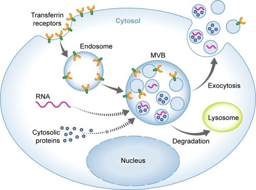
Exosomes were first defined as EVs arising from maturing reticulocytes and appreciated as a means to remove obsolete material. A role of exosomes in intercellular communication was suggested by Raposo and Stoorvogel,Citation9 indicating that B cell-derived exosomes can deliver functional antigen-presenting complexes. Exosomes consist of proteins, mRNA, and noncoding miRNA surrounded by a lipid bilayer membrane.Citation10–Citation13 Further RNA forms including transfer RNA, ribosomal RNA, nucleolar RNA, and long noncoding RNA (lncRNA) have also been identified in exosomes.Citation14 Topical studies have demonstrated that fragments of DNA may exist in exosomes as well.Citation15–Citation17 The exosome biogenesis process consists of four phases: initiation, endocytosis, multivesicular body (MVB) development, and release.Citation18 Initially, endocytic vesicles are produced from the plasma membrane, forming an early endosome that subsequently develops into late endosomes. The late endosomes undergo inward budding leading to formation and accumulation of intraluminal vesicles inside the lumen, termed MVBs. The MVBs will merge with lysosome for degradation; otherwise they will merge with the plasma membrane, releasing intraluminal vesicles into extracellular spaces, which are now called exosomes ().Citation19–Citation21 Secreted exosomes can subsequently be directed to other cells via a rather poorly determined mechanism, probably through proteins located at the surface such as tetraspanins.Citation7 The exosomes are taken up by the target tissues through membrane fusion, endocytosis, or receptor–ligand interaction, which later can be transferred to lysosomes in order to be degraded or can release its cargo within the receiver cells.
Figure 1 Exosome biogenesis and secretion.
Note: Reprinted from Schorey JS, Cheng Y, Singh PP, Smith VL. Exosomes and other extracellular vesicles in host–pathogen interactions. EMBO Rep. 2015;16(1):24–43, with permission from John Wiley and Sons.Citation21
Abbreviation: MVB, multivesicular body.
The protein content of exosomes has been well identified using different proteomic approaches.Citation22 Mass spectrometry indicated the presence of over 4,000 various proteins within exosomes while there appears to be a unique preserved protein collection in exosomes derived from certain kinds of cells.Citation23 Alix, heat shock proteins, and TSG101 proteins have been recognized in most of the exosomes.Citation24 Exosomes have tetraspanins, such as CD9, CD81, and CD63, which are involved in cellular interactions through binding with various proteins, such as integrins and major histocompatibility complex (MHC) molecules.Citation25 Rab, annexins, myosin, actin, tubulin, ribosomal proteins, and signal transduction molecules are demonstrated to be enriched in exosomes.Citation9,Citation24 The exosomal components differ based on various physiological/pathological conditions and certain kinds of cells. Furthermore, the exosomal content can be dissimilar from the cells of origin owing to the discriminating sorting of the cargo into exosomes.
Exosome isolation methods
Several accepted techniques, such as differential ultracentrifugation, density gradients, precipitation, filtration, and size exclusion chromatography, have been applied for exosome separation.Citation19 Traditional ultracentrifugation has become a reliable technique among researchers in exosome research. It consists of a series of centrifugation cycles of varying centrifugal force and duration to isolate exosomes according to their density and size differences. Recently, a number of commercial kits have been introduced to isolate exosomes for different purposes.Citation9 These kits are superior to ultracentrifugation due to being less time consuming, less technique sensitive, and more compatible with limited volumes of samples. The relative success of these different methods needs to be considered in terms of recovery and specificity.
Roles of MSC-derived EVs/exosomes in cancer
The microenvironment of a tumor is composed of different cell types, such as fibroblasts, immune cells, and endothelial cells. The crosstalk of this microenvironment with the tumor seems to be vital for growth and progression of tumor cells.Citation26,Citation27 Previous studies have revealed that MSCs produce exosomes, which could perform as paracrine mediators by transferring signaling molecules, which regulate tumor cell proliferation, angiogenesis, and metastasis via controlling a number of cellular pathways.Citation28,Citation29
Tumor growth
The impact of exosomes on tumor progression has been extensively reported in the past decade. MSC-derived exosomes affect tumor development in both supporting and suppressing ways. Among the proposed mechanisms, the miRNA content of exosomes has been widely investigated. As indicated by Vallabhaneni et al,Citation30 stressed MSC-derived EVs including exosomes promoted breast cancer cell proliferation and metastasis via transferring tumor-supportive miRNAs and proteins (). It was demonstrated that a variety of tumor-promoting miRNAs, such as miR-21 and miR-34a, have been enriched in MSC-derived EVs. This tumor-supportive function may rely on epigenetic changes induced under stress conditions. Along with miRNAs, several possible mechanisms have been proposed to be associated in the protumor or antitumor features of MSC-derived exosomes. According to Qi et al,Citation31 MSC-derived exosomes from bone marrow (BM MSCs) have stimulated the Hedgehog signaling pathway in osteosarcoma and gastric cancer cell lines, and thus promoted tumor growth. It was also demonstrated that exosomes derived from MSCs hold matrix metalloproteinase-2 that might result in tumor microenvironment reorganization and growth.Citation32 While MSC-derived exosomes largely mediate tumor-promoting effects in the tumor microenvironment, there is also evidence for antitumor activity of EVs derived from MSCs. Wu et alCitation33 reported that EVs from human umbilical cord Wharton’s jelly MSCs reversed the development of bladder carcinoma cells possibly by down-regulating phosphorylation of Akt protein kinase and up-regulating cleaved caspase-3 (). Similarly, adipose MSC-derived exosomes were demonstrated to inhibit prostate cancer via delivery of miR-145 by reducing the activity of Bcl-xL and promoting apoptosis through the caspase-3/7 pathway.Citation34 In the same way, EVs obtained from normal human BM MSCs were found to inhibit proliferation and to promote apoptosis in liver carcinoma, Kaposi’s sarcoma, and ovarian tumor cell lines.Citation35 The inconsistency between the results pointed to a need for additional research in developing a standardized condition for MSC culture as the MSC culture condition may affect the overall features of the secreted vesicles. In addition, the source of MSCs where exosomes were obtained was also demonstrated to be important for the final tumor-suppressive or tumor-promoting effects. Roccaro et alCitation36 noticed that EVs derived from BM MSCs of patients with multiple myeloma could support multiple myeloma tumor/cell progression while EVs isolated from normal individuals suppressed the development of multiple myeloma tumor/cells perhaps by transferring a lower content of miR-15a. Besides higher miRNA content, other factors such as superior amounts of cytokines and adhesion molecules in patient-derived exosomes might also be involved in the tumor-promoting effects.
Table 1 Natural or modified MSC EVs/exosomes that promote tumors or extend their metastatic effect
Table 2 Natural or modified MSC EVs/exosomes that have an inhibitory effect on tumors
Angiogenesis
Angiogenesis is an essential physiological multistep procedure involved in tumorigenesis. Exosomes hold several angiogenic factors that control tumor angiogenesis. Exosomes derived from MSCs were shown to stimulate angiogenesis through elevating vascular endothelial growth factor (VEGF) production in tumor cells and via stimulating ERK1/2 and p38 mitogen-activated protein kinase pathways.Citation37 MSCs secrete exosomes that transport mRNAs and miRNAs to target cells and induce endothelial cell proliferation, thus leading to angiogenic effects, superior blood flow restoration, and capillary network formation. Placental MSC-derived exosomes have been shown to facilitate placental microvascular endothelial cell migration and vascularization.Citation38 It was shown that adipose MSC-derived exosomes and MVs are internalized by human microvascular endothelial cells and lead to angiogenesis.Citation39 According to this study, platelet-derived growth factor enhances angiogenesis by causing adipose MSCs to secrete exosomes and MVs that are rich in proangiogenic factors. In a similar study, injection of MSC-derived exosomes into stroke rats was shown to lessen severe symptoms by stimulating angiogenesis, neurite remodeling, and neurogenesis.Citation40 Lee et al,Citation41 however, showed that exosomes derived from MSCs blocked vessel formation by delivery of miR-16 via downregulating VEGF within the tumor microenvironment. Similarly, Huang et alCitation42 revealed that the angiogenic function of MSCs was mostly mediated through MVs while exosomes and paracrine factors were shown to prevent HIMF and Smad2, which lead to anti-vascular remodeling. Although several reports have shown that MSCs play a key role in angiogenesis, the role of MSC exosomes in angiogenesis is still controversial. It seems that some miRNAs found in MSC exosomes are supposed to be specifically involved in tumor angiogenesis. According to a bioinformatic analysis conducted by Ferguson et al,Citation43 various genes targeted by the miRNA content of exosomes derived from MSCs were associated with angiogenesis and vessel formation, and thus the angiopoietin network might be a key target of these exosomes in angiogenesis induction. Other factors such as amounts of oncogenic proteins, cytokines, and adhesion molecules are thought to be involved in the exosome-mediated angiogenesis.
Metastasis/invasion
The role of exosomes derived from MSCs has also been examined in metastasis and the premetastatic niche. Exosomes derived from MSCs were reported to transfer miR-221 to HGC27 cells, thus enabling the growth and migration of tumor cells.Citation44 The results of this study showed that while MSC-derived exosomes from both normal and cancer tissue shared similarities in promoting tumor proliferation and migration, MSCs obtained from cancer tissue secreted exosomes enriched in tumor-supportive miR-221 and thus favored cancer promotion and migration. Similarly, exosomes isolated from MSCs induced Wnt signaling activation, hence facilitating the growth and migration of the breast tumor cells.Citation45 It has been shown that MSCs induced the invasiveness of breast cancer cells partly through MSC-derived MVs.Citation46 Furthermore, BM-derived MSCs were revealed to release exosomes that encompass miRNAs, stimulating dormancy in invasive breast cancer cells.Citation47 Exosomes are likewise made up of multi functional proteins. Lai et alCitation48 noticed that abundant amounts of all seven α chains and seven β chains of the 20S proteasome and the three β subunits of the immunoproteasome were present in MSC-derived exosomes, showing that exosomes were capable of targeting tumor cells via proteasome transfer.
In conclusion, even though exosomes hold a hopeful future as drug delivery vehicles, their application has been restricted by the lack of standardized approaches in the case of production, isolation, and purification. The protumor or antitumor features of stem cell exosomes may possibly rely on the settings utilized to cultivate the stem cells and promote vesicle formation and on the tumor model to be utilized, as both the tumor microenvironment and the systemic environment of the host can differ seriously from tumor suppression to tumor promotion. Therefore, the main outcomes in some studies are not adequately supported by the experiments performed and detailed data need to be given to allow replication. It is crucial to demonstrate all detailed procedures for reported parameters, such as culture media composition and harvesting condition as well as any supplements, such as antibiotics and growth factors, as these can influence EV production and the relevant content. Furthermore, the method of isolation needs to be stated as exosomes and other EVs mediate quite different or overlapping functions. The controversial effects of MSC-derived exosomes might be partially associated with the complexity of mechanisms applied by stem cells to sense and home to tumors that might overlap with strategies that direct them to sites of injury and inflammation. Besides, miRNAs are crucial players in the ultimate biological function of exosomes as these miRNAs are formed as a central fraction of the exosomal content.
Modification of MSC-derived exosomes for cancer drug delivery
In the previous section, we have highlighted the effect of natural (nonmodified) MSC-derived exosomes on cancer biology. In this section, we will summarize the application of modified MSC-derived exosomes as a novel therapeutic strategy for drug delivery in cancer. As demonstrated by Smyth et al,Citation49 internalization of exosomes within tumor cells is ten times greater than liposomes of comparable size, representing a superior specificity of exosomes for cancer targeting. In addition, cancer cells were demonstrated to internalize a greater percentage of exosomes when compared to normal cells.Citation50 In conventional therapies, the lack of selectivity to the diseased site is considered the major drawback. Many stem cells types, however, show intrinsic tropism toward tumors, making them attractive candidates for the targeted delivery of anticancer drugs. Additionally, by engineering/modifying these cells to express anticancer agents, they can effectively target tumor sites.Citation51 This novel exosome-based therapy might be an interesting alternative due to their benefits over the corresponding stem cells. They are smaller, less complex, and less immunogenic than their parent cells since they have a lower content of membrane-bound proteins.Citation52 Furthermore, production and storage of exosomes are easier than for their parental cells. Pascucci et alCitation53 demonstrated that MSCs treated with paclitaxel-mediated powerful anti-tumorigenic outcomes due to their potential to get the drug and subsequently packed it in EVs. Besides approaches utilized for loading drugs into exosomes, various loading methods can also be applied to encapsulate miRNA inside exosomes. According to Munoz et al,Citation54 anti-miR-9-loaded MSC-derived exosomes reversed the expression of multi-drug transporters in drug-resistant glioblastoma multiforme cells and reversed the chemoresistance. Alternative research showed that intratumoral injection of miR-146b-expressing MSC-derived exosomes resulted in considerable reduction in glioma xenograft development in a rat brain tumor model and decreased the growth, migration, and invasion of cells.Citation55 According to these findings, miRNAs could be packaged into MSC-derived exosomes and later suppressed glioma tumor cells, suggesting that engineered MSCs to secrete exosomes enriched with miRNAs could be an effective strategy for malignant glioma treatment. In a similar study, the obtained results indicated that the transfer of miR-143 by means of MSC exosomes decreased the in vitro migration of osteosarcoma cells.Citation56 Likewise, miR-122-transfected adipose MSCs can generate miR-122 encapsulated exosomes to deliver miR-122 into hepatocellular tumor cells, which elevated tumor cell sensitivity to chemotherapeutic agents via gene expression alternations and tumor proliferation in vitro and in vivo.Citation57 Transfected MSCs to release exosomes encapsulated with miR-379 have been administered for breast cancer therapy in vivo.Citation58 According to the results of this study, the modified exosomes were delivered to the tumor site and have therapeutic effects. Application of EVs for anticancer protein delivery by genetic manipulation of parental MSCs has also been investigated. Yuan et alCitation59 examined EV-mediated TRAIL delivery in various cancer cells. The modified MSC EVs expressing TRAIL promoted apoptotic death in 11 cancer cell lines in a dose-dependent manner. A similar result was observed in MSC-derived exosome-mediated small interfering RNA (siRNA) gene delivery.Citation50 Selective gene silencing of PLK-1 was achieved using MSC-derived exosomes as a vehicle to transfer PLK-1 siRNA to tumor cells of the bladder. The approach led to effective repression of PLK-1 mRNA and protein. Lee et alCitation60 noticed that exosomes derived from MSCs delivered particular miRNA mimics (miR-124 and miR-145) and reduced glioma cell migration and the stem cell properties of cancer cells. siRNA loading of exosomes was also investigated by an electro-poration approach. According to the study conducted by Kamerkar et al,Citation61 electroporated MSC-derived exosomes with siRNA against oncogenic Kras suppressed cancer in mouse pancreatic tumor models and remarkably improved overall survival. Bliss et alCitation62 demonstrated that breast cancer cells prime MSCs to secrete exosomes with a particular profile of miRNAs, which account for the cellular quiescence effect and drug resistance in some types of cancer cells. According to this finding, a beneficial approach to target dormant breast cancer cells was introduced by systemic application of MSCs encapsulated with antagomiR-222/223 resulting in chemosensitization and an increased survival rate.
In summary, these findings demonstrate the possible effectiveness of exosomes in drug and/or nucleic acid delivery. Several features including the selection of cells from which exosomes are obtained, yield of exosomes, cargo encapsulation process, choice of targeting biomolecules on the surface, biodistribution, and immune response are important elements that need to be considered for exosome-mediated drug transfer in upcoming clinical settings. Encapsulation of small-molecule drugs may be effective, although further investigation is essential for genetic materials encapsulation. The quantity and nature of RNA in exosomes may differ based on the type and origin of the parental cells; however, certain RNAs are enriched.Citation63,Citation64 This raises concepts that specific mechanisms may occur for RNA sorting into EVs; hence, discovering these mechanisms might be used to deliver selective therapeutic RNA cargoes. In addition, a full characterization and comparison of exosome contents of various sources will be essential to demonstrate whether stem cell-derived exosomes differ from others. Application of recent technologies such as a highly versatile single-cell assay offers a suitable and low-cost approach allowing quantitative and time-lapsed examination of single-cell features including secreted exosomes.Citation65
Strategies to modify exosomes for drug delivery
In this section, we highlight the exciting potential for exosomes as therapeutic vehicles for drug delivery and to develop strategies to engineer/modify the exosome surface and content.
Cargo loading
With regard to the intrinsic capability of exosomes to transfer genetic materials, it is possible to engineer vesicles for gene therapy through exosomal-mediated delivery of some important regulating miRNAs, various noncoding RNAs, or tumor suppressors.Citation66,Citation67 One of the most widely used methods is to bring about transgene expression in the parental cell, which is inspired by the regular action of the exosome for intercellular transfer. Using this manipulation strategy, mRNA or noncoding RNAs could be packed into exosomes using a donor cell transfection method.Citation55,Citation64,Citation68 Another approach for manufacturing exosome-based formulation is genetic fusions of polypeptides to the enriched protein in exosomes to guarantee the ideal localization of the expressed product. For example, application of poly A binding protein that attaches mature mRNAs to selectively recruit mRNAs into exosomes. Otherwise, a zip-like, approximately 25-nucleotide fragment present in the 3′-UTR of particular mRNAs can be fused into the 3′-UTR of the mRNA of interest and be recruited by Z-DNA binding protein 1 (ZBP1).Citation64 Similarly, to selectively enrich exosomes with miRNAs, Ago protein or catalytically dead Dicer/Drosha can be attached to EV-specific proteins such as tetraspanin CD63 to cargo miRNAs or pre-miRNAs and pri-miRNAs, respectively.Citation69 Likewise, polycomb repressive complex 2 (gene symbols EZH1 and EZH2) identifies specific lncRNAs selectively, and thus could be applied to enrich exosomal lncRNAs.Citation70 For drug incorporation into exosomes, one method is to introduce exogenous materials to the parental cells. The encapsulation efficiency normally depends on the quantity of material to be transported to the cell. Hence, in order to maximize cell loading, high concentrations and prolonged incubation periods are required. The main restriction of this method is that it depends on phagocytosis as an uptake mechanism. Therefore, it is challenging to obtain equivalent encapsulation. Another strategy for drug loading is the passive approach, in which the drug is incorporated into exosomes through post-isolation methods. This approach is not restricted to biological cargo and can include small-molecule drugs. The simplest passive approach of cargo loading is obtained by incubation of cargo with exosomes. This approach efficiently works with some hydrophobic molecules that affect lipid rearrangement and modify the lipid fluidity. For those large and charged molecules that cannot diffuse across the membrane, loading can be obtained via electroporation. Loading therapeutic RNAs into exosomes has several challenges as the process can be inefficient and generates RNA precipitates.Citation71 Regarding the exosomal-mediated protein delivery, incorporation of catalase into exosomes was practical by several encapsulation procedures, such as incubation at room temperature, freeze/thaw cycles, and sonication.Citation72 Recently, an exosome-based delivery system called EXPLORs has been developed for protein loading via optically reversible protein–protein interactions.Citation73 Taken together, these studies indicate the potential application of exosome-mediated nucleic acid, protein, and small-molecule drug delivery. For cargo loading, each method has its own pros and cons, and varies based on the therapeutic payload, site of the disease, and proper settings for a certain type of exosome-cargo vehicle.
Exosome display technology
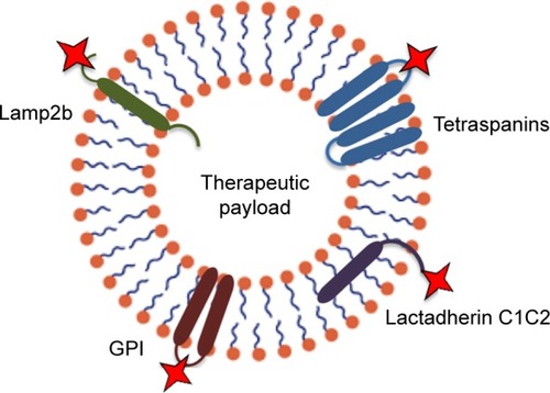
Despite the feasibility of exosomes as natural carriers for various types of RNAs, proteins, and artificial drugs,Citation74 systemically administered exosomes might be gathered in some other tissues. Exosome display technology is a procedure allowing re-engineering of the exosomal protein composition to modify exosomes with novel desired features. Using this technology, several forms of ligands such as a multi-meric antigen, which does not typically exist on exosomes, can be produced at the surface of exosomes in a natural conformation.Citation75 A popular application of this technology is to engineer exosomes with targeting ligands by transfection of the parental cells in order to obtain production of targeting moieties attached to exosome native membrane proteins (). Lysosomal-associated membrane protein 2 (Lamp2b) is a well-known exosome membrane protein that has been widely investigated for exosome targeting.Citation76–Citation78 Alvarez-Erviti et alCitation79 used the rabies virus glycoprotein (RVG) peptide to target exosomes to the mouse brain by manipulation the parental dendritic cells to express Lamp2b, fused to the neuron-specific peptide derived from RVG. Despite the effectiveness of the method, there are serious concerns about the longstanding stability of Lamp-2b hybrids;Citation77 hence, more stable substitutes to Lamp-2b such as glycosylphosphatidylinositol (GPI) have been introduced. As demonstrated by this group, EGF-expressing tumor cells were targeted by EVs displaying anti-EGF receptor nanobodies fused to GPI. Likewise, others have established a human embryonic kidney cell line that stably expressed EGF binding peptide fused with the transmembrane receptor of platelet-derived growth factor for targeted tumor therapy.Citation80 Another exosome membrane protein candidate is the C1C2 domain of lactadherin. Lactadherin has been indicated to localize to the lipid membrane of exosome through binding of its C1C2 domain.Citation81 Inspired from display technology, a group of researchers used the exosomal surface structure in order to discover potential sites on the tetraspanin CD63 for integration of fluorescent fusion proteins on both sides of the exosomal membrane.Citation82 Zhao et alCitation83 used the cell’s own machinery to engineer a chimeric multidomain transmem-brane targeting protein, which contained the intracellular and transmembrane domains of the transferrin receptor capable of targeting EVs to specific populations of cells.
Figure 2 Strategies for targeting extracellular vesicles to particular target cells can be achieved by genetic modification of exosomes to express targeting moieties fused with exosome native membrane proteins, such as lysosomal-associated membrane protein 2 (Lamp2b), tetraspanins, glycosylphosphatidylinositol (GPI), and lactadherin C1C2.
Taken together, this technology demonstrates an approach to display targeting of oligonucleotides and proteins on the surface of EVs. However, such strategies might be vulnerable since they require modifications of producer cells that are often time-consuming and challenging, particularly in the case of primary cells. Besides, a number of targeting moieties protein that attaches inappropriate expression and degradation that restricts their functional demonstration on EVs.
Hybrid membrane engineering
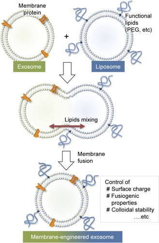
For further applications of exosomes in drug transfer, it may be essential to alter and tune the exosome interface to improve the surface features to reduce exosome immunogenicity, increase stability, and improve its half-life in blood. Recently, a new approach was proposed to provide stealth in addition to tumor cell-targeting features to pre-isolated exosomes. This group developed a technique for tailoring exosomes with targeting moieties attached to polyethylene glycol.Citation4 While exosomes without modification were quickly cleared from the bloodstream within a few minutes after systematic injection in mice, the engineered exosomes were detectable in plasma for prolonged circulation times. This approach enables a dual strategy to design targeted liposomes, where multiple ligands can be inserted into a variety of preformed liposomes containing a number of drugs, allowing the treatment to be personalized based on the needs of individuals. In an alternative approach to optimize the performance of exosomal carriers, a novel membrane-engineering strategy was introduced to modify the exosome interface using direct membrane fusion between synthetic liposomes and exosomes in pre-isolated vesicles ().Citation84 Furthermore, Goh et alCitation85 introduced a hybrid system called EXOPLEXs, obtained by fusion of EVs and liposomes as a novel drug delivery vehicle. Interestingly, similar loading efficiencies were obtained when doxorubicin was encapsulated into liposomes and EXOPLEXs, which recommends that the fusion procedure does not adversely affect the encapsulation capacity of EXOPLEXs. In conclusion, it is assumed that these characteristics would be favorable for the exosome-based drug transfer, as exogenously administered exosomes have been indicated to be cleared quickly from the bloodstream by the reticuloendothelial system, therefore preventing their accumulation in the target tissue.
Figure 3 Schematic of the procedure used to engineer the exosome–liposome hybrids. Reprinted from Sato YT, Umezaki K, Sawada S, et al. Engineering hybrid exosomes by membrane fusion with liposomes. Sci Rep. 2016;6:21933.Citation84
Surface functionalization
Surface modification is another strategy to improve the targeting capability of exosomes.Citation4,Citation5 Click chemistry is a method that enables the bioconjugation of functional ligands to the surfaces of exosomes by copper-catalyzed azidealkyne cycloaddition.Citation86 Furthermore, it is rapid and highly efficient in comparison with traditional cross-linking chemistries. Tian et alCitation87 recently proposed conjugation of the cyclo (Arg-Gly-Asp-d-Tyr-Lys) peptide, which displays high affinity toward integrin αvβ3 on the MSC-derived exosome surface. Another strategy for modification of exosomes is the application of the ApoA-I mimetic peptide (L-4F), which enables the interaction of EVs and a therapeutic/targeted peptide by attachment to the phospholipid vesicles. Ye et alCitation88 have shown that methotrexate-loaded EVs functionalized with a synthetic multifunctional peptide facilitated the membrane receptor-mediated internalization procedure both in vitro and in vivo in a glioma model. In summary, surface functionalization of exosomes using click chemistry could improve the targeting capability of exosomes. This approach seems to be superior compared to exosome display technology as it does not have the challenges of modifying producer cells.
Large-scale production and storage of exosomes for clinical use
One of the key issues in developing exosome-mediated drug delivery systems is to scale up the exosome production.Citation89 Large-scale production of therapeutic exosomes necessitates exosomal isolation and purification under controlled conditions. In order to manufacture therapeutic exosomes, the exosome yield per cell is important and has an impact on ultimate production cost and clinical applications. MSCs are superior in large-scale production of exosomes and can be applied on a clinical scale.Citation90 The MSC source is one of the key factors that should be considered in exosome-based drug delivery systems. The ideal source would be a high-exosome-yielding cell with a high expansion capacity. Another important consideration is the age of the donor tissues, as the exosome production might be inversely associated with the age of the donor tissue. The prolonged donor cell cultivation may result in significantly increased production. According to the literature, prolonged culture and maintaining cells at low pH improved exosomal release.Citation91,Citation92 In order to enhance vesicle release from cells, other strategies have also been proposed. Raising intracellular calcium, serum starvation, or endotoxin can increase vesicle production. Establishment of immortalized cells from MSCs is another strategy to scale up EV production.Citation93 Overexpression of oncogene c-myc, for instance, was reported to increase EV production in MSCs.Citation94 Extrusion or filtration of cells is another approach which might be easily scaled up to obtain high yields of exosomes. Isolation of exosome-like nanoparticles following the breakdown of parental cells encapsulated with antitumor drugs led to a 100-fold higher yield of the drug carriers.Citation95 Lastly, manufacturing bioreactors similar to those for tissue engineering might be employed for large-scale production of exosomes.Citation96 Particularly, MSCs may be cultured from different origins, propagated, and later loaded with the desired therapeutic biomolecules. The choice of the loading method and the therapeutic cargo are other features that need to be considered for successful exosome-based drug delivery. Strategies to preserve exosomes are challenges that need to be further addressed. Previous reports have demonstrated that the size of exosome drops by 60% after 2 days of storage at 37°C. Nevertheless, the initial size was maintained when exosomes are stored at −80°C for 2 days.Citation97 It is possible to preserve exosomes at −80°C for extended periods. Based on the promising preclinical studies, EV-based therapies are making their way into the clinic. Our knowledge of exosome-mediated therapies is rapidly expanding although exosomes have already been approved for application in several clinical trials.
Conclusion and future perspectives
In recent years, the rapid development of the MSC exosome field has attracted the attention of many researchers. MSC exosomes hold remarkable potential in therapeutic applications considering their relative ease of isolation and manipulation of both contents and surface. The lipid and protein composition of exosomes, which enhance exosomal stability and slow clearance in circulation are other features that make these vesicles as perfect carriers. Owing to their small size, lack of toxicity, and target specificity as well as being tolerated by the body, these vehicles might be regarded as the next-generation drug delivery system. Furthermore, exosomes could be generated on a large scale, are easier to handle, are less expensive, and do not raise potential ethical and legal concerns compared to MSCs. These characteristics demonstrate potentials of exosomes for upcoming therapeutic applications. Research in exosome biology has been in the early stage of development and much effort needs to be made in order to guarantee the safe and effective application of them for therapeutic use. In addition, component characterization, immune reactions, and loading of exosomes without modifying the natural characteristics of the donor cell require to be obviously understood. Translating therapy from the lab into the clinics requires scale up of exosome isolation. Therefore, adequate standards for exosome manipulation, isolation, and characterization need to be established to bring this exciting development a step closer to clinical reality.
Disclosure
The authors declare no conflicts of interest in this work.
References
- ShaoHImHCastroCMBreakefieldXWeisslederRLeeHNew technologies for analysis of extracellular vesiclesChem Rev201811841917195010.1021/acs.chemrev.7b0053429384376
- BoriachekKIslamMNMöllerABiological functions and current advances in isolation and detection strategies for exosome nanovesiclesSmall2018146170215310.1002/smll.201702153
- WangBLiPShangguanLA novel bacterial cellulose membrane immobilized with human umbilical cord mesenchymal stem cells-derived exosome prevents epidural fibrosisInt J Nanomedicine2018135257527310.2147/IJN.S16788030237713
- KooijmansSASchiffelersRMZarovniNVagoRModulation of tissue tropism and biological activity of exosomes and other extracellular vesicles: new nanotools for cancer treatmentPharmacol Res201611148750010.1016/j.phrs.2016.07.00627394168
- ArmstrongJPHolmeMNStevensMMRe-engineering extracellular vesicles as smart nanoscale therapeuticsACS Nano2017111698310.1021/acsnano.6b0760728068069
- TatischeffICell-derived extracellular vesicles open new perspectives for cancer researchCancer Res Front20151208224
- NevianiPFabbriMExosomic microRNAs in the tumor microenvironmentFront Med20152475310.3389/fmed.2015.00047
- ZhangH-GCaoPTengYIsolation, identification, and characterization of novel nanovesiclesOncotarget2016727413464136210.18632/oncotarget.932527191656
- RaposoGStoorvogelWExtracellular vesicles: exosomes, microvesicles, and friendsJ Cell Biol2013200437338310.1083/jcb.20121113823420871
- BraicuCTomuleasaCMonroigPCucuianuABerindan-NeagoeICalinGExosomes as divine messengers: are they the Hermes of modern molecular oncology?Cell Death Differ2015221344510.1038/cdd.2014.13025236394
- LeeB-RKimJ-HChoiE-SChoJ-HKimEEffect of young exosomes injected in aged miceInt J Nanomedicine2018135335534510.2147/IJN.S17068030254438
- KooijmansSAVaderPvan DommelenSMvan SolingeWWSchiffelersRMExosome mimetics: a novel class of drug delivery systemsInt J Nanomedicine201271525154122619510
- ZhangLHaoCYaoSExosomal miRNA profiling to identify nanoparticle phagocytic mechanismsSmall201814151704008
- HuangXYuanTTschannenMCharacterization of human plasma-derived exosomal RNAs by deep sequencingBMC Genomics201314131933310.1186/1471-2164-14-31923663360
- KahlertCMeloSAProtopopovAIdentification of double-stranded genomic DNA spanning all chromosomes with mutated KRAS and p53 DNA in the serum exosomes of patients with pancreatic cancerJ Biol Chem201428973869387510.1074/jbc.C113.53226724398677
- GuesciniMGenedaniSStocchiVAgnatiLFAstrocytes and glioblastoma cells release exosomes carrying mtDNAJ Neural Transm201011711410.1007/s00702-009-0288-819680595
- TakahashiAOkadaRNagaoKExosomes maintain cellular homeostasis by excreting harmful DNA from cellsNat Commun20178152871530110.1038/ncomms1528728508895
- ZhangXYuanXShiHWuLQianHXuWExosomes in cancer: small particle, big playerJ Hematol Oncol201581839610.1186/s13045-015-0181-x26156517
- El AndaloussiSMägerIBreakefieldXOWoodMJExtracellular vesicles: biology and emerging therapeutic opportunitiesNat Rev Drug Discov201312534735710.1038/nrd397823584393
- AlenquerMAmorimMExosome biogenesis, regulation, and function in viral infectionViruses2015795066508310.3390/v709286226393640
- SchoreyJSChengYSinghPPSmithVLExosomes and other extracellular vesicles in host–pathogen interactionsEMBO Rep2015161244310.15252/embr.20143936325488940
- WeltonJLKhannaSGilesPJProteomic analysis of bladder cancer exosomesMol Cell Proteomics2010961324133810.1074/mcp.M000063-MCP20120224111
- TicknerJAUrquhartAJStephensonS-ARichardDJOByrneKJFunctions and therapeutic roles of exosomes in cancerFront Oncol2014412713510.3389/fonc.2014.0012724904836
- Villarroya-BeltriCBaixauliFGutierrez-VazquezCSanchez-MadridFMittelbrunnMSorting it out: regulation of exosome loadingSemin Cancer Biol20142831310.1016/j.semcancer.2014.04.00924769058
- FrydrychowiczMKolecka-BednarczykAMadejczykMYasarSDworackiGExosomes-structure, biogenesis and biological role in non-small-cell lung cancerScand J Immunol201581121010.1111/sji.1224725359529
- CorcoranCRaniSOBrienKDocetaxel-resistance in prostate cancer: evaluating associated phenotypic changes and potential for resistance transfer via exosomesPLoS One2012712e5099910.1371/journal.pone.005099923251413
- WeiYLaiXYuSExosomal miR-221/222 enhances tamoxifen resistance in recipient ER-positive breast cancer cellsBreast Cancer Res Treat2014147242343110.1007/s10549-014-3037-025007959
- RatajczakJMiekusKKuciaMEmbryonic stem cell-derived microvesicles reprogram hematopoietic progenitors: evidence for horizontal transfer of mRNA and protein deliveryLeukemia200620584785610.1038/sj.leu.240413216453000
- GrangeCTapparoMCollinoFMicrovesicles released from human renal cancer stem cells stimulate angiogenesis and formation of lung premetastatic nicheCancer Res201171155346535610.1158/0008-5472.CAN-11-024121670082
- VallabhaneniKCPenfornisPDhuleSExtracellular vesicles from bone marrow mesenchymal stem/stromal cells transport tumor regulatory microRNA, proteins, and metabolitesOncotarget2015674953496710.18632/oncotarget.321125669974
- QiJZhouYJiaoZExosomes derived from human bone marrow mesenchymal stem cells promote tumor growth through hedgehog signaling pathwayCell Physiol Biochem20174262242225410.1159/00047999828817816
- YangYBucanVBaehreHVon Der OheJOtteAHassRAcquisition of new tumor cell properties by MSC-derived exosomesInt J Oncol201547124425210.3892/ijo.2015.300125963929
- WuSJuG-QDuTZhuY-JLiuG-HMicrovesicles derived from human umbilical cord Wharton’s jelly mesenchymal stem cells attenuate bladder tumor cell growth in vitro and in vivoPLoS One201384e6136623593475
- TakaharaKInamotoTIbukiN245 microRNA-145 mediates the inhibitory effect of adipose-derived stem cells on androgen-independent prostate cancerEur Urol Suppl2016153e24510.1016/S1569-9056(16)60247-6
- BrunoSCollinoFDeregibusMCGrangeCTettaCCamussiGMicrovesicles derived from human bone marrow mesenchymal stem cells inhibit tumor growthStem Cells Dev201222575877110.1089/scd.2012.030423034046
- RoccaroAMSaccoAMaisoPBM mesenchymal stromal cell–derived exosomes facilitate multiple myeloma progressionJ Clin Invest201312341542155510.1172/JCI6651723454749
- ZhuWHuangLLiYExosomes derived from human bone marrow mesenchymal stem cells promote tumor growth in vivoCancer Lett20123151283710.1016/j.canlet.2011.10.00222055459
- SalomonCRyanJSobreviaLExosomal signaling during hypoxia mediates microvascular endothelial cell migration and vasculogenesisPLoS One201387e6845123861904
- LopatinaTBrunoSTettaCKalininaNPortaMCamussiGPlatelet-derived growth factor regulates the secretion of extracellular vesicles by adipose mesenchymal stem cells and enhances their angiogenic potentialCell Commun Signal2014121263824725987
- XinHLiYCuiYYangJJZhangZGChoppMSystemic administration of exosomes released from mesenchymal stromal cells promote functional recovery and neurovascular plasticity after stroke in ratsJ Cereb Blood Flow Metab201333111711171510.1038/jcbfm.2013.15223963371
- LeeJ-KParkS-RJungB-KExosomes derived from mesenchymal stem cells suppress angiogenesis by down-regulating VEGF expression in breast cancer cellsPLoS One2013812e8425610.1371/journal.pone.008425624391924
- HuangLMaWMaYFengDChenHCaiBExosomes in mesenchymal stem cells, a new therapeutic strategy for cardiovascular diseases?Int J Biol Sci201511223824510.7150/ijbs.1072525632267
- FergusonSWWangJLeeCJThe microRNA regulatory landscape of MSC-derived exosomes: a systems viewSci Rep2018811419143110.1038/s41598-018-19581-x29362496
- WangMZhaoCShiHDeregulated microRNAs in gastric cancer tissue-derived mesenchymal stem cells: novel biomarkers and a mechanism for gastric cancerBr J Cancer201411051199121010.1038/bjc.2014.1424473397
- LinRWangSZhaoRCExosomes from human adipose-derived mesenchymal stem cells promote migration through Wnt signaling pathway in a breast cancer cell modelMol Cell Biochem20133831–2132010.1007/s11010-013-1746-z23812844
- MaffeyAStoriniCDiceglieCMesenchymal stem cells from tumor microenvironment favour breast cancer stem cell proliferation, cancerogenic and metastatic potential, via ionotropic purinergic signallingSci Rep201771131621317110.1038/s41598-017-13460-729030596
- OnoMKosakaNTominagaNExosomes from bone marrow mesenchymal stem cells contain a microRNA that promotes dormancy in metastatic breast cancer cellsSci Signal20147332ra6310.1126/scisignal.200523124985346
- LaiRCTanSSTehBJProteolytic potential of the MSC exosome proteome: implications for an exosome-mediated delivery of therapeutic proteasomeInt J Proteomics2012201297190797192110.1155/2012/97190722852084
- SmythTJRedzicJSGranerMWAnchordoquyTJExamination of the specificity of tumor cell derived exosomes with tumor cells in vitroBiochim Biophys Acta Biomembr20141838112954296510.1016/j.bbamem.2014.07.026
- GrecoKAFranzenCAForemanKEFlaniganRCKuoPCGuptaGNPLK-1 silencing in bladder cancer by siRNA delivered with exosomesUrology201691241241e1241.e710.1016/j.urology.2016.01.030
- StuckeyDWShahKStem cell-based therapies for cancer treatment: separating hope from hypeNat Rev Cancer2014141068369110.1038/nrc379825176333
- LouGChenZZhengMLiuYMesenchymal stem cell-derived exosomes as a new therapeutic strategy for liver diseasesExp Mol Med2017496e34610.1038/emm.2017.6328620221
- PascucciLCocceVBonomiAPaclitaxel is incorporated by mesenchymal stromal cells and released in exosomes that inhibit in vitro tumor growth: a new approach for drug deliveryJ Control Release201419226227025084218
- MunozJLBlissSAGrecoSJRamkissoonSHLigonKLRameshwarPDelivery of functional anti-miR-9 by mesenchymal stem cell-derived exosomes to glioblastoma multiforme cells conferred chemosensitivityMol Ther Nucleic Acids20132e12610.1038/mtna.2013.6024084846
- KatakowskiMBullerBZhengXExosomes from marrow stromal cells expressing miR-146b inhibit glioma growthCancer Lett2013335120120410.1016/j.canlet.2013.02.01923419525
- ShimboKMiyakiSIshitobiHExosome-formed synthetic microRNA-143 is transferred to osteosarcoma cells and inhibits their migrationBiochem Biophys Res Commun2014445238138710.1016/j.bbrc.2014.02.00724525123
- LouGSongXYangFExosomes derived from miR-122-modified adipose tissue-derived MSCs increase chemosensitivity of hepatocellular carcinomaJ Hematol Oncol20158112213310.1186/s13045-015-0220-726514126
- O’BrienKKhanSGilliganKEmploying mesenchymal stem cells to support tumor-targeted delivery of extracellular vesicle (EV)-encapsulated microRNA-379Oncogene201837162137214910.1038/s41388-017-0116-929367765
- YuanZKolluriKKGowersKHJanesSMTRAIL delivery by MSC-derived extracellular vesicles is an effective anticancer therapyJ Extracell Vesicles201761126529110.1080/20013078.2017.126529128326166
- LeeHKFinnissSCazacuSMesenchymal stem cells deliver synthetic microRNA mimics to glioma cells and glioma stem cells and inhibit their cell migration and self-renewalOncotarget20134234636110.18632/oncotarget.86823548312
- KamerkarSLeBleuVSSugimotoHExosomes facilitate therapeutic targeting of oncogenic KRAS in pancreatic cancerNature2017546765949850310.1038/nature2234128607485
- BlissSASinhaGSandifordOAMesenchymal stem cell-derived exosomes stimulate cycling quiescence and early breast cancer dormancy in bone marrowCancer Res201676195832584410.1158/0008-5472.CAN-16-109227569215
- Koppers-LalicDHogenboomMMMiddeldorpJMPegtelDMVirus-modified exosomes for targeted RNA delivery; a new approach in nanomedicineAdv Drug Deliv Rev201365334835610.1016/j.addr.2012.07.00622820525
- BolukbasiMFMizrakAOzdenerGBmiR-1289 and “Zipcode”-like sequence enrich mRNAs in microvesiclesMol Ther Nucleic Acids20121e1e1010.1038/mtna.2011.123344618
- ChiuYJCaiWShihYRVLianILoYHA single-cell assay for time lapse studies of exosome secretion and cell behaviorsSmall201612273658366610.1002/smll.20160072527254278
- SempereLFKetoJFabbriMExosomal microRNAs in breast cancer towards diagnostic and therapeutic applicationsCancers (Basel)20179771
- Yanez-MoMSiljanderPR-MAndreuZBiological properties of extracellular vesicles and their physiological functionsJ Extracell Vesicles2015412706625979354
- MaguireCABalajLSivaramanSMicrovesicle-associated AAV vector as a novel gene delivery systemMol Ther201220596097110.1038/mt.2011.30322314290
- ShurtleffMJTemoche-DiazMMKarfilisKVRiSSchekmanRY-box protein 1 is required to sort microRNAs into exosomes in cells and in a cell-free reactionElife20165e1927610.7554/eLife.1927627559612
- RiazifarMPoneEJLotvallJZhaoWStem cell extracellular vesicles: extended messages of regenerationAnnu Rev Pharmacol Toxicol20175712515410.1146/annurev-pharmtox-061616-03014627814025
- MarcusMELeonardJNEngineering exosomes as therapeutic delivery vehiclesMol Ther201422S96
- HaneyMJKlyachkoNLZhaoYExosomes as drug delivery vehicles for Parkinson’s disease therapyJ Control Release2016207183010.1016/j.jconrel.2015.03.033
- YimNRyuS-WChoiKExosome engineering for efficient intracellular delivery of soluble proteins using optically reversible protein–protein interaction moduleNat Commun201671227710.1038/ncomms1227727447450
- SafdarASaleemATarnopolskyMAThe potential of endurance exercise-derived exosomes to treat metabolic diseasesNature Rev Endocrinol201612950451710.1038/nrendo.2016.7627230949
- LuMXingHXunZExosome-based small RNA delivery: progress and prospectsAsian J Pharm Sci2017131111
- TianYLiSSongJA doxorubicin delivery platform using engineered natural membrane vesicle exosomes for targeted tumor therapyBiomaterials20143572383239010.1016/j.biomaterials.2013.11.08324345736
- HungMELeonardJNStabilization of exosome-targeting peptides via engineered glycosylationJ Biol Chem2015290138166817210.1074/jbc.M114.62138325657008
- NaseriZOskueeRKJaafariMRMoghadamMFExosome-mediated delivery of functionally active miRNA-142-3p inhibitor reduces tumorigenicity of breast cancer in vitro and in vivoInt J Nanomedicine2018137727774710.2147/IJN.S18238430538455
- Alvarez-ErvitiLSeowYYinHBettsCLakhalSWoodMJDelivery of siRNA to the mouse brain by systemic injection of targeted exosomesNat Biotechnol201129434134510.1038/nbt.180721423189
- OhnoS-ITakanashiMSudoKSystemically injected exosomes targeted to EGFR deliver antitumor microRNA to breast cancer cellsMol Ther201321118519110.1038/mt.2012.18023032975
- DelcayreAEstellesASperindeJExosome display technology: applications to the development of new diagnostics and therapeuticsBlood Cells Mol Dis200535215816810.1016/j.bcmd.2005.07.00316087368
- StickneyZLosaccoJMcDevittSZhangZLuBDevelopment of exosome surface display technology in living human cellsBiochem Biophys Res Commun20164721535910.1016/j.bbrc.2016.02.05826902116
- ZhaoCBuschDJVershelCPStachowiakJCMultifunctional transmembrane protein ligands for cell-specific targeting of plasma membrane-derived vesiclesSmall201612283837384810.1002/smll.20160049327294846
- SatoYTUmezakiKSawadaSEngineering hybrid exosomes by membrane fusion with liposomesSci Rep201662193310.1038/srep2193326911358
- GohWJZouSLeeCKEXOPLEXs: chimeric drug delivery platform from the fusion of cell-derived nanovesicles and liposomesBiomacromolecules2017191223010.1021/acs.biomac.7b0117629172449
- HeinCDLiuX-MWangDClick chemistry, a powerful tool for pharmaceutical sciencesPharm Res200825102216223010.1007/s11095-008-9616-118509602
- TianTZhangH-XHeC-PSurface functionalized exosomes as targeted drug delivery vehicles for cerebral ischemia therapyBiomaterials201815013714910.1016/j.biomaterials.2017.10.01229040874
- YeZZhangTHeWMethotrexate-loaded extracellular vesicles functionalized with therapeutic and targeted peptides for the treatment of glioblastoma multiformeACS Appl Mater Interfaces20181015123411235010.1021/acsami.7b1813529564886
- NordinJZLeeYVaderPUltrafiltration with size-exclusion liquid chromatography for high yield isolation of extracellular vesicles preserving intact biophysical and functional propertiesNanomedicine201511487988310.1016/j.nano.2015.01.00325659648
- YeoRWYLaiRCZhangBMesenchymal stem cell: an efficient mass producer of exosomes for drug deliveryAdv Drug Deliv Rev201365333634110.1016/j.addr.2012.07.00122780955
- BanJ-JLeeMImWKimMLow pH increases the yield of exosome isolationBiochem Biophys Res Commun20154611767910.1016/j.bbrc.2015.03.17225849885
- ChenLCharrierAZhouYEpigenetic regulation of connective tissue growth factor by microRNA-214 delivery in exosomes from mouse or human hepatic stellate cellsHepatology20145931118112910.1002/hep.2676824122827
- BatrakovaEVKimMSUsing exosomes, naturally-equipped nano-carriers, for drug deliveryJ Control Release201521939640510.1016/j.jconrel.2015.07.03026241750
- ChenTSArslanFYinYEnabling a robust scalable manufacturing process for therapeutic exosomes through oncogenic immortalization of human ESC-derived MSCsJ Transl Med201191475710.1186/1479-5876-9-4721513579
- JangSCKimOYYoonCMBioinspired exosome-mimetic nanovesicles for targeted delivery of chemotherapeutics to malignant tumorsACS Nano2013797698771010.1021/nn402232g24004438
- PortnerRNagel-HeyerSGoepfertCAdamietzPMeenenNMBioreactor design for tissue engineeringJ Biosci Bioeng2005100323524510.1263/jbb.100.23516243271
- ShaoYShenYChenTXuFChenXZhengSThe functions and clinical applications of tumor-derived exosomesOncotarget20167376073610.18632/oncotarget.v7i3727517627