Abstract
Background
Escherichia coli O157:H7 (E. coli O157:H7) is an important pathogenic bacterium that threatens human health. A rapid, simple, highly sensitive, and specific method for the detection of E. coli O157:H7 is necessary.
Methods
In the present study, immunomagnetic nanoparticles (IMPs) were prepared with nanopure iron as the core, coated with E. coli O157:H7 polyclonal antibodies. These IMPs were used in combination with immunochromatographic assay (ICA) and used to establish highly sensitive and rapid kits (IMPs+ICA) to detect E. coli O157:H7. The kits were then used to detect E. coli O157:H7 in 150 food samples and were compared with conventional ICA to evaluate their efficacy.
Results
The average diameter of IMPs was 56 nm and the amount of adsorbed antibodies was 106.0 μg/mg. The sensitivity of ICA and IMPs+ICA was 105 colony-forming units/mL and 103 CFUs/mL, respectively, for purified E. coli O157:H7 solution. The sensitivity of IMPs+ICA was increased by two orders, and its specificity was similar to ICA.
Conclusion
The kits have the potential to offer important social and economic benefits in the screening, monitoring, and control of food safety.
Introduction
Enterohemorrhagic Escherichia coli O157:H7 is a new enteropathogenic bacterium that can cause human diarrhea, hemorrhagic enteritis, thrombotic thrombocytopenic purpura, and hemolytic uremic syndrome. Since the USA reported food poisoning caused by this strain of bacteria in 1982,Citation1 multiple epidemic outbreaks have occurred throughout the world. It has been estimated that 73,000 individuals are infected by E. coli O157:H7 per year in the USA. Of those 73,000 individuals, approximately 62,000 are infected through food transmission and 11,000 through water transmission. Citation2 Since the first case of infection was reported in Xuzhou, China in 1986,Citation3 E. coli O157:H7 has been successively isolated from humans, livestock, and other animals in Fujian, Gansu, Zhejiang, Jiangsu, and Anhui. The threat of this pathogenic bacterium is growing in China.Citation4 Improving protocol for the detection of E. coli O157:H7 in animal food and the environment will play an important role in epidemiological investigation and the prevention and control of this disease. The method of conventional isolation and culture takes a few days to produce results. There are many methods for the detection of E. coli O157:H7, including polymerase chain reaction, gene chip, phage typing, biosensor technique, and enzyme-linked immunosorbent assay.Citation5–Citation7 Most of these methods, however, require special equipment and a long detection time.
Immunochromatographic assay (ICA) has been widely used in biological detection, including for a variety of pathogenic microorganisms. ICA is simple, rapid, highly sensitive, specific, and does not require special equipment or reagents. The results of ICA can be judged by the naked eye and are readily preserved. However, false negatives occur easily in ICA, and the sensitivity of ICA is generally less than 1 × 10⁵ colony-forming units (CFUs)/mL.Citation8
Immunomagnetic nanoparticles (IMPs) enrichment is an advanced tool for detecting pathogenic organisms, and IMPs are characterized by high specificity, ability to form high-concentration suspensions, high separation rates, and noninfluence on organism activity. In this study, a new, modified IMPs were prepared with nanopure iron (Fe) as the core, coated with E. coli O157:H7 polyclonal antibodies, in combination with ICA technology. Sensitivity increased by two orders. The protocol was used to detect E. coli O157:H7 in 150 food samples, including milk, purified water, and beef, and was compared with conventional ICA to evaluate its advantages and disadvantages.
Materials and methods
Preparation of IMPs with E. coli O157:H7
One hundred milligrams of sodium alginate (Sigma- Aldrich, St Louis, MO) solution was dissolved in 4 mL of water. Then, 2 mL of 5% pure Fe magnetic fluid solution was added (average diameter 10 ± 5 nm, purity 99.99%, specific saturation magnetization ≥1800 Am2/kg, intrinsic coercivity ≥34.8 KA/m; provided by Shenzhen Junye Nano Material Co Ltd, Shenzhen, China). The magnetic nanoparticles were prepared and samples vacuum freeze- dried for storage as previously described.Citation9
One milligram of prepared magnetic nanoparticles was taken out and washed three times with phosphate buffer solution (PBS). Then, 0.01 mol/L PBS (pH 7.0) was added to the final 4 mL volume 5 mg carbodiimide (Sigma-Aldrich, St Louis, MO), and 7.5 mg sulfo-N- hydroxysuccinimide (Sigma-Aldrich) was added and mixed thoroughly for 15 minutes at room temperature. A total of 50 mg of 6-aminocaproic acid was then added and stirred for 3 hours. Following this, 2 μg of E. coli O157:H7 polyclonal antibodies (Abcam, Cambridge, MA) was added and stirred for 1 hour. The mixture was then sealed with 1 mL of 0.2 mol/L glycine solution containing 0.2% bovine serum albumin (BSA) (Gibco, Carlsbad, CA). The mixture was preserved at 4°C followed by magnetic separation and addition of storage solution. The morphology was observed with a transmission electron microscope (TECHAI-10; Philips, Amsterdam, The Netherlands) and light microscope (TE2000-U; Nikon, Tokyo, Japan).
Preparation of colloidal gold
Colloidal gold was prepared as previously described by Frens.Citation10 HAuCl (100 mL, 0.01% [W/V]) (Sigma-Aldrich) was heated to boiling, then 5 mL trisodium citrate (1% [W/V]) (Sigma-Aldrich) was rapidly added while the mixture was stirred at high speed and heated for 30 minutes. After natural cooling, colloidal gold was filtered through a 0.22 μm membrane and stored in the dark at 4°C for future use.
Preparation of immune colloidal gold
Choice of optimal protein content
E. coli O157:H7 monoclonal antibodies (ViroStat, Portland, ME) were diluted to 4 μg/mL, 6 μg/mL, 8 μg/mL, 10 μg/mL, 12 μg/mL, 14 μg/mL, 16 μg/mL, and 18 μg/mL with 0.01 M pH 7.4 PBS. E. coli O157:H7 monoclonal antibodies of different concentrations (1 mL) were each added into 1 mL of colloidal gold, and another 1 mL of colloidal gold without protein served as control. After 5 minutes, this was added to 0.1 mL 10% NaCl. Two hours later, the concentration that stabilized the colloidal gold, plus 20% was served as the optimal protein-labeled content.
Choice of optimal pH
Colloidal gold (1 mL) was then added into each of eight test tubes and these were adjusted to pH values of 3, 4, 5, 6, 7, 8, 9, and 10 with K2CO3 (0.1 mol/L). E. coli O157:H7 monoclonal antibodies of optimal protein content were added and mixed. After 5 minutes, 0.1 mL 10% NaCl was added. Two hours later, test tubes were observed to determine the optimal pH value.
Colloidal gold labeled with antibodies
Colloidal gold (1 mL) was adjusted to pH 8.0 followed by the addition of E. coli O157:H7 monoclonal antibodies (16.8 μg/mL) with stirring for 2–3 minutes. It was sealed with 10% BSA. The mixture was then centrifuged at 1500 r/min for 20 minutes at 4°C. After the removal of sediment, the supernatant was centrifuged at 10,000 r/min for 30 minutes at 4°C. The sediment was resuspended with TBS (0.005 mol/L, pH 7.6, 0.1% BSA, and 0.05% sodium azide) and then stored at 4°C for future use.
Preparation of ICA to E. coli O157:H7
Preparation of the colloidal gold-antibody pads
The colloidal gold-labeled antibody was diluted 1:1, 1:1.5, and 1:2 with PBS (0.01 mol/L) and then placed on fiberglass membrane (GF-06; Gold Bio-Pharmaceutical Technology Co, Ltd, Shanghai, China) at 37°C for 30 minutes. When other conditions were unchanged, phalanx titrimetry was performed to determine the optimal dilution.
Concentrations of polyclonal antibody for test line and rabbit anti-mouse IgG for control line
E. coli O157:H7 polyclonal antibodies and rabbit antimouse immunoglobulin G (IgG) (Tingguo, Beijng, China) were diluted with PBS (0.01 mol/L, pH 7.6) and spotted onto nitrocellulose (NC) membrane (Hi-Flow Plus HF135; EMD Millipore, Billerica, MA) with different flow rates to be served as test (T) line and control (C) line. After drying at room temperature, immunochromatographic strips were made. ICA was performed with a pure culture of E. coli O157:H7 as antigens to determine the optimal concentrations according to coloration and chromatographic velocity.
Assembly of ICA to E. coli O157:H7
The NC membrane containing E. coli O157:H7 polyclonal antibodies and rabbit anti-mouse IgG secondary antibodies, absorption pad, colloidal gold-labeled antibody pad (glass-fiber membrane), and sample pad (absorbent paper) (Gold Bio-Pharmaceutical Technology Co, Ltd) were assembled to form the strip and attached to a plastic scaleboard. The assembly was cut into strips 60 mm long and 3 mm wide. The ICA for detection of E. coli O157:H7 was obtained.
Characteristics of ICA to E. coli O157:H7
Specificity of ICA to E. coli O157:H7
The specificity of ICA was observed in E. coli of ten different serotypes and non-E. coli of eleven different strains. Each strain was incubated in Luria-Bertani (LB) broth (HKM, Guangzhou, China) (E. coli O157:H7 EDL933; E. coli O157:H7 86–24; E. coli O157:H7 [F25]; E. coli O157:H6p; E. coli O26:H11; E. coli O148:H28; E. coli O3:H2; E. coli O15:H−; E. coli O103:H−; E. coli LE392), Selenite Cystine broth (HKM, Guangzhou, China) to increase bacterium fluid (Salmonella typhi, Salmonella paratyphi A, and S. paratyphi B), 3.5% NaCl sodium chloride violet purple enrichment (Vibrio parahaemolyticus), 7.5% NaCl sodium chloride broth (Staphylococcus aureus and Staphylococcus epidermidis), glucose-increased bacterium fluid (β Streptococcus, Streptococcus pneumoniae, and Enterococcus faecalis), and glycerin broth (Malleomyces and Burkholderia pseudomallei) at 37°C overnight. After inactivation at 100°C for 15 minutes, 250 μL from each inactivated liquid was placed into a 1.5 mL centrifuge tube. The ICA was soaked in the inactivated liquid, and 10 minutes later the results were observed.
Sensitivity of ICA to E. coli O157:H7
After incubating in LB broth at 37°C overnight, the E. coli O157:H7 medium was centrifuged and resuspended with PBS (0.01 mol/L, pH 7.4). E. coli O157:H7 was adjusted to 103–108 CFUs/mL. After inactivation at 100°C for 15 minutes, 250 μL from each concentration solution was placed into a 5 mL centrifuge tube, then the ICA was soaked in the solution. Ten minutes later, the results were observed.
Repetitiveness of ICA to E. coli O157:H7
Strips from different batches were used to detect two positive samples and two negative samples, and each sample was detected four times to observe the repetitiveness.
Stability of ICA to E. coli O157:H7
These strips were stored either at room temperature or 4°C. The strips at room temperature and 4°C were detected with strong positive, weak positive, and negative samples every 30 days.
Detection of IMPs combined with ICA
IMPs enriched for E. coli O157:H7
E. coli O157:H7 was diluted 1:10 with sterile saline. One milliliter of bacillus solution from different concentrations (103–106 CFUs/mL) was placed into Eppendorf tubes. IMPs and E. coli O157:H7 (10 μL, 30 μL, 50 μL, 70 μL, 100 μL) were also added into the EP tubes and mixed at room temperature for 10 minutes, separated using a magnetic separator before removal of supernatant, and washed twice to obtain the extracts containing E. coli O157:H7. The IMPs enriched for E. coli O157:H7 were resuspended with 0.1 mL of PBS, placed in a 60°C water bath for 15 minutes, and then underwent magnetic separation. The supernatant contained the enriched E. coli O157:H7.
Detection of IMPs combined with ICA to E. coli O157:H7
Sensitivity
IMPs were used to gather E. coli O157:H7 in 102–108 CFUs/mL bacillus solutions. After inactivation at 100°C for 15 minutes, the ICA was soaked in the bacillus solutions. Ten minutes later, the results were observed.
Specificity
IMPs were used to gather E. coli of ten different serotypes and non-E. coli of eleven different strains. The obtained enrichment bacteria were resuspended with sterile PBS and then cultured in chromogenic E. coli O157:H7 agar (HKM) at 37°C for 24 hours to observe whether specific bacterium colonies would occur.
Detection of IMPs combined with ICA in food samples
Food sample
There were 50-aliquot samples each of milk, purified water, and beef. Each aliquot (10 mL) was added to 90 mL of sterile saline, following centrifugation at 8000 r/min for 1 minute, and was made into a 1:10 homogeneous dilution. E. coli O157:H7 strains were made into 107–105 CFUs/mL bacillus solutions. A 0.9 mL food sample (45 milk, 45 purified water, and 45 beef) was contaminated with 0.1 mL of E. coli O157:H7 (107–105 CFUs/mL) (each concentration contaminated 15 food samples) to give the final concentrations of the solution as 105–103 CFUs/mL. The other 15 food samples (five milk, five purified water, and five beef) were used as negative control.
Detection of E. coli O157:H7
IMPs (30 μL) were added into each contaminated food sample (1 mL) with rocking for 10 minutes, then separated using a magnetic separator. The gathering E. coli O157:H7 was incubated in a 60°C water bath for 15 minutes. The ICA was soaked in the medium. Ten minutes later, the results were observed.
Results and discussion
Characteristics of IMPs
When viewed under a transmission electron microscope, the IMPs were of regular spherical shape, showed good dispersion properties, and had a mean diameter of 56 nm (ranging from 34 nm to 86 nm). The antibody cross-linked on the surface of magnetic microspheres usually does not have full contact between the biological macromolecules and ligands as a result of steric hindrance, reducing the space utilization of the ligands. Therefore, in this study, a spacer arm of 6-amino hexanoic acid was linked with a carbon chain length of six covered with active groups to the microspheres. Following coupling to the corresponding antibodies using EDC, high-quality IMPs were successfully prepared.Citation11 There were up to 108.6 μg antibodies per milligram of IMPs, and flow cytometry showed that 97.4% of the IMPs possessed immunoactive antibodies. Large IMPs do not necessarily have a good separation effect and may produce false positive results. IMPs with a mean diameter of 50 nm can obtain the best separation effect.
Preparation of colloidal gold
Transmission electron microscope and a laser light scattering instrument showed that uniform colloidal gold particles had an average diameter of 20 ± 3 nm. The diameter of colloidal gold particles is strongly associated with the amount of trisodium citrate. When other conditions are unchanged, different amounts of trisodium citrate may obtain colloidal gold particles of different color and size.
Preparation of immunecolloidal gold
The minimum protein content of stable colloidal gold was 14 μg/mL. On this basis, 14 μg/mL plus 20% was used as the actual protein-labeled content (16.8 μg/mL). When the protein-labeled content was 16.8 μg/mL and the pH of colloidal gold was 7.0–9.0, immunecolloidal gold was stable and brightly colored with a pH of 8.0. Nonmodified colloidal gold is unstable in the high-concentration salt solutions due to the layer of negative charge around colloidal gold formed by residual anion in solution.Citation12 The surface of colloidal gold is modified with antibodies to make colloidal gold stable in high-concentration salt solution so coagulation cannot occur.
Preparation of ICA
The concentration of rabbit anti-E. coli O157:H7 polyclonal antibody serving as T line was identified as 1.0 mg/mL, and the concentration of rabbit antimouse IgG antibody serving as C line was identified as 5.0 mg/mL. NC membrane was coated by an automatic spray dot instrument (XYZ-3000D; BioDot, Irvine, CA) with the parameters 1 μL/cm and 5 cm/s, following drying, and was then sealed with PBS (0.01 mol/L, pH7.6, 1% BSA). The background was clear after ICA. Whether preparation of ICA is successful depends on the characteristics of antibodies. In this study, E. coli O157:H7 monoclonal antibody protein was identified by sodium dodecyl sulfate polyacrylamide gel electrophoresis, and results indicated highly pure antibody and only two clear bands of heavy- and light-chain Ig. Strip material is also closely related to experimental results. Two NC membranes of different pore size and flow rate were compared and the HF135 NC membrane from EMD Millipore was more ideal. The concentrations of E. coli O157:H7 polyclonal antibody (T line) and rabbit antimouse IgG antibody (C line) were optimized and it was found that NC membrane was not sealed. A light red background may occur outside the T line and C line. Therefore, the composition and concentration of the sealing solution was also determined.
Specificity of ICA
After the strips are inserted into samples, colloidal gold-labeled antibodies combine with E. coli O157:H7-specific antigens. The antigen-antibody-colloidal gold particles move up by syphonage and combine with E. coli O157:H7 polyclonal antibodies in T line to form a microscopic red line. The remaining colloidal gold-labeled antibodies continue to rise and combine with rabbit antimouse IgG secondary antibodies in C line. If there is no E. coli O157:H7 in the samples, the red line will be formed only in the C line. The authors’ results showed that E. coli O157:H7 of three strains were all positive and that other strains were negative. E. coli of ten different serotypes and non-E. coli of eleven different strains were enriched with IMPs and cultured with increased bacterium fluid. E. coli O157:H7 strains were positive, whereas other tested strains were negative. The results show that the immune chromatography dipsticks have very good specificity.
Sensitivity of ICA
E. coli O157:H7 fluid of concentration gradient 103–108 CFUs/mL was detected with ICA. The sensitivity of ICA was 105 CFUs/mL. This result was consistent with the sensitivity reported by Jung et al,Citation13 but the result was one order higher than that of the sensitivity of Fratamico and Bagi.Citation14 This may be caused by E. coli O157 polyclonal antibodies in this study.
Reproducibility and stability of ICA
The dipsticks from different batches, different storage temperatures, and different storage time were used to detect samples, and results indicated good reproducibility and a validity of at least 6 months.
Detection of IMPs combined with ICA
IMPs enriching E. coli O157:H7
IMPs of 10 μL, 30 μL, 50 μL, 70 μL, and 100 μL were each mixed with 1 mL E. coli O157:H7 of concentration gradient 102–108 CFUs/mL at room temperature for 10 minutes. Following magnetic separation and removal of supernatant, they were washed twice to obtain E. coli O157:H7. The E. coli O157:H7 underwent a colony count. When IMPs were more than 30 μL, the efficiency of IMPs gathering bacteria was basically maintained at about 105 CFUs/mL, which demonstrated that, by combining anti-E. coli O157:H7 antibody and IMPs close to saturation, the efficiency of IMPs gathering bacteria was not obviously increased. The efficiencies of IMPs gathering bacteria for 106, 105, and 104 E. coli O157:H7 were 6%, 22%, and 31%, respectively, demonstrating that the concentration of bacillus solution is lower, and the efficiency of IMPs gathering bacteria is higher. The optimal amount of IMPs for bacillus solution of different concentrations was 30 μL. If E. coli O157:H7 enrichment with IMPs is directed by ICA, there will be two problems. Firstly, IMPs can be delayed in the NC membrane due to their larger size, affecting ICA. Secondly, partial combining sites of antigens are occupied by a combination of IMPs with target bacteria, affecting the coloration produced by the combination of colloidal gold-labeled monoclonal antibodies with antigens. In this study, target bacteria were isolated with IMPs by heating to ensure the experiment was successfully carried out.
Sensitivity and specificity of IMPs combined with ICA
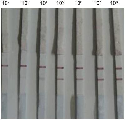
Dipsticks were used to detect the bacillus solutions mixed by 102–108 CFUs/mL E. coli O157:H7 and 30 μL of IMPs. The results indicated that 103 CFUs/mL and 104 CFUs/mL bacillus solutions were weakly positive, whereas 105–108 CFUs/mL bacillus solutions were positive and strongly positive. With the increase in the concentration of bacillus solution, the color above T line is increasingly clear (). The data showed that the sensitivity of IMPs combined with ICA reached 103 CFUs/mL.
Figure 1 The sensitivity of Escherichia coli O157:H7 (1 × 10⁸ to 1 × 10² CFUs/mL) with IMPs+ICA.
Abbreviations: CFUs, colony-forming units; ICA, immunochromatographic assay; IMPs, immunomagnetic nanoparticles.
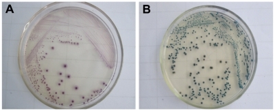
E. coli of ten different serotypes and non-E. coli of eleven different strains were enriched by IMPs. Following magnetic separation and removal of supernatant, the obtained bacteria were coated on E. coli O157:H7 chromogenic medium at 37°C for 24 hours. The results indicated that there was colony growth of E. coli O157:H7 (, aubergine). There was no other E. coli or bacteria (, blue-green color). It showed that IMPs failed to gather other strains and could only gather the target bacteria ().
Figure 2 The specificity of IMP enrichment for Escherichia coli O157:H7. (A) E. coli O157:H7 will appear as aubergine colonies. (B) Non-E. coli O157:H7 will appear as blue-green colonies and no growth.
Detection in food samples
When ICAs were used to detect E. coli O157:H7 (detection range ≥105 CFUs/mL) in milk, purified water, and beef, the positive rate was 100% in milk, 86.7% in purified water, and 80% in beef, showing high sensitivity. However, with the outside test range (≤105 CFUs/mL), sensitivity was greatly reduced. Because IMPs enriched E. coli O157:H7, the positive rate of IMPs combined with ICA was still ≥80% when the concentration of bacillus solution was 103 CFUs/mL (). A total of 150 food samples (135 positive and 15 negative samples) were detected (). The sensitivity and specificity of ICA were 29.6% and 93.3%, respectively, and the sensitivity and specificity of IMPs combined with ICA were 95.5% and 100%, respectively.
Table 1 Comparison of two detection methods for Escherichia coli O157:H7
Table 2 The sensitivity and specificity of the two detection methods for Escherichia coli O157:H7
Conclusion
A rapid and highly sensitive method for detection of E. coli O157:H7 was developed. When IMPs enriched for E. coli O157:H7 are combined with ICA they increase the sensitivity from ≥105 CFUs/mL to ≥103 CFUs/mL, and test results can be obtained within 1 hour. The method is simple, rapid, highly sensitive, specific, and does not require special equipment. The method potentially has a range of practical applications.
Acknowledgments
This work was financially supported by National 863 High Technology Project of China (No. 2003BA310A23), Technology Project of Shenzhen (No. 2004A110), and Technology Project of Guangdong (No. 2006B14701001).
Disclosure
The authors report no conflicts of interest in relation to this work.
References
- RileyLWRemisRSHelgersonSDHemorrhagic colitis associated with a rare Escherichia coli serotypeN Engl J Med19833086816856338386
- MeadPSSlutskerLDietzVFood-related illness and death in the United StatesEmerg Infect Dis1999560762510511517
- QuanTai-shuLiWeiFanTian-ruiFirst separation of E. coli O157:H7 from patients with colitis haemorrhagicChinese Journal of Epidemiology198992428
- WangHMaoXDingHZouQPengXEpidemiological survey on Escherichia coli O157 in Chongqing and Three-Gorge Reservoir Areas of ChinaVet Res Commun200832644946118481192
- ValiLHamoudaAPearceMCDetection of genetic diversity by pulsed-field gel electrophoresis among Escherichia coli O157 isolated from bovine faecal samples by immunomagnetic separation techniqueLett Appl Microbiol200744192317209809
- BruunGMWernerssonRJunckerASImproving comparability between microarray probe signals by thermodynamic intensity correctionNucleic Acids Res200735e4817337437
- DeCoryTRDurstRAZimmermanSJDevelopment of an immunomagnetic bead-immunoliposome fluorescence assay for rapid detection of Escherichia coli O157:H7 in aqueous samples and comparison of the assay with a standard microbiological methodAppl Environ Microbiol2005711856186415812012
- JelinekTWastlhuberJPröllSInfluence of rheumatoid factor on the specificity of a rapid immunochromatographic test for diagnosing dengue infectionEur J Clin Microbiol Infect Dis20001955555610968330
- LiQQiHZhouHXDetect of micrometastases in peripheral blood of non-small cell lung cancer with a refined immunomagnetic nanoparticles enrichment assayInt J Nanomed2011620752181
- FrensGControlled nucleation for the regulation of the particle size in monodisperse gold solutionNat Phys Sci19732412022
- KongXLQiHZhouHXA novel sensitive immunoassay by nucleic acid barcode dot and its application in the detection of prostate-specific antigenClin Chem Lab Med20104827928320001442
- KanarasAGWangZBatesADTowards multistep nanostructure synthesis: programmed enzymatic self-assembly of DNA/gold systemsAngew Chem Int Ed Engl20034219119412532347
- JungBYJungSCKweonCHDevelopment of a rapid immunochromatographic strip for detection of Escherichia coli O157J Food Prot2005682140214316245720
- FratamicoPMBagiLKComparison of an immunochromatographic method and the TaqMan E. coli O157:H7 assay for detection of Escherichia coli O157:H7 in alfalfa sprout spent irrigation water and in sprouts after blanchingJ Ind Microbiol Biotechnol20012712913411641772