
Anand N, Sehgal R, Kanwar R, Dubey M, Vasishta RK, Kanwar J. Int J Nanomedicine. 2015;10:6355–6369.
The authors have advised due to an error that occurred inadvertently at the time of figure assembly the incorrect images were selected by the first author for on page 6361.
The correct is shown below.
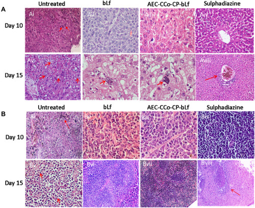
Figure 2 Histopathological analysis in liver (A) and spleen (B) at day 10 and 15 post infection.
Notes: (Ai) Liver histopathology of untreated group of mice at day 10 post infection showing the presence of tachyzoites (marked with red arrows). (Aii–iv) Liver histopathology of treatment groups showing no sign of inflammation or parasite at day 10 post infection. (Av) Untreated control group showing huge number of infected macrophages marked by red arrows. (Avi–vii) bLf and NCs treatment groups showing development of bradyzoites or tissue cyst inside the macrophages (Kupffer cells) marked by red arrows. (Aviii) Sulphadiazine treatment group showing no infection but sign of inflammation marked by red arrow. (Bi) Untreated Spleen cells showing site of tachyzoite multiplication (red arrow). (Bii–iv) Treatment groups showing no sign of inflammation or infection. (Bv) Spleen histopathology at day 15 post infection showing multiple sites (50%) with tachyzoites marked with red arrows. (Bvi–vii) No parasite infection shown in treatment groups, however, inflammation and reactive spleen was seen in sulphadiazine group (Bviii) marked as red arrow.
Abbreviations: AEC-CCo-CP-bLf, alginate chitosan calcium phosphate bovine lactoferrin; bLf, bovine lactoferrin.
The authors apologize for this error and advise it does not affect the results and conclusions of the paper.