
Abstract
Background
Therapeutic selectivity and drug resistance are critical issues in cancer therapy. Currently, zinc oxide nanoparticles (ZnO NPs) hold considerable promise to tackle this problem due to their tunable physicochemical properties. This work was designed to prepare SnO2-doped ZnO NPs/reduced graphene oxide nanocomposites (SnO2-ZnO/rGO NCs) with enhanced anticancer activity and better biocompatibility than those of pure ZnO NPs.
Materials and Methods
Pure ZnO NPs, SnO2-doped ZnO (SnO2-ZnO) NPs, and SnO2-ZnO/rGO NCs were prepared via a facile hydrothermal method. Prepared samples were characterized by field emission transmission electron microscopy (FETEM), energy dispersive spectroscopy (EDS), field emission scanning electron microscopy (FESEM), X-ray diffraction (XRD), ultraviolet-visible (UV-VIS) spectrometer, and dynamic light scattering (DLS) techniques. Selectivity and anticancer activity of prepared samples were assessed in human breast cancer (MCF-7) and human normal breast epithelial (MCF10A) cells. Possible mechanisms of anticancer activity of prepared samples were explored through oxidative stress pathway.
Results
XRD spectra of SnO2-ZnO/rGO NCs confirmed the formation of single-phase of hexagonal wurtzite ZnO. High resolution TEM and SEM mapping showed homogenous distribution of SnO2 and rGO in ZnO NPs with high quality lattice fringes without any distortion. Band gap energy of SnO2-ZnO/rGO NCs was lower compared to SnO2-ZnO NPs and pure ZnO NPs. The SnO2-ZnO/rGO NCs exhibited significantly higher anticancer activity against MCF-7 cancer cells than those of SnO2-ZnO NPs and ZnO NPs. The SnO2-ZnO/rGO NCs induced apoptotic response through the upregulation of caspase-3 gene and depletion of mitochondrial membrane potential. Mechanistic study indicated that SnO2-ZnO/rGO NCs kill cancer cells through oxidative stress pathway. Moreover, biocompatibility of SnO2-ZnO/rGO NCs was also higher against normal breast epithelial (MCF10A cells) in comparison to SnO2-ZnO NPs and ZnO NPs.
Conclusion
SnO2-ZnO/rGO NCs showed enhanced anticancer activity and better biocompatibility than SnO2-ZnO NPs and pure ZnO NPs. This work suggested a new approach to improve the selectivity and anticancer activity of ZnO NPs. Studies on antitumor activity of SnO2-ZnO/rGO NCs in animal models are further warranted.
Introduction
According to a World Health Organization (WHO) report, cancer is the second leading cause of death worldwide.Citation1 It has also been estimated that the total number of cancer cases will be doubled by year 2030 from 12.4 million new cases reported in 2008.Citation2 In spite of the incredible effort to treat cancer, a lot needs to be done in cancer therapy. Recent cancer chemotherapies often fail to provide a complete anticancer response due to the development of drug resistance or their inability to efficiently differentiate between cancerous and noncancerous cells.Citation3 This indiscriminate action normally leads to systemic toxicity in the human body.
Cancer nanotechnology can play a crucial role in the diagnosis and treatment of cancer to improve human health.Citation4 A growing number of studies demonstrated that certain types of metal oxide nanoparticles (NPs) can selectively kill cancer cells with low toxicity to their normal counterparts.Citation5–Citation8 Particularly, ZnO NPs showed great promise for cancer therapy. Due to unique physiochemical characteristics ZnO NPs have shown inherent selective toxicity against cancer cells while posing minimum effects to the normal cells.Citation9–Citation11 ZnO NPs induce apoptosis in cancer cells through the generation of ROS and depletion of mitochondrial membrane potential (MMP)Citation12,Citation13 Manipulation in intracellular ROS generation is a probable way to irreversibly damage cancerous cells selectively without applying much harm to normal cells. The ROS-generating potential of ZnO NPs is associated with its optical properties.Citation14 Optical behavior of ZnO NPs can be tuned by several methods including metal ion doping and synthesis of ZnO-based nanocomposites.Citation14,Citation15
Alternatively, studies reported some degree of toxicity of ZnO NPs in a wide range of organisms such as bacterial, microalgae, yeast, protozoa, zebrafish, and mice.Citation16–Citation20 Hence, there is an urgent need to tailor ZnO NPs with improved selectivity and anticancer activity.Citation21 Semiconductor tin oxide (SnO2) and reduced graphene oxide (rGO) have shown great potential for biomedical applications.Citation5,Citation22 SnO2 and rGO can also be applied to tune physicochemical properties of metal oxide NPs including ZnO.Citation23,Citation24
Keeping the above points in mind, we prepared SnO2-doped ZnO NPs/reduced graphene oxide nanocomposites (SnO2-ZnO/rGO NCs) with improved anticancer potential and higher biocompatibility as compared to pure ZnO NPs. Pure ZnO NPs, SnO2-ZnO NPs, and SnO2-ZnO/rGO NCs were prepared through a facile hydrothermal method. Prepared samples were characterized by field emission electron microscopy (FETEM), energy dispersive spectroscopy (EDS), field emission scanning electron microscopy (FESEM), X-ray diffraction (XRD), ultraviolet-visible (UV-VIS) spectrometer, and dynamic light scattering (DLS) techniques. Anticancer activity of pure ZnO NPs, SnO2-ZnO NPs, and SnO2-ZnO/rGO NCs was examined in human breast cancer (MCF-7) cells. Possible mechanism of anticancer potentials of SnO2-ZnO/rGO NCs was delineated through ROS pathway. Biocompatibility of prepared samples were investigated in human normal breast epithelial (MCF10A) cells. Breast cancer cell lines were chosen in the present study because female breast cancer is the most commonly diagnosed cancer and the third leading cause of cancer death globally after colorectal and lung cancer.Citation2
Materials and Methods
Synthesis of ZnO NPs, SnO2-ZnO NPs, and SnO2-doped-ZnO/rGO NCs
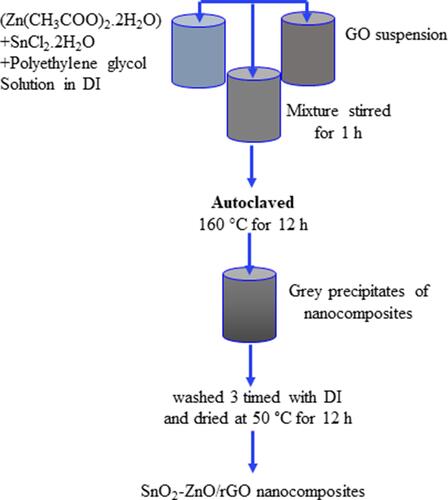
GO was prepared from pure graphite powder (<150 μm, 99.99% trace metals basis) (Millipore-Sigma, St Louis, MO, USA) applying modified Hummers' protocol.Citation25 The SnO2-doped ZnO/rGO nanocomposite were synthesized through a facile hydrothermal method. Firstly, 0.02 g of GO was dispersed into 20 mL of ethanol (C2H5OH) (Millipore-Sigma) and sonicated for three hours in an ultrasonic bath to get uniform GO suspension. Zinc acetate dihydrate (Zn(CH3COO)2.2H2O) (99.999% trace metals basis, Millipore-Sigma) (2 mM), tin (II) chloride dihydrate (SnCl2.2H2O) (≥99.995% trace metals basis, Millipore-Sigma) (0.1 mM), and polyethylene glycol (PEG) (Millipore-Sigma) (0.2 g) were mixed in 20 mL distilled water under continuous stirring for one hour at room temperature to get homogenous solution. Then, GO suspension was mixed with homogenous solution and stirred for one hour, and pH of solution was maintained at 9 by adding aqueous ammonia solution. Then, solution transferred into a 50 mL autoclave and heated at 160°C for 12 h. Finally, the grey precipitates were washed and dried at 50°C for 12 h to get SnO2-ZnO/rGO nanocomposites. SnO2-doped ZnO NPs was prepared in similar procedure without adding GO, and pure ZnO NPs was synthesized without addition of tin chloride and GO during hydrothermal process. A schematic diagram of SnO2-ZnO/rGO NCs synthesis is given in .
Figure 1 A schematic diagram of SnO2-ZnO/rGO NCs synthesis.
Characterization
Absorption spectra of prepared samples were recorded at the wavelength of 300–900 nm with a resolution of 0.5 nm (Shimadzu UV-1800 spectrometer, Reinach BL, Switzerland). Crystallinity and phase-purity were analyzed at powder X-ray diffraction (PXRD) (PanAnalytic X’Pert Pro, Malvern Instruments, UK) with Cu-Kα radiation (λ=0.15405 nm, at 45 kV and 40 mA). Structural characterization was further assessed through FETEM (JEM-2100F, JEOL, Inc., Tokyo, Japan). Elemental analysis was done by energy dispersive X-ray spectroscopy (EDS). Mapping of elemental distribution of SnO2-ZnO/rGO NCs was further done through FESEM (JSM-7600F, JEOL, Inc.). Hydrodynamic size and zeta potential in distilled water and culture medium were determined by DLS (ZetaSizer, Nano-HT, Malvern Instruments).
Cell Culture and Exposure Procedure
MCF-7 cells (ATCC HTB-22, ATCC, Virginia, USA) were cultured in DMEM (Invitrogen, Carlsbad, CA, USA) with 10% FBS and antibiotics (100 µg/mL streptomycin+100 U/mL penicillin). MCF10A (ATCC CRL-10317) cells were grown in mammary epithelial cell growth basal medium (MEBM) (Lonza Group Ltd, Basel, Switzerland) supplemented with mammary epithelial cell growth medium (MEGM) kit (Lonza Group Ltd) and 100 ng/mL cholera toxin. Cells were maintained in a humidified incubator under control conditions (37°C and 5% CO2). First, we exposed the cells with a range of concentrations (0–200 µg/mL) of ZnO NPs, SnO2-ZnO NPs, and SnO2-ZnO/rGO NCs for 24 h for cell viability assay. On the basis of cell viability results, we have chosen three moderate concentrations (10, 25, and 50 µg/mL) of these samples for further experiments. For microscopic study we have chosen one concentration (50 µg/mL) of prepared samples. In some experiments, cells were exposed to ZnO NPs, SnO2-ZnO NPs, and SnO2-ZnO/rGO NCs with or without N-acetyl-cysteine (NAC) (2 mM) (Millipore-Sigma). Briefly, NPs/NCs were suspended in culture medium (DMEM+10% FBS) and diluted to desired concentration (0–200 µg/mL). Different dilutions of NPs were further sonicated utilizing sonicator bath at room temperature for 10 min at 40 W to avoid agglomeration of NPs/NCs before exposure to cells. Cells without NPs/NCs served as control in each experiment.
Biochemical Assays
Cell viability was determined through MTTCitation26 and NRUCitation27 assays with some specific changes.Citation28 Morphology of treated and control cells was grabbed through phase-contrast inverted microscope (Leica Microsystems, GmbH, Germany). Expression level of mRNA of caspase-3 gene (CASP3) was determined through real-time PCR (ABI PRISM, 7900HT Sequence Detection System) (Applied Biosystems, Foster City, CA, USA) according to the procedures described earlier.Citation28,Citation29 Colorimetric assay of caspase-3 enzyme was performed using BioVision kit (Milpitas, CA, USA). MMP was assayed using rhodamine-123 probe (Rh-123) (Millipore-Sigma).Citation30 MMP level was quantitatively assayed through a microplate reader (Synergy-HT, BioTek Winooski, VT, USA) and intracellular images were grabbed using a DMi8 fluorescent microscope (Leica Microsystems). Intracellular level of ROS was assayed using 2ʹ-7ʹ-dichlorodihydrofluorescein diacetate (H2DCFDA, Millipore-Sigma) probe.Citation8 ROS level was also determined both quantitatively (Microplate reader, Synergy-HT, BioTek) and qualitatively (DMi8 fluorescent microscope, Leica Microsystems). Fluorometric assay of intracellular hydrogen peroxide (H2O2) was done applying a commercial kit (MAK164 green fluorescence, Millipore-Sigma). Glutathione (GSH) level was quantified using Ellman’s method.Citation31 Malondialdehyde (MDA), one of the end products of lipid peroxidation was estimated using procedures of Ohkawa et al.Citation32 Glutathione peroxidase (GPx) enzyme activity was assayed applying methods of Rotruck et al.Citation33 Procedures of Sinha were utilized to assay the catalase (CAT) enzyme activityCitation34 Colorimetric assay of superoxide dismutase (SOD) enzyme was done using kit from Cayman Chemical Company (Michigan, OH, USA). Protein content was quantified using Bradford’s protocol.Citation35 Brief description of each biochemical procedure with necessary steps is provided in supplementary information.
Statistical Analysis
One-way ANOVA followed by Dunnett’s multiple comparison tests were applied for analysis of results. The p<0.05 was assigned as statistically significant.
Results and Discussion
Morphological and Microstructural Study
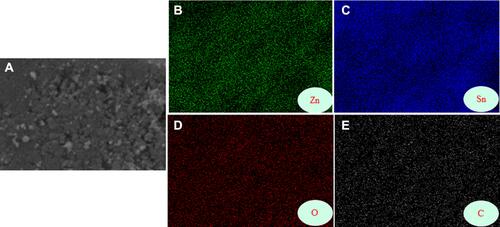
Morphology, particle size, and crystal structure of ZnO NPs, SnO2-ZnO NPs, and SnO2-ZnO/rGO NCs were determined by FETEM. showed that ZnO NPs were almost spherical shaped with size of 14 nm. It was noticed that the shape of nanoparticles remains same, but the size of NPs increases after SnO2 doping (19 nm) () and rGO integration (29 nm) (). Increment of NP size after doping and rGO incorporation was also reported by other studies.Citation15 – showed the HRTEM of ZnO NPs, SnO2-ZnO NPs, and SnO2-ZnO/rGO NCs, respectively. These images depict the presence of ZnO, SnO2 and rGO with high quality lattice fringes without any distortion. The calculated interplanar spacing of adjacent lattice fringes of ZnO NPs, SnO2-ZnO NPs, and SnO2-ZnO/rGO NCs were 0.345 nm, 0.342 nm, and 0.340 nm that correspond to the (101) plane of hexagonal wurtzite structure of ZnO.Citation36 The lattice fringes of SnO2 were 0.334 nm and 0.338 nm corresponding to the (110) planes of tetragonal phase of SnO2.Citation23 The lattice fringes were in agreement with XRD spectra. TEM images were also suggested all grown particles were highly crystalline that confirm the incorporation of SnO2 and rGO into the crystalline matrix of ZnO without creating lattice defects. EDS spectra of prepared SnO2-ZnO/rGO NCs indicated the stoichiometric presence of Zn, Sn, O, and C without any impurities (). The Cu peak came from copper grid of TEM. The SEM mapping of elemental distribution of SnO2-ZnO/rGO NCs showed uniform distribution of SnO2 and rGO in ZnO NPs ().
Figure 2 (A–C) Low resolution TEM images and (D–F) high resolution TEM images of ZnO NPs, SnO2-ZnO NPs, and SnO2-ZnO/rGO NCs. Arrows represent rGO.
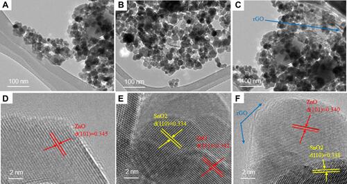
Figure 3 Elemental composition of SnO2-ZnO/rGO NCs assessed through EDS.
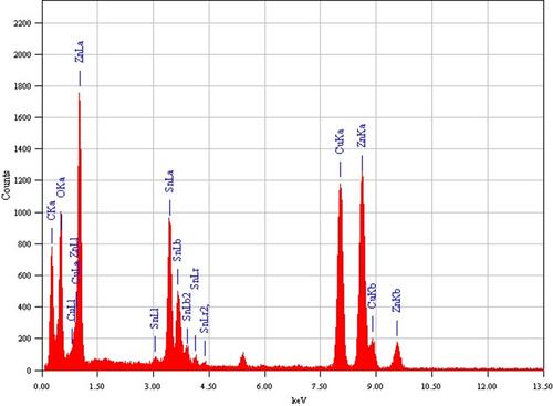
Figure 4 SEM elemental mapping of SnO2-ZnO/rGO NCs. (A) SEM image. (B) Zinc. (C) Tin. (D) Oxygen. (E) Carbon mapping.
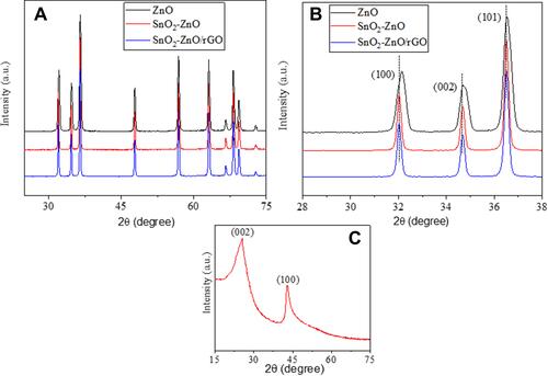
Crystal structure, phase purity, and size of ZnO NPs, SnO2-ZnO NPs, and SnO2-ZnO/rGO NCs were also analyzed by XRD technique. The XRD spectra of all samples were presented in . The indexing of diffraction peaks represents the formation of single-phase hexagonal wurtzite ZnO according to JCPDS card number 36–1451. No clear diffraction peak of SnO2 indicated that homogeneous doping of SnO2 throughout the ZnO lattice and ionic radii of Zn (0.74°A) and Sn (0.71°A) are almost same.Citation37 Absence of diffraction peak of rGO in XRD spectra of SnO2-ZnO/rGO NCs also indicate the uniform distribution of SnO2-ZnO NPs inhibited the restacking of rGO sheets.Citation38 Besides, XRD spectra show narrow and sharp diffraction peaks depicting excellent crystallinity of prepared samples. Crystallite size of synthesized samples were determined corresponding to highest intensity peak (101) by Scherrer’s formula and provided in . Results showed that crystallite size increases from 15 nm to 31 nm after SnO2 doping and rGO integration, which was well matched with to size calculated from TEM. We further noticed slight shifting of XRD peaks toward a lower value as compared to pure ZnO (). This shifting also indicates successful integration of SnO2 and rGO into ZnO NPs. represent XRD spectra of prepared rGO, which shows intense reflection planes (002) and (100).Citation24
Table 1 Structural and Optical Properties
Figure 5 (A) XRD spectra of ZnO NPs, SnO2-ZnO NPs, and SnO2-ZnO/rGO NCs. (B) XRD spectra of same samples in the diffraction region of 28–38. (C) XRD spectra of rGO.
Optical Study
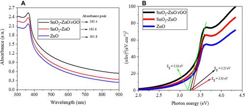
Optical properties of all prepared samples were examined through optical absorption study. Absorption spectra of ZnO NPs, SnO2-ZnO NPs, and SnO2-ZnO/rGO NCs, indicates SnO2 doping and rGO integration shifted the absorption edge of ZnO toward higher wavelength (). The band gap energy (Eg) for ZnO NPs, SnO2-ZnO NPs, and SnO2-ZnO/rGO NCs were determined by Tauc’s formula.Citation36
Figure 6 (A) optical absorption spectra of ZnO NPs, SnO2-ZnO NPs, and SnO2-ZnO/rGO NCs. (B) Tauc’s plot (αhν)2 vs (hν) of the same samples.
αhν = A(hν-Eg)1/2
where hν is the photon energy and A is the constant that does not depend on photon energy. The band gap energy (Eg) was determined by the extrapolation of linear portion of (αhν)2 vs hν curve to photon energy (hν) axis corresponding to α=0 (). The band gap values are provided in , which represent a decrement in band gap of ZnO NPs after SnO2 doping and rGO integration (3.30 eV-3.18 eV). It is reported that band gap energy of ZnO NPs could be tuned by doping metals oxides and/or integration of other semiconducting materials. Our results are in agreement with previous studies.Citation39–Citation41 Band gap tuning of ZnO NPs might be useful in cancer therapy. Tuning of band gap of semiconductor NPs might increase the threshold of ROS generation and oxidative stress in cancer cells through the generation of higher holes (h+) and electrons (e−) on the surface of NPs.Citation8,Citation42
Dynamic Light Scattering Study
It is important to assess the colloidal stability and surface charge of NPs in physiological media to understand the interaction of NPs with biological systems. DLS was applied to examine the hydrodynamic size and zeta potential of ZnO NPs, SnO2-ZnO NPs, and SnO2-ZnO/rGO NCs in distilled water and culture medium (). Hydrodynamic size of these samples in distilled water and culture medium were in the range of 55 nm to 96 nm, which were around three-to-five times higher than those of powder size calculated from TEM and XRD. Higher hydrodynamic size could be due to the agglomeration of particles in aqueous suspension and also reported in earlier studies.Citation5,Citation8,Citation43 Zeta potential data showed that particles surface charge were ranging from 25–30 eV in distilled water and culture medium (DMEM+FBS). Zeta potential data suggested that colloidal suspension of prepared NPs and NCs were fairly stable. The higher value of zeta potential (positive or negative), the greater is the colloidal stability of nanoscale materials.Citation44,Citation45 Positive surface charge of prepared NPs and NCs under physiological medium provides favorable condition for their interaction with cancerous cells, which frequently carry negative surface charge.Citation45
Table 2 Dynamic Light Scattering Characterization
Cytotoxicity
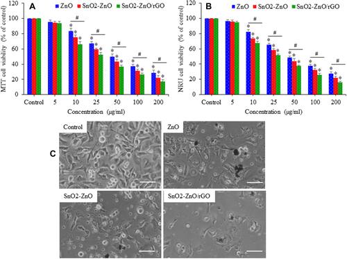
Anticancer potential of ZnO NPs can be further improved by tuning of its physicochemical properties.Citation3 In this study, we tried to enhance the cytotoxic potential of ZnO NPs against cancer cells through SnO2 doping and integration of rGO to prepare ZnO nanocomposites. Cytotoxicity of ZnO NPs, SnO2-ZnO NPs, and SnO2-ZnO/rGO NCs in breast cancer MCF-7 cells was assessed through MTT, NRU, and morphological examination. MCF-7 cells were exposed to different concentrations (0–200 µg/mL) of these samples for 24 h. MTT results demonstrated that all three samples were found to reduce cell viability in a dose-dependent manner in the concentration range of 10–200 µg/mL (). Furthermore, we observed that cytotoxicity of SnO2-ZnO/rGO NCs were significantly higher in comparison to pure ZnO NPs. MTT cell viability data were utilized to calculate the IC50s of prepared samples in MCF-7 cells. IC50s for ZnO NPs, SnO2-ZnO NPs, and SnO2-ZnO/rGO NCs were 54.61, 38.73, and 27.86 µg/mL, respectively (Figure S1 and Table S1 of supplementary information). NRU data on cell viability was according to MTT results (). demonstrated that cell morphology after exposure to 50 µg/mL of ZnO, SnO2-ZnO, and SnO2-ZnO/rGO for 24 h. These images suggested that a significant number of cell death occurs (rounded morphology and low cell density) after exposure to ZnO NPs, SnO2-ZnO NPs, and SnO2-ZnO/rGO NCs compared to controls. Similar to cell viability results, cell death induced by SnO2-ZnO/rGO NCs were greater than those of SnO2-ZnO NPs and pure ZnO NPs. Cytotoxic potential of ZnO NPs against different types of cancer cell lines has also been reported by other investigators.Citation46–Citation48 For example, a recent study reported anticancer activity of ZnO NPs against human small-cell lung cancer in an orthotopic mouse model.Citation10
Figure 7 Cytotoxic potential of ZnO NPs, SnO2-ZnO NPs, and SnO2-ZnO/rGO NCs in MCF-7 cells. Cells were treated for 24 h to different concentration of these samples (0–200 µg/mL). (A) MTT cell viability. (B) NRU cell viability. (C) Cell morphology after exposure to 50 µg/mL of ZnO NPs, SnO2-ZnO NPs, and SnO2-ZnO/rGO NCs for 24 h. (Scale bar presents 50 µm.) Data represented as mean ±SD of five independent experiments (n=5). *p<0.05 control vs treated groups and #p<0.05 pure ZnO NPs vs SnO2-ZnO/rGO NCs.
Apoptosis
Apoptosis (a form of programmed cell death) might be stimulated by various internal or external factors such as injury, stress, starvation, and known apoptotic agents.Citation49 ZnO NPs induce apoptosis in cancer cells through caspase-activation and MMP depletion.Citation12,Citation13 Caspase-3 is a protease that is present in mitochondria and critically involved in apoptotic pathway.Citation50 Apoptotic response of prepared samples was determined by measuring the regulation of caspase-3 gene and MMP level in MCF-7 cells. showed that in comparison to control, ZnO NPs, SnO2-ZnO NPs, and SnO2-ZnO/rGO NCs upregulated the mRNA expression level of the CASP3 gene in a dose-dependent manner (10–50 µg/mL). To confirm the mRNA data, we further examined the activity of caspase-3 enzyme (protein level). Results showed that activity of caspase-3 enzyme was dose-dependently higher in ZnO NPs, SnO2-ZnO NPs, and SnO2-ZnO/rGO NCs treated groups compared to control group (). Moreover, effect of SnO2-ZnO/rGO NCs on activation of the caspase-3 gene was significantly higher than those of pure ZnO NPs.
Figure 8 Apoptotic potential of ZnO NPs, SnO2-ZnO NPs, and SnO2-ZnO/rGO NCs in MCF-7 cells. Cells were treated for 24 h to different concentration of these samples (10–50 µg/mL). (A) mRNA expression level of CASP3 gene. (B) Activity of caspase-3 enzyme. (C) Quantitative data MMP. (D) Fluorescent cellular images of Rh-123 (MMP indicator) probe after exposure to 50 µg/mL of same samples for 24 h. (Scale bar presents 50 µm.) Data represented as mean ±SD of five independent experiments (n=5). *p<0.05 control vs treated groups and #p<0.05 pure ZnO NPs vs SnO2-ZnO/rGO NCs.
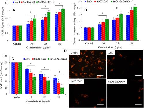
MMP level of cells is compromised under stressful condition and is an excellent indicator of apoptosis.Citation51 showed that dose-dependent reduction in MMP level after exposure to ZnO NPs, SnO2-ZnO NPs, and SnO2-ZnO/rGO NCs. Fluorescent microscopy data also indicated that brightness of Rh-123 probe decreases (indicator of MMP loss) in all three samples ZnO NPs, SnO2-ZnO NPs, and SnO2-ZnO/rGO NCs compared to control (). Similarly, MMP loss caused by SnO2-ZnO/rGO NCs was significantly higher in comparison to pure ZnO NPs. Apoptosis mediated anticancer activity of ZnO NPs was also reported by other investigators. Duan et al observed the anticancer activity of ZnO NPs in human melanoma (A375) cells was mediated through MMP depletion and caspase activation.Citation52 Another recent report also suggested that ZnO NPs induced caspase-dependent apoptosis in gingival squamous cell carcinoma cells mediated through the mitochondrial pathway.Citation53
Oxidative Stress
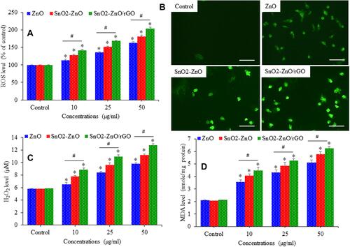
Intracellular ROS generation and oxidative stress have been suggested as a possible mechanism to eliminate cancer cells.Citation54 Recent studies indicated that ZnO NPs destroy cancer cells through ROS generation.Citation14,Citation55 Oxidative stress generating potential of ZnO NPs, SnO2-ZnO NPs, and SnO2-ZnO/rGO NCs in MCF-7 cells were explored through measuring several markers of pro-oxidants and antioxidants. depicted that all three samples induce ROS generation dose-dependently. Fluorescent microscopic images also suggested that brightness of DCF probe (indicator of ROS generation) were higher in ZnO NPs, SnO2-ZnO NPs, and SnO2-ZnO/rGO NCs treated cells than those of control cells (). Intracellular H2O2 level () and MDA level () were also significantly higher in ZnO NPs, SnO2-ZnO NPs, and SnO2-ZnO/rGO NCs treated cells as compared to controls (p<0.05). Importantly, pro-oxidants generating potential of SnO2-ZnO/rGO NCs were significantly higher in comparison to pure ZnO NPs.
Figure 9 Pro-oxidants generating potential of ZnO NPs, SnO2-ZnO NPs, and SnO2-ZnO/rGO NCs in MCF-7 cells. Cells were treated for 24 h to different concentration of these samples (10–50 µg/mL). (A) Quantitative data ROS level. (B) Fluorescent cellular images of DCF (ROS indicator) probe after exposure to 50 µg/mL of same samples for 24 h. (Scale bar presents 50 µm.) (C) Quantitative analysis of intracellular H2O2 level (D) MDA level. Data represented as mean ±SD of five independent experiments (n=5). *p<0.05 control vs treated groups and #p<0.05 pure ZnO NPs vs SnO2-ZnO/rGO NCs.
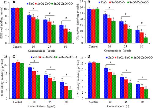
Cell possesses antioxidant defense systems to get rid of ROS by inducing antioxidant molecule GSH along with several antioxidant enzymes.Citation56 GSH scavenge ROS and free radicals to protect cells from oxidative damage.Citation57 Main antioxidant enzymes are GPx, SOD, and CAT. GPx is mainly responsible for removal of hydroperoxides, hence, it may protect membranes, lipids, and proteins from oxidation. SOD provides a first line of defense by converting highly reactive superoxide anion (O2•–) into H2O2. CAT further breaks H2O2 into water and molecular oxygen.Citation58 Antioxidant defense potential of MCF-7 cells was assessed following exposure for 24 h to ZnO NPs, SnO2-ZnO NPs, and SnO2-ZnO/rGO NCs. (A–) showed that all three samples reduced the GSH level and decreased the activity of several antioxidant enzymes (GPx, SOD, and CAT) in a dose-dependent manner. Again, effects of SnO2-ZnO/rGO NCs on antioxidant markers were significantly higher than those of pure ZnO NPs.
Figure 10 Antioxidants depleting potential of ZnO NPs, SnO2-ZnO NPs, and SnO2-ZnO/rGO NCs. In MCF-7 cells. Cells were treated for 24 h to different concentration of these samples (10–50 µg/mL). (A) GSH level. (B) GPx activity. (C) SOD activity. (D) CAT activity. Data represented as mean ±SD of five independent experiments (n=5). *p<0.05 control vs treated groups and #p<0.05 pure ZnO NPs vs SnO2-ZnO/rGO NCs.
Possible Mechanism of Anticancer Activity
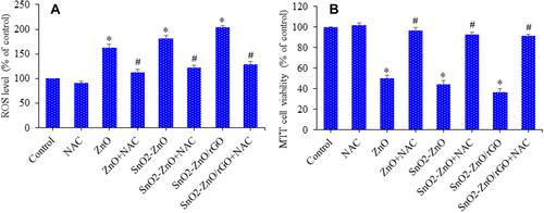
Higher ROS production that surpasses the antioxidant defense capacity of the cells causes oxidative stress that induces oxidative damage of cellular components such DNA, lipid, and proteins.Citation9 Recent studies indicated that ROS generation induces enzyme deactivation, lipid peroxidation, and membrane damage, which are the core mechanisms accountable for anticancer activity of ZnO NPs.Citation59,Citation60 Hence, in this study, we further examined the role of ROS and oxidative stress in anticancer activity of SnO2-ZnO/rGO NCs in MCF-7 cancer cells. Cells were exposed for 24 h to 50 µg/mL of ZnO NPs, SnO2-ZnO NPs, and SnO2-ZnO/rGO NCs in the presence and absence of NAC. demonstrated that NAC significantly alleviated the ROS generation induced by ZnO NPs, SnO2-ZnO NPs, and SnO2-ZnO/rGO NCs. We further found that NAC, efficiently reverted that cytotoxicity induced by ZnO NPs, SnO2-ZnO NPs, and SnO2-ZnO/rGO NCs (). These results confirm that SnO2-ZnO/rGO NCs induced cytotoxicity in MCF-7 cancer cells was mediated through ROS generation. Altogether, our data suggested that SnO2-ZnO/rGO NCs-induced toxicity in MCF-7 cells might be caused by ROS generation via mitochondrial pathway.
Figure 11 ROS mediated cytotoxicity. Cells were exposed for 24 h to 50 µg/mL of ZnO NPs, SnO2-ZnO NPs, and SnO2-ZnO/rGO NCs in the presence or absence of NAC. (A) ROS level with or without NAC. (B) Cell viability with or without NAC. Data represented as mean ±SD of five independent experiments (n=5). *Significantly different from the control (p<0.05). #Significantly different from ZnO NPs, SnO2-ZnO NPs, and SnO2-ZnO/rGO NCs groups (p<0.05).
Effect of ZnO NPs, SnO2-ZnO NPs, and SnO2-ZnO/rGO NCs on Normal Human Mammary Epithelial (MCF10A) Cells
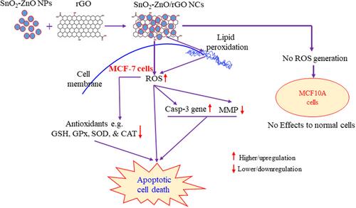
It is very important for anticancer drugs to have minimum or no effects on noncancerous normal cells. Benign nature of ZnO NPs against noncancerous normal cells have been reported previously.Citation11,Citation14,Citation61 Hence, at the end of this study, we examined the effects of ZnO NPs, SnO2-ZnO NPs, and SnO2-ZnO/rGO NCs in normal breast cells (MCF10A). Cells were exposed to different concentrations (0–200 µg/mL) of these samples for 24 h. showed that ZnO NPs were not toxic to normal MCF10A cells up to 25 µg/mL concentration. However, cytotoxicity of pure ZnO NPs increases with increasing the concentrations from 50–200 µg/mL. In contrast, SnO2-ZnO/rGO NCs were not toxic to normal MCF10A cells even at a higher concentration (200 µg/mL). These results suggested that in comparison to ZnO NPs and SnO2-ZnO NPs, SnO2-ZnO/rGO NCs have greater biocompatibility toward normal cells and higher cytotoxicity toward cancer cells. As we observed that ROS generation due to SnO2-ZnO/rGO NCs exposure was the primary cause of killing of MCF-7 cancer cells. Hence, we further examined the ROS-generating potential of ZnO NPs, SnO2-ZnO NPs, and SnO2-ZnO/rGO NCs in MCF10A cells. As we can see that SnO2-ZnO/rGO NCs did not induce intracellular ROS generation in normal human mammary epithelial cells even at 200 µg/mL (). Consequently, SnO2-ZnO/rGO NCs showed improved anticancer activity and better biocompatibility compared to pure ZnO NPs. Possible mechanism of anticancer potential of SnO2-ZnO/rGO NCs is presented as a schematic diagram ().
Figure 12 Effect of ZnO NPs, SnO2-ZnO NPs, and SnO2-ZnO/rGO NCs on normal human mammary epithelial (MCF10A) cells. Cells were treated for 24 h to different concentration of these samples (0–200 µg/mL), and cell viability and ROS level were determined. (A) MTT cell viability assay and (B) intracellular ROS level. Data represented as mean ±SD of five independent experiments (n=5). *Significantly different from the control (p<0.05).
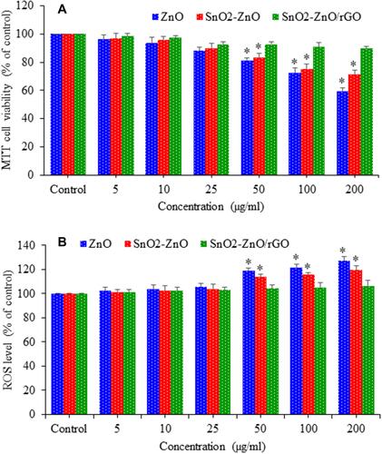
Figure 13 Possible mechanism of anticancer activity of SnO2-ZnO/rGO NCs.
Conclusion
In summary, SnO2-ZnO/rGO NCs were successfully synthesized by a simple hydrothermal procedure. XRD data confirmed the formation of single-phase of hexagonal wurtzite ZnO. HRTEM and SEM mapping showed homogenous distribution of SnO2 and rGO in ZnO NPs with high quality lattice fringes without any distortion. Band gap energy of SnO2-ZnO/rGO NCs decreases after SnO2 doping and rGO integration. The SnO2-ZnO/rGO NCs demonstrated strong anticancer activity against MCF-7 cells compared to pure ZnO NPs. The SnO2-ZnO/rGO NCs induced apoptosis in MCF-7 cancer cells through the upregulation of the caspase-3 gene and depletion of MMP. Mechanistic approach showed that SnO2-ZnO/rGO NCs kill breast cancer cells through ROS generation via mitochondrial pathway. Moreover, biocompatibility of SnO2-ZnO/rGO NCs was also greater in normal breast epithelia MCF10A cells than those of pure ZnO NPs. Overall our results provided a new approach to improve the selectivity and anticancer activity of ZnO in human breast cancer cells by tailoring its physicochemical properties. This work warrants further research on antitumor activity of SnO2-ZnO/rGO NCs in animal breast cancer models.
Author’s Contribution
All authors made substantial contributions to conception and design, acquisition of data, or analysis and interpretation of data; took part in drafting the article or revising it critically for important intellectual content; agreed to submit to the current journal; gave final approval of the version to be published; and agree to be accountable for all aspects of the work.
Acknowledgments
This work was supported by the National Plan for Science, Technology, and Innovation (MAARIFAH), King Abdulaziz City for Science and Technology, Kingdom of Saudi Arabia, under Award 13-NAN908-02.
Disclosure
The authors report no conflicts of interest in this work.
References
- Wiesmann N, Tremel W, Brieger J. Zinc oxide nanoparticles for therapeutic purposes in cancer medicine. J Mater Chem B. 2020;8(23):4973–4989. doi:10.1039/d0tb00739k
- Bray F, Ferlay J, Soerjomataram I, Siegel RL, Torre LA, Jemal A. Global cancer statistics 2018: GLOBOCAN estimates of incidence and mortality worldwide for 36 cancers in 185 countries. CA Cancer J Clin. 2018;68(6):394–424. doi:10.3322/caac.21492
- Rasmussen JW, Martinez E, Louka P, Wingett DG. Zinc oxide nanoparticles for selective destruction of tumor cells and potential for drug delivery applications. Expert Opin Drug Deliv. 2010;7(9):1063–1077. doi:10.1517/17425247.2010.502560
- Wicki A, Witzigmann D, Balasubramanian V, Huwyler J. Nanomedicine in cancer therapy: challenges, opportunities, and clinical applications. J Controlled Release. 2015;200:138–157. doi:10.1016/j.jconrel.2014.12.030
- Ahamed M, Akhtar MJ, Majeed Khan MA, Alhadlaq HA. Oxidative stress mediated cytotoxicity of tin (IV) oxide (SnO2) nanoparticles in human breast cancer (MCF-7) cells. Coll Surf B Biointerfaces. 2018;172:152–160. doi:10.1016/j.colsurfb.2018.08.040
- Akhtar MJ, Alhadlaq HA, Kumar S, Alrokayan SA, Ahamed M. Selective cancer-killing ability of metal-based nanoparticles: implications for cancer therapy. Arch Toxicol. 2015;89:11. doi:10.1007/s00204-015-1570-1
- Ahamed M, Alhadlaq HA, Khan MAM, Akhtar MJ. Selective killing of cancer cells by iron oxide nanoparticles mediated through reactive oxygen species via p53 pathway. J Nanoparticle Res. 2013;15:1. doi:10.1007/s11051-012-1225-6
- Ahamed M, Khan MAM, Akhtar MJ, Alhadlaq HA, Alshamsan A. Ag-doping regulates the cytotoxicity of TiO2 nanoparticles via oxidative stress in human cancer cells. Sci Rep. 2017;7(1):1–14. doi:10.1038/s41598-017-17559-9
- Akhtar MJ, Ahamed M, Kumar S, Majeed Khan MA, Ahmad J, Alrokayan SA. Zinc oxide nanoparticles selectively induce apoptosis in human cancer cells through reactive oxygen species. Int J Nanomedicine. 2012;7. doi:10.2147/IJN.S29129
- Tanino R, Amano Y, Tong X, et al. Anticancer activity of ZnO nanoparticles against human small-cell lung cancer in an orthotopic mouse model. Mol Cancer Ther. 2020;19(2):502–512. doi:10.1158/1535-7163.MCT-19-0018
- Premanathan M, Karthikeyan K, Jeyasubramanian K, Manivannan G. Selective toxicity of ZnO nanoparticles toward Gram-positive bacteria and cancer cells by apoptosis through lipid peroxidation. Nanomedicine. 2011;7(2):184–192. doi:10.1016/j.nano.2010.10.001
- Ahamed M, Akhtar MJ, Raja M, et al. ZnO nanorod-induced apoptosis in human alveolar adenocarcinoma cells via p53, survivin and bax/bcl-2 pathways: role of oxidative stress. Nanomedicine. 2011;7:6. doi:10.1016/j.nano.2011.04.011
- Perera WPTD, Dissanayake RK, Ranatunga UI, et al. Curcumin loaded zinc oxide nanoparticles for activity-enhanced antibacterial and anticancer applications. RSC Adv. 2020;10(51):30785–30795. doi:10.1039/d0ra05755j
- Akhtar MJ, Alhadlaq HA, Alshamsan A, Majeed Khan MA, Ahamed M. Aluminum doping tunes band gap energy level as well as oxidative stress-mediated cytotoxicity of ZnO nanoparticles in MCF-7 cells. Sci Rep. 2015;5. doi:10.1038/srep13876
- Majeed Khan MA, Siwach R, Kumar S, Ahmed M, Ahmed J. Investigations on microstructure, optical, magnetic, photocatalytic, and dielectric behaviours of pure and Co-doped ZnO NPs. J Mater Sci. 2020;31(8):6360–6371. doi:10.1007/s10854-020-03192-2
- Wang D, Li H, Liu Z, Zhou J, Zhang T. Acute toxicological effects of zinc oxide nanoparticles in mice after intratracheal instillation. Int J Occup Environ Health. 2017;23(1):11–19. doi:10.1080/10773525.2016.1278510
- Verma SK, Panda PK, Jha E, Suar M, Parashar SKS. Altered physiochemical properties in industrially synthesized ZnO nanoparticles regulate oxidative stress; Induce in vivo cytotoxicity in embryonic zebrafish by apoptosis. Sci Rep. 2017;7(1):1. doi:10.1038/s41598-017-14039-y
- Zhang W, Bao S, Fang T. The neglected nano-specific toxicity of ZnO nanoparticles in the yeast Saccharomyces cerevisiae. Sci Rep. 2016;6(1):1–11. doi:10.1038/srep24839
- Franklin NM, Rogers NJ, Apte SC, Batley GE, Gadd GE, Casey PS. Comparative toxicity of nanoparticulate ZnO, bulk ZnO, and ZnCl2 to a freshwater microalga (Pseudokirchneriella subcapitata): the importance of particle solubility. Environ Sci Technol. 2007;41(24):8484–8490. doi:10.1021/es071445r
- Mortimer M, Kasemets K, Kahru A. Toxicity of ZnO and CuO nanoparticles to ciliated protozoa Tetrahymena thermophila. Toxicology. 2010;269(2–3):182–189. doi:10.1016/j.tox.2009.07.007
- Mishra PK, Mishra H, Ekielski A, Talegaonkar S, Vaidya B. Zinc oxide nanoparticles: a promising nanomaterial for biomedical applications. Drug Discov Today. 2017;22(12):1825–1834. doi:10.1016/j.drudis.2017.08.006
- Ahamed M, Akhtar MJ, Khan MAM, Alaizeri ZM, Alhadlaq HA. Evaluation of the Cytotoxicity and Oxidative Stress Response of CeO2-RGO Nanocomposites in Human Lung Epithelial A549 Cells. Nanomaterials. 2019;9(12):1709. doi:10.3390/nano9121709
- Wang J, Liu S, Cao X, et al. One-pot synthesis and gas sensitivity of SnO2 nanoparticles prepared using two Sn salts of SnCl4·5H2O and SnCl2·2H2O. Appl Phys a Mater Sci Process. 2020;126(1):44. doi:10.1007/s00339-019-3230-4
- Khan MAM, Khan W, Ahamed M, Alhazaa AN. Investigation on the structure and physical properties of Fe 3 O 4/RGO nanocomposites and their photocatalytic application. Mater Sci Semiconductor Proc. 2019;99:44–53. doi:10.1016/j.mssp.2019.04.005
- Yu H, Zhang B, Bulin C, Li R, High-efficient XR. Synthesis of Graphene Oxide Based on Improved Hummers Method. Sci Rep. 2016;6. doi:10.1038/srep36143
- Mosmann T. Rapid colorimetric assay for cellular growth and survival: application to proliferation and cytotoxicity assays. J Immunol Methods. 1983;65(1–2):55–63. doi:10.1016/0022-1759(83)90303-4
- Borenfreund E, Puerner JA. A simple quantitative procedure using monolayer cultures for cytotoxicity assays (HTD/NR-90). J Tissue Culture Methods. 1985;9(1):7–9. doi:10.1007/BF01666038
- Ahamed M, Akhtar MJ, Siddiqui MA, et al. Oxidative stress mediated apoptosis induced by nickel ferrite nanoparticles in cultured A549 cells. Toxicology. 2011;283(2–3):101–108. doi:10.1016/j.tox.2011.02.010
- Mohammad A, Saini RV, Kumar R, et al. A curious case of cysteines in human peroxiredoxin I. Redox Biol. 2020;37:101738. doi:10.1016/j.redox.2020.101738
- Siddiqui MA, Alhadlaq HA, Ahmad J, Al-Khedhairy AA, Musarrat J, Ahamed M. Copper Oxide Nanoparticles Induced Mitochondria Mediated Apoptosis in Human Hepatocarcinoma Cells. PLoS One. 2013;8(8):8. doi:10.1371/journal.pone.0069534
- Ellman GL. Tissue sulfhydryl groups. Arch Biochem Biophys. 1959;82(1):70–77. doi:10.1016/0003-9861(59)90090-6
- Ohkawa H, Ohishi N, Yagi K. Assay for lipid peroxides in animal tissues by thiobarbituric acid reaction. Anal Biochem. 1979;95(2):351–358. doi:10.1016/0003-2697(79)90738-3
- Rotruck JT, Pope AL, Ganther HE, Swanson AB, Hafeman DG, Hoekstra WG. Selenium: biochemical role as a component of glatathione peroxidase. Science. 1973;179(4073):588–590. doi:10.1126/science.179.4073.588
- Sinha AK. Colorimetric assay of catalase. Anal Biochem. 1972;47(2):389–394. doi:10.1016/0003-2697(72)90132-7
- Bradford MM. A rapid and sensitive method for the quantitation of microgram quantities of protein utilizing the principle of protein-dye binding. Anal Biochem. 1976;72(1–2):248–254. doi:10.1016/0003-2697(76)90527-3
- Khan MAM, Kumar S, Alhazaa AN, Al-Gawati MA. Modifications in structural, morphological, optical and photocatalytic properties of ZnO:Mn nanoparticles by sol-gel protocol. Mater Sci Semiconductor Proc. 2018;87:134–141. doi:10.1016/j.mssp.2018.07.016
- Faisal M, Ibrahim AA, Harraz FA, Bouzid H, Al-Assiri MS, Ismail AA. SnO2 doped ZnO nanostructures for highly efficient photocatalyst. J Mol Catal a Chem. 2015;397:19–25. doi:10.1016/j.molcata.2014.10.027
- Zhu S, Fan L, Lu Y. Highly uniform Fe3O4 nanoparticle-rGO composites as anode materials for high performance lithium-ion batteries. RSC Adv. 2017;7(87):59939–59946. doi:10.1039/c7ra11779e
- Hamrouni A, Moussa N, Parrino F, Di Paola A, Houas A, Palmisano L. Sol-gel synthesis and photocatalytic activity of ZnO-SnO2 nanocomposites. J Mol Catal a Chem. 2014;390:133–141. doi:10.1016/j.molcata.2014.03.018
- Dargahi Z, Asgharzadeh H, Maleki-Ghaleh H. Synthesis of Mo-doped TiO2/reduced graphene oxide nanocomposite for photoelectrocatalytic applications. Ceramics Int. 2018;44(11):13015–13023. doi:10.1016/j.ceramint.2018.04.120
- Khan MAM, Kumar S, Ahamad T, Alhazaa AN. Enhancement of photocatalytic and electrochemical properties of hydrothermally synthesized WO3 nanoparticles via Ag loading. J Alloys Compd. 2018;743:485–493. doi:10.1016/j.jallcom.2018.01.343
- Zhang H, Pokhrel S, Ji Z, et al. PdO Doping Tunes Band-Gap Energy Levels as Well as Oxidative Stress Responses to a Co 3 O 4 p -Type Semiconductor in Cells and the Lung. J Am Chem Soc. 2014;136(17):6406–6420. doi:10.1021/ja501699e
- Mittal S, Kumar V, Dhiman N, Chauhan LKS, Pasricha R, Pandey AK. Physico-chemical properties based differential toxicity of graphene oxide/reduced graphene oxide in human lung cells mediated through oxidative stress. Sci Rep. 2016;6. doi:10.1038/srep39548
- Ahamed M, Akhtar MJ, Alhadlaq HA. Influence of silica nanoparticles on cadmium‐induced cytotoxicity, oxidative stress, and apoptosis in human liver HepG2 cells. Environ Toxicol. 2020;22895. doi:10.1002/tox.22895
- Jiang J, Oberdörster G, Biswas P. Characterization of size, surface charge, and agglomeration state of nanoparticle dispersions for toxicological studies. J Nanoparticle Res. 2009;11(1):77–89. doi:10.1007/s11051-008-9446-4
- Wang J, Lee JS, Kim D, Zhu L. Exploration of Zinc Oxide Nanoparticles as a Multitarget and Multifunctional Anticancer Nanomedicine. ACS Appl Mater Interfaces. 2017;9(46):39971–39984. doi:10.1021/acsami.7b11219
- Khorsandi L, Farasat M. Zinc oxide nanoparticles enhance expression of maspin in human breast cancer cells. Environ Sci Pollution Res. 2020;27(30):38300–38310. doi:10.1007/s11356-020-09986-5
- Zhang T, Du E, Liu Y, et al. Anticancer Effects of Zinc Oxide Nanoparticles Through Altering the Methylation Status of Histone on Bladder Cancer Cells. Int J Nanomedicine. 2020;15:1457–1468. doi:10.2147/IJN.S228839
- Zhuang C, She Y, Zhang H, et al. Cytoprotective effect of deferiprone against aluminum chloride-induced oxidative stress and apoptosis in lymphocytes. Toxicol Lett. 2018;285:132–138. doi:10.1016/j.toxlet.2018.01.007
- Balakireva AV, Zamyatnin AA. Cutting Out the Gaps Between Proteases and Programmed Cell Death. Front Plant Sci. 2019;10. doi:10.3389/fpls.2019.00704
- Chang SY, Lee MY, Chung PS, et al. Enhanced mitochondrial membrane potential and ATP synthesis by photobiomodulation increases viability of the auditory cell line after gentamicin-induced intrinsic apoptosis. Sci Rep. 2019;9:1. doi:10.1038/s41598-019-55711-9
- Duan X, Liao Y, Liu T, et al. Zinc oxide nanoparticles synthesized from Cardiospermum halicacabum and its anticancer activity in human melanoma cells (A375) through the modulation of apoptosis pathway. J Photochem Photobiol B. 2020;202:111718. doi:10.1016/j.jphotobiol.2019.111718
- Wang S-W, Lee C-H, Lin M-S, et al. ZnO Nanoparticles Induced Caspase-Dependent Apoptosis in Gingival Squamous Cell Carcinoma through Mitochondrial Dysfunction and p70S6K Signaling Pathway. Int J Mol Sci. 2020;21(5):1612. doi:10.3390/ijms21051612
- Perillo B, Di Donato M, Pezone A, et al. ROS in cancer therapy: the bright side of the moon. Exp Mol Med. 2020;52(2):192–203. doi:10.1038/s12276-020-0384-2
- Yang Y, Song Z, Wu W, Xu A, Lv S, Ji S. ZnO Quantum Dots Induced Oxidative Stress and Apoptosis in HeLa and HEK-293T Cell Lines. Front Pharmacol. 2020;11. doi:10.3389/fphar.2020.00131
- Akhtar MJ, Ahamed M, Alhadlaq HA, Alshamsan A. Mechanism of ROS scavenging and antioxidant signalling by redox metallic and fullerene nanomaterials: potential implications in ROS associated degenerative disorders. Biochimica et Biophysica Acta. 2017;1861:4. doi:10.1016/j.bbagen.2017.01.018
- Kuang F, Liu J, Tang D, Oxidative Damage KR. Antioxidant Defense in Ferroptosis. Front Cell Dev Biology. 2020;8:969. doi:10.3389/fcell.2020.586578
- Hasanuzzaman M, Bhuyan MHMB, Zulfiqar F, et al. Reactive oxygen species and antioxidant defense in plants under abiotic stress: revisiting the crucial role of a universal defense regulator. Antioxidants. 2020;9(8):1–52. doi:10.3390/antiox9080681
- Sivakumar P, Lee M, Kim YS, Shim MS. Photo-triggered antibacterial and anticancer activities of zinc oxide nanoparticles. J Mater Chem B. 2018;6(30):4852–4871. doi:10.1039/c8tb00948a
- Bai DP, Zhang XF, Zhang GL, Huang YF, Gurunathan S. Zinc oxide nanoparticles induce apoptosis and autophagy in human ovarian cancer cells. Int J Nanomedicine. 2017;12:6521–6535. doi:10.2147/IJN.S140071
- Ostrovsky S, Kazimirsky G, Gedanken A, Brodie C. Selective cytotoxic effect of ZnO nanoparticles on glioma cells. Nano Res. 2009;2(11):882–890. doi:10.1007/s12274-009-9089-5