 ?Mathematical formulae have been encoded as MathML and are displayed in this HTML version using MathJax in order to improve their display. Uncheck the box to turn MathJax off. This feature requires Javascript. Click on a formula to zoom.
Abstract
Purpose
Awareness of the harmful effects of ultraviolet radiation has led to the increasing use of sunscreens, thus, the development of safe and effective antisolar preparations is important. The inclusion of sunscreen molecules in different release systems, like liposomes (lipo) and cyclodextrins (CD) is therefore required.
Methods
The in vivo sun protection factor (SPF), water resistance, and in vitro transdermal penetration test of octyl p-methoxycinnamate (OMC) in different dispersions, such as OMC encapsulated in liposomes (lipo/OMC), OMC encapsulated in β-cyclodextrins (β-CD/OMC), OMC encapsulated in both release systems (lipo/OMC and β-CD/OMC), and an OMC-free formulation were determined.
Results
Although the formulation containing only the lipo/OMC system revealed high value of in vivo SPF (11.0 ± 1.3) and water resistance (SPF = 10.3 ± 2.2), the formulation containing both release systems (lipo/OMC + β-cyclodextrin/OMC) showed the best result in the in vivo SPF test (11.6 ± 1.6). In the penetration test, the formulation containing the lipo/OMC system had better performance, since a high amount of OMC in the epidermis (18.04 ± 1.17 μg) and a low amount of OMC in the dermis (9.4 ± 2.36 μg) were observed. These results suggest that liposomes interact with the cells of the stratum corneum, promoting retention of OMC in this layer.
Conclusion
According to our study, the lipo/OMC system is the most advantageous release system, due to its ability to both increase the amount of OMC in the epidermis and decrease the risk of percutaneous absorption.
Introduction
The harmful effects of ultraviolet (UV) radiation from sunlightCitation1–Citation3 have led to the widespread use of topical sunscreen preparations.Citation4,Citation5 The most common active ingredients in these preparations are organic sunscreen agents, which absorb UV radiation.Citation6,Citation7 A sunscreen can be classified according to its optical and physical characteristics. Optically, an ideal sunscreen should absorb light over a broad spectrum and, physically, cover, protect, and adhere well to the skin, as well as resist removal by water and sweat.Citation8
Further, for a sunscreen to be effective, UV absorbers must remain in the outermost layer of the skin. Additionally, an ideal sunscreen should exhibit high skin accumulation of UV absorbers, with minimal permeation to systemic circulation. Once UV absorbers penetrate the systemic circulation, photo-protection is lost and the skin becomes susceptible to sun damage.Citation9
Octyl p-methoxycinnamate (OMC), an organic UV-B filter originally developed in the 1950s, has been one of the most widely used sunscreens for decades, and its use in pharmaceutical formulations is allowed by the US Food and Drug Administration, the European Cosmetics, Toiletry and Perfumery Association (COLIPA), the Agência Nacional de Vigilância Sanitária, Brazil, and the Australian Register of Therapeutic Goods. Recent studies have shown that OMC can be detected in deep layers of the skin, urine, and milk after application on the skin.Citation7,Citation10 Several approaches have been studied to ensure adequate efficacy for this sunscreen agent, for example, nanocapsules,Citation10 microspheres,Citation11 cyclodextrins,Citation12,Citation13 liposomesCitation13,Citation14 and nanoparticles.Citation14 The advantage of encapsulating UV filters into nanostructured delivery systems is that they significantly enhance the accumulation of the sunscreen at the administration site to increase water resistance, sun protection factor (SPF), and the photostability of these agents.Citation13–Citation15
Cyclodextrins (CD) are cyclic oligosaccharides composed of α-D-glucopyranoside linked by glycosidic bonds (α-1,4). They exist in three natural forms, α, β, and γ. They can entrap lipophilic drugs into the hydrophobic cavity of their ringed structures. CD are used to increase the aqueous solubility of hitherto poorly soluble drugs, protect against oxidation, and improve photostability.Citation12,Citation13
Liposomes are vesicles consisting of one or more concentric phospholipidic bilayers organized around an aqueous inner compartment, and can be produced from nontoxic phospholipids and cholesterol. These vesicles enable the incorporation of both water-soluble and lipid-soluble substances.Citation15–Citation17 Liposomes with a high content of soy phosphatidylcholine were used in this study, since they are able to both transport sunscreen agents into the horny layer and form a depot, increasing the amount of OMC in the skin.
This work aimed to: develop a novel formulation containing OMC loaded in liposomes and β-CD with a targeted synergic effect; compare the in vivo SPF, water resistance and in vitro transdermal penetration test of OMC, a usually lipid-soluble sunscreen, in different formulations, such as OMC encapsulated in liposomes (lipo/OMC), OMC encapsulated in β-cyclodextrins (β-CD/OMC), OMC encapsulated in both release systems (lipo/OMC and β-CD/OMC), and an OMC free formulation; and prove the separation of the epidermis of the skin by light microscopy.
Methods and materials
Materials
The following materials were used in the formulations: UV-B filter octyl p-methoxycinnamate (OMC) purchased from Merck (Merck KGaA, Darmstadt, Germany), β-cyclodextrin (β-CD) from Wacker-Chemie (Wacker-Chemie GmbH, Munich, Germany), cholesterol and Tris[hydroxymethyl]aminomethane (TRIS) buffer from Sigma-Aldrich (St Louis, MO), Phenova® (phenoxyethanol and parabens) from Croda International, PLC (East Yorkshire, England), Lipoid S 100® (phospholipid) from Idealfarma (Anápolis, Goiás, Brazil), Eumulgin® VL 75 (polyglyceryl-2 Dipolyhydroxystearate, lauryl glucoside, glycerin) from Cognis (Monheim, Germany), Structure® XL (hydroxypropyl starch phosphate) from Akzonobel Corporation (Amsterdam, The Netherlands), butylated hydroxytoluene (BHT) from Via Farma Importandora Ltda (São Paulo, SP, Brazil). In addition, chloroform PA and ethanol PA were purchased from Vetec (Vetec Ltda, Rio de Janeiro, Brazil).
Formulation containing free OMC (formulation 1)
A cream formulation containing 8% of free OMC was prepared to establish a comparison among other formulations. The formulation components and their respective concentrations are listed in .
Table 1 Composition of formulations 1–4
Formulation containing liposome/OMC (lipo/OMC) (formulation 2)
The liposomal sunscreen preparation was obtained by hydration of a phospholipids/OMC film. For this, the lipid film was formed when a formulation of 280 mM phospholipon, 80 mM cholesterol, and 72 mM OMC in chloroform was evaporated (Heidolph Rotary Evaporator, Laborota 4000G1, Schwabach, Germany) and deposited in a round-bottom flask. The film was detached with TRIS buffer by vortex (Marconi, Piracicaba, SP, Brazil) shaking for 15 minutes.Citation18 The incorporated OMC corresponded to 20% of the total phospholipids. A sequential extrusion through polycarbonate membranes (0.4 μm), under nitrogen pressure, was performed to homogenize liposome size.Citation18 The microdispersion system was characterized using a transmission electron microscope (Morgani 268; FEI, Hillsboro, OR) (under 80 kV). Images were captured using a Megaview G2 digital camera from Olympus (Tokyo, Japan). Incorporation efficiency was measured by UV spectrophotometer (Shimadzu UV-1601, Kyoto, Japan). Particle size distribution, polydispersity index, and zeta potential were obtained using a particle size analyzer (Zetasizer® Nano Z; Malvern Instruments, Worcestershire, UK).
The lipo/OMC inclusion complex was manually incorporated in a cream with an OMC concentration of 8%. The formulation components are described in .
Formulation containing β-CD/OMC (formulation 3)
The Kneading method was employed to include the OMC in the cavity of the β-CD. This method is suitable for complexation of water-insoluble compounds.Citation18 β-CD and OMC at a ratio of 1.7:1 were mixed together for 5 minutes. After that, 1.0 mL of ethanol:water (70:30) solution was added to the formulation, and a mass was formed.Citation18–Citation20 The product (total mass 20 g) was then transferred to a ball mill (11 cm long and 8 cm diameter), containing 27 g of porcelain balls (average diameter 1 cm). The mixture was kept in the mill for 1 hour. It was then purified with a 50% methanol:water solution and dried at 55°C for 30 minutes.Citation19,Citation20 Complex formation was characterized by 1H-nuclear magnetic resonance (NMR) spectroscopy (Gemini-200 MHZ; Varian, Palo Alto, CA) and X-ray diffractometry (Rigaku-Miniflex, Tokyo, Japan); complex yield was determined by UV spectrophotometer (UV-1601; Shimadzu, Kyoto, Japan).Citation20
The β-CD/OMC inclusion complex was manually incorporated in a cream with an OMC concentration of 8%. The formulation components are described in .
Formulation containing β-CD/OMC and lipo/OMC (formulation 4)
Both β-CD/OMC and lipo/OMC inclusion complexes were incorporated in a cream with an OMC concentration of 8%. The formulation components are listed in .
In vitro SPF determination
SPF values were determined according to the method described by Mansur et al,Citation21 which was corroborated by Santos et alCitation22 and Freitas et alCitation23 using similar experimental conditions. Sunscreen formulations containing both free OMC and OMC included in the delivery systems were prepared.
Absorbance values of each preparation at a final concentration of 2 μL/mL diluted in ethanol (in triplicate) were determined under the emission spectrum of 290–320 nm, with 5 nm intervals, using a Shimadzu UV-1601 UV-visible spectrophotometer. The SPF determination, Equationequation 1(1) (mathematical expression of Mansour et al),Citation21 and the correlation between the erythemogenic effect (EE) and the radiation intensity at each wavelength (EE × I) were adjusted according to Sayre et al Citation24 ().
Table 2 Weight employed to determine sun protection factor by spectrophotometry
where CF = 10 (correction factor), EE (λ) = erythemogenic effect of radiation of wavelength λ, I (λ) = intensity of solar light with wavelength λ, abs (λ) = spectrophotometric absorbance value of a solution of the preparation at wavelength λ.
In vivo SPF determination
The SPF value (Equationequation 2(2) ) is defined as the ratio between the UV energy required to produce a minimum erythemal dose (MED) – or redness – on protected skin and the UV energy required to produce MED on unprotected skin.
SPFs were determined in vivo according to the COLIPA method.Citation25–Citation27 Ten individuals with skin phototypes (SPT) I, II, and III, aged 18–42 years, were used as volunteers. The back of each volunteer was exposed to UV, using a 601 Multiport UV Solar Simulator (Solar Light Company, Glenside, PA). Volunteers were informed about the protocols, agreed to participate in the study, and gave written consent. All studies were made by Alergisa (Campinas, SP, Brazil) according to Brazilian ethical protocol.
SPFs were obtained using the following in vivo method. On the first day, the volunteers’ backs were exposed to a UV simulator; the exposure times varied according to the skin types. After about 20 hours, the MEDs of each volunteer were observed. On the second day, the MEDs without sunscreen were verified and then the tested sunscreen was applied. The sunscreen samples and standard formulation were applied in the amount of 2 mg/cm2 to an adjacent area of the same back of each volunteer. After application, the product was left to dry for 15 minutes before irradiation. Each test area was exposed to controlled amounts of simulated sunlight using a solar simulator with continuous emission spectrum of 290–400 nm. An area of 5 × 6 cm was irradiated in six points (diameter of 1 cm) in a series of geometrically increasing doses. The standard formulation was a lotion with 7% of octyl dimethyl PABA and 3% of benzophenone-3. On the third day, the MEDs of the sunscreens were again analyzed and then the SPF for the product was calculated using the arithmetic average of all the individual SPF values obtained from each volunteer.Citation25–Citation26
SPF determination after in vivo immersion
The SPF subsequent in vivo immersion was determined according to COLIPA with ten volunteers aged 18–42 years with SPT I, II, and III, whose backs were exposed to UVCitation25 using a 601 Multiport UV Solar Simulator.
The term “water resistant” means that the sunscreen photo-protective effect remains 40 minutes after exposure to water. The water resistance component was determined by evaluating the volunteers after two cycles of 20-minute immersion intervals, with moderate activity in water, each followed by a 20-minute rest/air dry period until the total water exposure time was reached. The test areas were air dried without toweling at the end of the last water-immersion period.Citation25–Citation27
Experimental data are presented as mean ± standard deviation. To compare both the absorbance and in vivo SPF values of the sunscreen preparations, data were analyzed by one-way analysis of variance (P = 0.05), using the SigmaStat for Windows software (v 3.11; Cranes Software, Chicago, IL).
In vitro cutaneous penetration test
Skin preparation
Skins from the ears of 4-month-old female pigs were obtained at the Federal Rural University of Rio de Janeiro, Brazil. An internationally validated and accepted animal-free test method (SCCNFP/0119/99) is available. Pig ear skin is a very attractive model for in vitro percutaneous absorption studies since it mimics human skin.
Immediately following slaughter, the pig ears were removed before receiving a hot bath. After this, they were taken as quickly as possible under cooling to the laboratory, where they were shaved and washed under running water. Later, the ear skins of the dorsal region were cut with scalpel and scissors to eliminate excess adipose tissue from the ventral region.Citation28,Citation29 Then rounded pieces of skin approximately 11 cm2 were maintained in airtight conditions within a film of polyvinyl chloride (PVC) and foil and stored at −20°C for no longer than 4 weeks before use (only intact skin discs with an effective permeation area of 1.96 cm2 were kept). Prior to the in vitro cutaneous penetration test, the skin surface (epidermal side) was cleaned with 0.5 mL aqueous solution of sodium lauryl sulfate (1%; 1 g sodium lauryl sulfate/99 g water) impregnated in cotton. The remaining fat on the dermal side was entirely removed with ether impregnated in cotton. Then the skin was washed five times with distilled water and dried with dry cotton to maintain skin integrity.Citation28,Citation29
Histology of pig skin
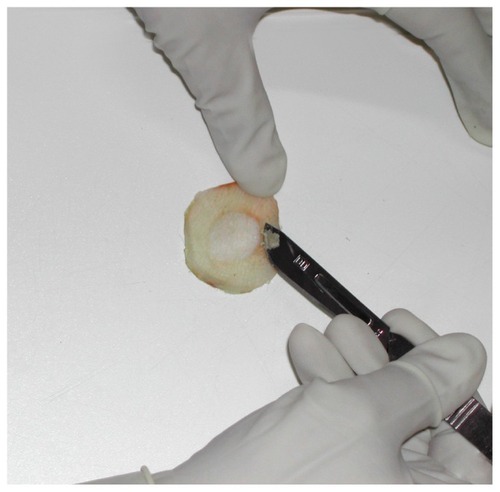
To analyze the in vitro distribution of OMC into the skin layers, the epidermis was separated from the dermis. Histological analysis was then performed to evaluate the procedure that allowed the separation of the skin layers by scalpel. The skins were heated at 60°C for 30 seconds. After that, from a half segment the epidermis was removed using a scalpel. illustrates the procedure of the epidermis removal used in the present study.
Figure 1 Removal of the epidermis using a scalpel.
The rounded pieces were cut into small fragments (5 × 2 cm), fixed with 10% buffered formaldehyde and/or Bouin’s liquid, and processed according to standard histological techniques for paraffin embedding. Five-micrometer thick slices, obtained using a Leica 2025 microtome (Wetzlar, Germany), were stained with hematoxylin-eosin.Citation30
Diffusion cell preparation
Initially, to establish sink conditions, the solubility of OMC was studied in different receptor media.Citation9,Citation31 Four receptor solutions were studied: (1) phosphate buffer pH 7.4 and phosphate buffer pH 7.4 containing (2) 2%, (3) 4%, or (4) 5% polysorbate 80. OMC in excess was added to 5 mL of each receptor medium. The samples were shaken for 24 hours at 25°C and were then centrifuged (8000 g) for 15 minutes, filtered through a Millipore filter (pore size 0.45 μm; Millex HV, Millipore, Brazil), and diluted with receptor media. The concentration of the OMC was determined by absorption spectroscopy using a wavelength of 310 nm.
The skin samples were mounted in modified Franz diffusion cells with a surface of 1.96 cm2 and a receptor volume of 7.5 mL, allowing the dermal side of the skin to be exposed to the receptor fluid and the stratum corneum exposed to the air so that the receptor fluid would not adversely affect the superficial skin layers or the physical–chemical properties of OMC.
The receptor fluid (pH 7.4) consisted of 120 mM/L phosphate-buffered saline solutions containing sodium chloride (NaCl), potassium chloride (KCl) 2.7 mM/L, and 10 mM/L phosphate buffer, containing 5% Tween® 80. The solubility of OMC in the receptor fluid was checked prior to commencement of experiments since OMC under these conditions is readily soluble in the receptor fluid.
Products were applied to the skin at a finite dose of 400 μg/cm2 using an automatic pipette so that the formulation film could cover the entire skin surface with a uniform and homogenous spread without determining its thickness. The cell was filled with 7.5 mL of receptor fluid through a lateral inlet using a burette. Visible air bubbles, between the bottom side of the skin and the receptor fluid, were eliminated through a lateral inlet.
The diffusion cells were placed in a thermostat-controlled water bath with horizontal agitation (speed of 900 strokes per minute), assuring both the homogeneity of the receptor fluid and the mean skin temperature of 32°C ± 0.1°C. Four replicates were used for the study. A 1 mL aliquot was taken for analysis at predetermined intervals (1 hour and 2, 3, 4, 5, and 6 hours), then the volume was replaced with the receiver solution. After that, OMC levels were analyzed by high-performance liquid chromatography (HPLC).Citation18
In vitro distribution of OMC into the pig skin layers
Six hours later, the skin was removed from the cell using tweezers and the formulation excess was removed from the skin surface, which was cleaned twice with moistened cotton and once with dried cotton. After that, the epidermis was separated from the dermis using a scalpel.
To determine the quantity of OMC, the remaining product on the skin surface was dissolved using methanol and each sample was shaken with a vortex mixer, before being agitated for 1 minute, submitted to ultrasound for 12 minutes, and centrifuged at 6400 rpm for 10 minutes. Subsequently, an aliquot of this last solution was dissolved in methanol to determine the amount of OMC remaining on the skin surface. OMC levels were analyzed by HPLC.Citation18
HPLC analysis
The concentrations of OMC in the formulations, in the skin layers (epidermis and dermis), and in the receptor fluid, were chromatographically determined by an isocratic HPLC system. The HPLC system consisted of a Waters model 510 pump with a Waters model 486 UV-Vis detector, a Rheodyne injector (Waters 717) with a 20 μL loop and a Waters 486 recorder (Waters Corporation, Milford, MA). Data acquisition and processing were accomplished with a Waters data model 7466, Hamilton 80665 Integrator (Sigma-Aldrich).
OMC was determined using a Hibar® LiChrosorb, a 5 × 25 cm reverse-phase column (C18) (Merck) operated at room temperature, and eluted with a mobile phase consisting of methanol/water (87:13) at a flow-rate of 1.0 mL minute–1. The column effluent was continuously monitored at 310 nm to detect OMC.Citation18 Quantification of the compound was performed by measuring the peak areas in relation to those of chromatographed standards under the same conditions. The sensitivity of the HPLC method was 0.1 μg mL−1 for OMC.
A standard stock solution of OMC (20 μg/mL) was prepared by dissolving the filter in methanol. The calibration curve was prepared with methanol solutions of OMC at concentrations ranging from 1 to 20 μg/mL. The standard curves were linear (r = 0.999). In addition, the validation results were established for five concentrations through three injections per concentration. The reproducibility of the method was expressed by the relative standard deviation.
Statistics
Experimental data are presented as mean ± standard deviation. The difference in the concentrations of the sunscreen agent in the receptor fluid, epidermis, and dermis for each formulation, was compared by one-way analysis of variance (P = 0.05), using the SigmaStat software. A P value of <0.05 was considered statistically significant.
Results
Lipo/OMC complex characterization
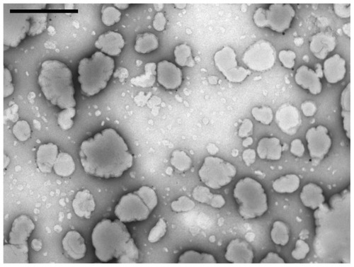
The liposomes obtained from the dry phospholipid film revealed spherical vesicles, which were morphologically homogeneous (). The roughness of the vesicles was due to the treatment of the sample prior to electron microscopy. The sample was diluted, stained with uranyl acetate and dried before microscopy, and this drying caused the liposomes to become rough. Average diameters of liposomes containing OMC and empty liposomes were 957.5 ± 12.1 (polydispersity index [PI] 0.45) and 941.2 nm ± 27.3 (PI 0.23) (n = 3), respectively. PI evaluates the quality of the sample. PI < 0.1 indicates that the range of size distribution is narrow and monomodal. PI < 0.5 indicates a wide range. Our results provide PI < 0.5, showing that the quality is satisfactory. The lipsomes’ incorporation efficiency for containing OMC was 88.88% (n = 3). The zeta potential of liposomes containing OMC and empty liposomes was −11.8 ± 0.2 mV and −10.2 ± 0.1 (n = 3), respectively.
Figure 2 Electron micrograph of liposomes formed after the hydration of the dry phospholipids film containing OMC.
Note: Scale bar = 500 nm.
Abbreviation: OMC, octyl p-methoxycinnamate.
β-CD/OMC complex characterization
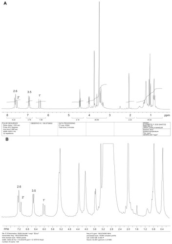
shows the up-field shifts (negative Δδ values) observed for the OMC aromatic proton signals, using 1H-NMR spectral studies (), suggesting that the aromatic portion of the sunscreen molecule () is located inside the cyclodextrin cavity (), with the ester group close to the external surface of the macrocycle.Citation12
Table 3 1H-nuclear magnetic resonance chemical shift changes (Δδ, ppm) for OMC in cyclodextrins
Figure 3 1H-NMR spectrum of the OMC (A), 1H-NMR spectrum of the β-CD/OMC (B), molecular structure of the sunscreen (C) and complexation reaction (D).
Abbreviations: OMC, octyl p-methoxycinnamate; β-CD, β-cyclodextrins.
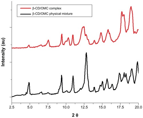
The molecular volume is the main restrictive factor of the inclusion process of sunscreens in the cavity of cyclodextrins.Citation32 The molecular volume of OMC, calculated by molecular modeling, was larger (367.53 Å3) than the cavity of the β-CD (cavity volume = 262 Å3).Citation20 The inclusion of OMC in α-CD (174 Å3) produces low reproducibility and yield; however, the use of γ-CD (472 Å3) produces an increase in the yield (20%). The results show that molecular volume is the major restrictive parameter of the inclusion process of sunscreens in CD.Citation20 Additionally, as illustrated in , the peaks of OMC of the physical sunscreen formulation with cyclodextrin observed through the X-ray diffraction pattern were absent in the diffractogram of the complex. These results provide evidence of the inclusion of OMC in the β-CD cavity. Moreover, the incorporation efficiency of OMC in the β-CD cavity was 89.0% (n = 3).
Figure 4 Powder X-ray diffraction patterns of β-CD/OMC (2:1) physical formulation (black) and β-CD/OMC (2:1) complex (red).
Abbreviations: OMC, octyl p-methoxycinnamate; β-CD, β-cyclodextrins.
In vitro SPF
The absorbance and SPF values for formulations 1–4 are shown in . There was no significant statistical difference between the in vitro SPF values of the developed sunscreen preparations (P = 0.600).
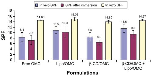
Figure 5 In vivo values after immersion and in vitro SPF of cream with free OMC (A), cream with lipo/OMC complex (B), cream with β-CD/OMC complex (C), and cream with both complexes (D) (mean ± standard deviation).
Abbreviations: β-CD, β-cyclodextrins; OMC, octyl p-methoxycinnamate; SPF; sun protection factor.
In vivo SPF
Erythema development at the analyzed areas was regarded as acceptable. Further, after UV radiation exposure, an area without erythema between areas of increasing erythema extension was observed. Calculated SPF values for formulations 1–4 are shown in . Statistical analysis showed that the in vivo SPF values for sunscreen preparations were significantly different (P < 0.001), since formulations with liposomes (lipo/OMC and β-CD/OMC + lipo/OMC) provided different SPFs. However, no significant statistical difference was observed between the preparation with both complexes (β-CD/OMC + lipo/OMC) and lipo/OMC complex, or between preparations containing free OMC and β-CD/OMC complex (P > 0.05).
The formulations containing the lipo/OMC + β-CD/OMC systems and the lipo/OMC release system exhibited the highest in vivo SPF values ().
After immersion in vivo SPF
The SPF values for formulations 1–4 are shown in . After in vivo immersion, the SPF values showed a significant statistical difference among preparations with complexes (β-CD/OMC + lipo/OMC), free OMC and β-CD/OMC complex (P < 0.05). There was also a significant difference among formulations with lipo/OMC complex. However, there was no significant statistical variation between the preparation containing both complexes (β-CD/OMC + lipo/OMC) and lipo/OMC complex and between the preparation with free OMC and β-CD/OMC complex (P > 0.05).
reveals that the liposomes incorporated with OMC increased the in vivo SPF (P > 0.05) and water resistance (P > 0.05). The inclusion of OMC in β-CD did not promote an increase of the in vivo SPF value (P > 0.05) and did not show any increase in water resistance (P < 0.05). The formulation with the lipo/OMC complex showed the best result.
In vitro cutaneous penetration test
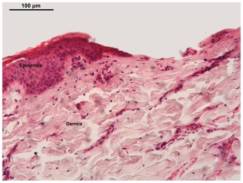
Histological analysis confirmed that the method used separates the epidermis from the dermis. shows the skin of a pig ear and the epidermis layers, including the stratum corneum. Additionally, when the scalpel was used, the epidermis was entirely removed and the dermis remained intact. The separation of both skin layers is important for penetration tests, which measure the amount of OMC in each layer, and avoid tissue contamination.
Figure 6 Light micrograph of the pig ear skin. The method totally removed the epidermis (E), remaining the dermis.
Notes: Staining with hematoxylin and eosin. Scale bar = 20 μm.
OMC solubility in the receptor media is shown in . Sink conditions were established to prevent the formation of saturated solution of the drug during the in vitro permeation study. Thus, the maximum concentration of the OMC released in the receptor media was five times smaller than the saturation concentration of sunscreen in this medium.Citation33
Table 4 Octyl p-methoxycinnamate solubility in the receptor medium
The solubility of the sunscreen in phosphate buffer pH 7.4 was 37.2 μg/mL. OMC is lipophilic and has low solubility in aqueous media. The addition of 2%, 4%, or 5% polysorbate 80 as a surfactant increased the solubility of the OMC in the receptor medium (). Phosphate buffer pH 7.4 containing 5% polysorbate 80 was chosen as the receptor medium because it provided the highest solubility (271.1 μg/mL). The concentration of sunscreen in the receptor medium should not exceed 54.22 μg /mL (20% of the saturation concentration of the OMC = 271.1 μg/mL) to maintain sink conditions. Thus, the permeation studies were performed in sink conditions, as 50 μg/mL corresponds to the maximum concentration of OMC able to be released during the permeation study.
The amounts of OMC per μg from both epidermis and dermis for formulations 1–4 are presented in . The statistical analysis revealed that the amounts of OMC in the epidermis (P = 0.066) and in the dermis (P = 0.251) for the four formulations were similar. However, a statistical difference (P = 0.037) in the amount of OMC between the epidermis and dermis in the lipo/OMC formulation was detected.
Figure 7 In vitro skin distribution of OMC in the epidermis and the dermis, 6 hours after application of cream with free OMC, cream with lipo/OMC complex, cream with β-CD/OMC complex and cream with both complexes (mean ± standard deviation, n = 4).
Abbreviations: OMC, octyl p-methoxycinnamate; β-CD, β-cyclodextrins.
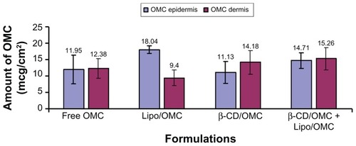
The amounts of OMC in epidermis and dermis for the free OMC formulation were 11.95 ± 4.41 μg and 12.38 ± 2.98 μg, respectively. The amounts of OMC for the β-CD/OMC formulation were 11.13 ± 3.36 μg and 14.18 ± 3.59 μg, respectively, and the amounts of OMC for the lipo/OMC formulation were 18.04 ± 1.17 μg and 9.4 ± 2.36 μg, respectively. Furthermore, the concentrations of OMC in the epidermis and dermis showed some statistical difference (P = 0.037).
Nevertheless, the amounts of OMC in the pig skin for the β-CD/OMC + lipo/OMC formulation were 14.71 ± 2.39 μg and 15.26 ± 3.47 μg, respectively – that is, they were similar in both epidermis and dermis.
Discussion
OMC () is a chemical sunscreen derived from the cinnamate class, which absorbs UV-B and UV-A rays from the sun, protecting the skin from damage. In the molecular structure of cinnamates, there is an unsaturation conjugated with the aromatic ring and the carbonyl group, which allows better electronic distribution. The energy required to produce the electronic transition corresponds to a wavelength of 305 nm.Citation27 It is an oily liquid, transparent, slightly yellow, odorless, and insoluble in water (water solubility <0.1 g/100 mL at 27°C), although soluble in ethanol, propylene glycol, and mineral oil. Its molecular weight is 290.4 and boiling point is in the range of 185°C–195°C. Its density is 1.01 g/cm3.Citation34 The octanol-water partition coefficient (Po/w) for OMC is 5.96, indicating that it has high lipid solubility. Octanol may mimic the solvent properties of the stratum corneum;Citation9 thus, OMC has an affinity for the lipid layer of the stratum corneum.
The American Association of Pharmaceutical Scientists recommends the use of phosphate buffer pH 7.4 as a receptor medium in in vitro release studies by using diffusion cells for hydrophilic drugs, but the addition of surfactants in the buffer is allowed for lipophilic actives to maintain the sink conditions.Citation31
Physical–chemical characterization shows that both empty liposomes and liposomes containing sunscreen have similar size distribution, PI, and zeta potential characteristics.
In this study, OMC sunscreen was included in two different release systems: liposomes and cyclodextrins. The lipo/OMC complex and β-CD/OMC complexes were developed, characterized, and incorporated in gel cream. A third formulation was developed using the combination lipo/OMC complex and β-CD/OMC complex. The ratio of the mixture was to obtain a synergistic effect. The formulation was 8% sunscreen, 4% of which was included in the cyclodextrin, and 4% in the liposomes. In vivo SPF, water resistance, and in vitro transdermal penetration tests were undertaken.
The in vitro SPF values of the sunscreen preparations were similar. These results may be due to the limitations of this method in assessing the interaction of the formulation, or the release system, with the skin. Thus, the method only determined the total content of OMC in the formulation. The major advantage of the in vitro test is its rapid and cost-effective screening methodology. In addition, in vitro tests can provide a formulation tool to identify new filters, optimize combinations of old ones, and pre-screen protective formulas prior to human in vivo tests.
Including OMC in liposomes increased the in vivo SPF values and water resistance, since the formulations with the lipo/OMC + β-CD/OMC complex and the lipo/OMC complex exhibited the highest SPF values. The main phospholipid in the liposome preparation was soybean lecithin, which is similar to the phospholipids in biological membranes. This suggests a major interaction between the liposomes and the stratum corneum lipids that creates a reasonable environment for OMC to reservoir. Possibly due to these mechanisms, the sunscreen remains for longer on the skin’s outermost layers, thereby increasing the SPF value.Citation35 The inclusion of OMC in β-CD did not promote the increase of the in vivo SPF value, nor did it show any increase in water resistance. The in vivo test performed with formulations showed that the cyclodextrin complex had little effect on producing water resistance. The cyclodextrin formulation displayed a lower SPF value when compared with the cream control; additionally, it was found that cyclodextrin did not increase formulation adherence but enhanced the solubility of OMC in the water, when associated with OMC.Citation18,Citation20 β-CD/OMC is more hydrophilic than the sunscreen-free preparation, so it is less able to interact with the lipid layer of the skin. Further, as this complex was more hydrophilic, it was more easily removed from the skin surface by water, as seen in . Also, the complex has the lowest SPF, due to low water resistance.
The use of pig ears in studies of skin care products has been widely discussed; the epidermis and dermis are important structures for these studies.Citation36,Citation37
Before permeation and retention studies, the skin samples were cleaned with SDS. The solution of 1% SDS was used to remove excess fat from the skin surface, as excess fat could interfere with the permeation and retention studies. After cleaning, the skin was washed with distilled water to remove the SDS and gently dried with cotton to maintain the integrity of the skin.
The free OMC formulation demonstrated a similar amount of OMC in the pig epidermis and dermis after the in vitro cutaneous penetration test. The amount of OMC in the pig epidermis can be related to its high lipophilic nature. As a result, the affinity of OMC for the stratum corneum and remaining epidermis increased. This high affinity is particularly important for sunscreen, as the amount of sunscreen in the stratum corneum can be related to SPF. However, amounts of OMC in the dermis were found, demonstrating that OMC is able to penetrate the skin despite its high affinity for the stratum corneum.
In the β-CD/OMC formulation, there was a higher amount of OMC found in the dermis. This can be attributed to cyclodextrin, which can be considered a permeation enhancer since both components interact with lipophilic compounds in the skin. Moreover, cyclodextrin can increase the topical transportation of some compounds by increasing their availability on the skin surface. The compound in the cyclodextrin cavity can be released by replacing other molecules with cavities with similar dimensions, such as skin lipids. If β-CD/OMC is associated with the lipophilic region of biological membranes, like the cell membranes of the stratum corneum. These substances can be transferred to the extracellular space with more affinity. Buffer solutions containing β-CD, RM-βCD, and HP-βCD are able to extract lipids from the stratum corneum.Citation38 Irie and Uekama found that natural α, β, and γ cyclodextrins can cause skin irritation, which can be related to the ability of cyclodextrin to extract lipophilic compounds from the skin, like cholesterol and phospholipids, thereby increasing skin permeability and flow.Citation39
The lipo/OMC formulation showed a higher amount of OMC in the epidermis, which is extremely significant for anti-solar formulations, and can be related to the liposome structure, which is similar to that of the cellular membrane. In this study, the liposomes’ affinity for the stratum corneum resulted in a reservoir effect of OMC, since liposomes are able to interact with the lipid/water interfaces of the stratum corneum, spreading among the cells of the horny layer. Liposomes have been used as a vehicle for the controlled release of some substances, increasing their amount in the epidermis, and avoiding systemic absorption.Citation40
The β-CD/OMC + lipo/OMC formulation showed that the amounts of OMC in the epidermis and dermis were similar, which can be attributed to the presence of both complexes in the same formulation. The β-CD most likely acted as a permeation enhancer, releasing OMC from its cavity and allowing its incorporation with the phospholipids and cholesterol in the cell membranes of the stratum corneum. Thus, this skin layer was disorganized, allowing the diffusion of OMC for the dermis.
Conclusion
The formulation containing the lipo/OMC complex had the highest in vivo SPF and water resistance, as well as the best penetration test results. This suggests that the lipo/OMC systems promote a more effective anti-solar action than other formulations and demonstrates that the lipo/OMC system enhanced skin protection and remained on the skin for a reasonable period, conserving its effectiveness after exposure to water. Further, the lipo/OMC system demonstrated a significant increase of the amount of OMC in the epidermis, without increasing its penetration due to the development of OMC storage. The in vivo SPF and water resistance tests confirmed that the greatest retention of OMC in the epidermis enabled the increase of the in vivo SPF and the water resistance of the liposomal preparations, which consequently ensured a more efficient photoprotection.
Acknowledgments
MSSB Monteiro gratefully acknowledges the fellowship from Coordenação de Aperfeiçoamento de Pessoal de Nível Superior (CAPES), Farmácia Universitária/UFRJ, and the technical support provided by LabCQ/UFRJ.
Disclosure
The authors report no conflicts of interest in this work.
References
- National Institutes of HealthNational Institute of Health Consensus Development Conference Statement [web page on the Internet] Available from: http://cool.conservation-us.org/bytopic/health/uvnih.htmlAccessed October 1, 2010
- KullavanijayaPLimHWPhotoprotectionJ Am Acad Dermatol200552693795815928611
- MerwaldHKlosnerGKokeschCDer-PetrossianMHonigsmannHTrautingerFUVA-induced oxidative damage and cytotoxicity depend on the mode of exposureJ Photochem Photobiol B200579319720715896646
- SarveiyaVStaceyRBensonHALiquid chromatographic assay for common sunscreen agents: application to in vivo assessment of skin penetration and systemic absorption in human volunteersJ Chromatography B20048032225231
- SimeoniSScaliaSBensonHAInfluence of cyclodextrins on in vitro human skin absorption of the sunscreen, butyl-methoxydibenzoylmethaneInt J Pharm20042801–216317115265556
- NohynekGJSchaeferHBenefit and risk of organic ultraviolet filtersRegul Toxicol Pharm2001333285299
- ScaliaSTursilliRIanuccelliVComplexation of the sunscreen agent, 4-methylbenzylidene camphor with cyclodextrins: effect on the photostability and human stratum corneum penetrationJ Pharm Biomed Anal2007441293417291707
- GodwinDAKimNHFeltonLAInfluence of Transcutol CG on the skin accumulation and transdermal permeation of ultraviolet absorbersEur J Pharm Biopharm2002531232711777749
- JiménezMMPelletierJBobinMFMartiniMCInfluence of encapsulation on the in vitro percutaneous absorption of octyl methoxycinnamateInt J Pharm20042721–2455515019068
- Alvarez-RománRBarréGGuyRHFessiHBiodegradable polymer nanocapsules containing a sunscreen agent: preparation and photoprotectionEur J Pharm Biopharm200152219119511522485
- AnselmiCCentiniMRossiCNew microencapsulated sunscreens: technology and comparative evaluationInt J Pharm20022421–220721112176248
- ScaliaSCasolariAIaconinotoASimeoniSComparative studies of the influence of cyclodextrines on the stability of the sunscreen agent, 2-ethylhexyl-p-methoxycinnamateJ Pharm Biomed Anal20023041181118912408908
- MorabitoKShapleyNCSteeleyKGTripathiAReview of sunscreen and the emergence of non-conventional absorbers and their applications in ultraviolet protectionInt J Cosmt Sci2011335385390
- MorgantiPUse and potential of nanotechnology in cosmetic dermatologyClin Cosmet Investig Dermatol20103513
- BetzGAeppliAMenshutinaNLeuenbergerHIn vivo comparison of various liposome formulations for cosmetic applicationInt J Pharm20052961–2445415885454
- MukherjeeBPadraBLayekBMukherjeeASustained release of acyclovir from nanoliposome and nanoniosomes: an in vitro studyInt J Nanomed200722213225
- ElsayedMMAbdallahOYNaggarVFKhalafallahNMLipids vesicles for skin delivery of drugs: reviewing three decades of researchInt J Pharm20073321–211617222523
- MotaACVolpatoNMFreitasZMSantosEPEstudo de liberação in vitro do filtro solar p-metoxicinamato de octila incluso em lipossoma e beta ciclodextrina [In vitro release study of the sunscreen octyl p-methoxycinnamate and included in beta cyclodextrin liposome]Rev Ciênc Farma Básica Apli200829285289 Portuguese
- MarquesHMCPreparations of complexes: evidence for complex formationRev Port Farm19944157163
- CoelhoGLDornelasCBSoaresKCPreparation and evaluation of inclusion complexes of commercial sunscreens in cyclodextrins and montmorillonites: performance and substantivity studiesDrug Dev Ind Pharm200834553654618473236
- MansurJSBrederMNRMansurMCADeterminação do fator de proteção solar por espectrometria [Determination of sun protection factor spectrometry]Ann Bras Derm198661121124 Portuguese
- SantosEPFreitasZMSouzaKRGarciaSVergnaniniAIn vitro and in vivo determinations of sun protection factors of sunscreen lotions with octylmethoxycinnamateInt J Cosm Sci199921115
- FreitasZMGonçalvesJCSantosEPVergnaniniAGlyceridic esters of p-methoxycinnamic acid. A new sunscreen of the cinnamate classInt J Cosm2001233147152
- SayreRMAginPPLeVeeGJMarloweEComparison of in vivo and in vitro testing of sunscreening formulasPhotochem Photobiol1979293559566441130
- COLIPAThe European Cosmetic, Toiletry and Perfumery Association – International Sun Protection Factor (SPF) Test Method2006 Available from: www.colipa.eu/downloads/86.htmlAccessed October 1, 2010
- Ruvolo-JúniorECProteção solar: comparação dos métodos de determinação por testes em humanos (in vivo), FDA, COLIPA, SAA [Sunscreen: comparison of measurement methods for testing in humans (in vivo), FDA, COLIPA, SAA]Cosmetics Online1997191053746 Portuguese
- ShaathNAEvolution of modern sunscreen chemicalsLoweNJShaathNAPathakMASunscreens Development, Evaluation, and Regulatory AspectsNew YorkMarcel Dekker1997589600
- Organization for Economic Co-operation and Development (OECD)OECD Guideline for the Testing of Chemicals: Skin Absorption: In Vitro Method Test number 428ParisOECD2004 Available from: http://iccvam.niehs.nih.gov/SuppDocs/FedDocs/OECD/OECDtg428.pdfAccessed March 5, 2012
- BarthABPereiraRLde VargasBAVolpatoNMA simple and rapid method to assess butenafine hydrochloride in skin samples and a comparative cutaneous retention study of two marketed formulationsBiomed Chromatogr201125191132113721337350
- LillieRDFullmerHMHistopathological Technique and Practical Histochemistry4th edNew YorkMcGraw-Hill1976
- SiewertMDressmanJBrownCKShahVPFIP, AAPSCIP/AAPS guidelines to dissolution/in vitro release testing of novel/special dosage formsAAPS Pharm Sci Tech200341E7
- DodziukHRigidity versus flexibility. A review of experimental and theoretical studies pertaining to the cyclodextrin nonrigidityJ Mol Struct20026143345
- ZatzJLDrug release from semisolids: effect of membrane permeability on sensitivity to product parametersPharm Research1995125787789
- O’NeilMJSmithAHeckelmanPEObenchainJRGallipeauJRD’AreccaMAThe Merck Index: An Encyclopedia of Chemicals, Drugs, and Biologicals13th edWhitehouse Station, NJMerck Co Inc2001
- BhatiaKSGaoSSinghJEffect of penetration enhancers and iontophoresis on FT-IR spectroscopy and LHRH permeability through porcine skinJ Control Rel1997478189
- BhattiASScottRCDyerAIn vitro percutaneous absorption: pig epidermal membranes as a model for human skinJ Pharm Pharmacol1989414045
- MeyerWZschemischNHDie Hautschichten am Ohr des Hausschweins, mit besonderer Berücksichtigung der Nutzung des Ohrinteguments in der humandermatologischen Forschung [Skin layer thickness at the ear of the domesticated pig, with special reference to the use of the ear integument for human dermatological research]Berl Münch Tierärztl Wochenschr200211511–12401406 German
- SerponeNDondiDAlbiniAInorganic and organic UV filters: their role and efficacy in sunscreen and suncare productsInorg Chim Acta20063603794802
- IrieTUekamaKPharmaceutical applications of cyclodextrins. III Toxicological issues and safety evaluationJ Pharm Sci19978621471629040088
- TerraRSMininMMChorilliMDevelopment and evaluation of physical – chemistry formulation stability increased with liposome containing synephrine and caffeineRev Bras Farm2009904303308