Abstract
Purpose
The currently available drug repertoire against lymphatic filariasis, a major health hazard in the developing world, is inadequate and is fraught with serious limitations. Thus, the development of an effective antifilarial strategy has become a global research thrust mandated by the World Health Organization. Nanoparticles of silver endowed with antibacterial potency are known to induce apoptosis in eukaryotic cells. The present study was designed to investigate the possible microfilaricidal efficacy of silver nanoparticles and to establish the validity of apoptotic rationale in antifilarial drug designing.
Methods
This report analyzed the effect of nanoparticles of silver as well as gold (size range: 10–15 nm) on the microfilariae of Brugia malayi obtained from the lavage of peritoneal cavities of infected jirds (Meriones unguiculatus). The study included a microfilarial motility assay, a trypan blue exclusion test, a poly(adenosine diphosphate-ribose) polymerase activity study, ethidium bromide/acridine orange differential staining, and transmission, as well as scanning electron microscopic evaluation of ultrastructural changes in microfilariae.
Results
The study demonstrates that nanoparticles of silver, but not of gold, elicited significant loss in microfilarial motility. Differential staining of parasites with ethidium bromide and acridine orange, poly(adenosine diphosphate-ribose) polymerase activity in microfilarial lysate, and electron microscopic findings underscored apoptotic death of parasites attributable to nanosilver. In a trypan blue exclusion test, the 50% lethal dose of nanosilver was measured to be 101.2 μM, which was higher than the recorded complete inhibitory concentration value (50.6 μM), thus supporting nanosilver as a potential drug candidate against lymphatic filariasis.
Conclusion
The present report provides the first ever conclusive proof in support of apoptosis as a novel stratagem in antifilarial drug designing and nanoscale silver as a valid lead in research on antifilarial therapeutics. The main embargo about the current drug diethylcarbamazine citrate is its empirical use without rationale. Effective microfilaricidal activity of nanosilver at relatively low concentrations as reported in this study, with evidence of the induction of apoptosis in microfilariae, projects nanosilver as a potential drug adjuvant against lymphatic filariasis. The much higher 50% lethal dose value of nanosilver compared to the complete inhibitory concentration value reported in this study argues in favor of a safe therapeutic window of this agent in its antifilarial efficacy.
Introduction
Human lymphatic filariasis is a major vector-borne disease in countries of tropical and subtropical regions caused by the nematode parasites Wuchereria bancrofti and Brugia malayi. The adult forms of the parasites harbor in host lymphatic tissue, whereas the microfilarial forms (Mf) circulate in the blood as a reproductive product. The latter are transmitted to the mosquito vector where the larval (infective) stage is generated. Passage of infective larvae into humans and subsequent development into adult worms complete the life cycle. The disease has varied presentations depending on the host immune status. Despite accelerated research to develop an effective therapeutic regimen against the disease, the antifilarial drug inventory still remains limited to diethylcarbamazine citrate (DEC).Citation1 Since last century, DEC has been almost the sole antifilarial drug, and its inherent disadvantages such as unwanted side effects, lack of patient compliance, and poor macrofilaricidal effectivenessCitation2 have warranted research on new antifilarial drug development. The World Health Organization has placed special emphasis on the development of novel drugs against human lymphatic filariasis, realizing the severe socioeconomic and emotional burden of this disease on developing nations.Citation3 An estimated populace of approximately 120 million clinical cases of lymphatic filariasis, with another 751 million people living in endemic areas, are bare statistics of this harsh fact.Citation4 According to the Indian Council of Medical Research, more than 550 million people are exposed to filarial infection in India, which is estimated to be 40% of the total global burden of the disease.Citation5
The natural response of the host to the microbial pathogen is inflammation.Citation6 Oxidative stress is a major contributing factor to innate immunity,Citation7 and hence generation of a pro-oxidant state provides a premise for antifilarial drug development. The authors have earlier reported potential antifilarial properties of traditional therapeutic herbal extracts composed of polyphenolicsCitation8 and synthetic inhibitors of dihydrofolate reductase.Citation9 Nanoparticles of silver (AgNPs) endowed with antibacterial potencyCitation10,Citation11 induced p53 expression and apoptosis in eukaryotic cells.Citation12,Citation13 Strikingly, free radical generation has been implicated in the apoptosis induced by AgNPs.Citation14–Citation16 DEC was also shown to induce microfilarial apoptosis.Citation17 DEC acts in an innate response-mimetic manner by recruitment of macrophages and by invoking proinflammatory status.Citation6 The importance of apoptosis in the host–parasite relationship is not itself a unique concept;Citation18 however, it has never been explored as an antifilarial therapeutic strategy. With this perspective, the present study was designed to investigate the possible microfilaricidal efficacy of AgNPs and to establish the validity of apoptotic rationale in antifilarial drug designing.
Materials and methods
Synthesis and characterization of biocompatible nanoparticles
AgNPs were synthesized following the procedure described in earlier reports.Citation11,Citation19,Citation20 In short, 0.017 g silver nitrate was dissolved in deionized water along with sodium hydroxide (0.01 M) and liquid ammonia (2%) to form a 0.01 M solution of stable soluble complex of silver ions. D-glucose and hydrazine (each at 0.01 M concentration) were added to the solution of silver ions to ensure its complete reduction at a final concentration of 0.005 M. The pH of the solution was adjusted to 7.4 with citric acid. The final solutions were carefully stored in glass vials at 4°C for further characterization. The size, morphology, and distribution of AgNPs were characterized using a transmission electron microscope (Tecnai 12; Philips, Eindhoven, the Netherlands) and an ultraviolet–visible spectrophotometer (GE Life Sciences, Uppsala, Sweden). Nanoparticles were found to be spherical in shape with average size of 10–15 nm (). Before each experiment, the solution containing the nanoparticles was sonicated (Labsonic® 2000; B Braun Melsungen AG, Melsungen, Germany) for about 2 minutes and passed through filters of 0.2 μm pore size (Sartorius Stedim Biotech, Gottingen, Germany). Gold nanoparticles (AuNPs) (G1527; 10 nm diameter) were procured from Sigma-Aldrich Corporation (Bangalore, India).
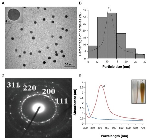
Figure 1 Characterization of silver nanoparticles. (A) Silver nanoparticles showing spherical, monodispersed particles (scale bar, 50 nm). The inset shows one single particle of silver (scale bar, 5 nm). (B) Particle size distribution showing preponderance of particles in the size range of 10–15 nm. (C) Electron diffraction pattern of nanoparticles showing various crystallographic planes. (D) Optical spectra of silver before (1) and after reduction (2). The inset shows the corresponding change in color.
Collection and preparation of B. malayi microfilariae
Microfilariae of B. malayi were obtained by lavage from the peritoneal cavities of infected jirds (Meriones unguiculatus). The Mf used for in vitro experiments were collected, washed with Roswell Park Memorial Institute (RPMI) 1640 medium supplemented with antibiotics (gentamicin 20 mg; penicillin 100 mg; streptomycin 100 mg), 15 mM 4-(2-hydroxyethyl)-1-piperazineethanesulfonic acid, organic acids (malic acid, α-ketoglutaric acid, D-succinic acid, and fumaric acid at concentrations of 670, 370, 60, and 55 mg/L, respectively) and sugars (sucrose and fructose at concentrations of 26.7 g/L and 0.4 g/L, respectively). The use of animals for the study was approved by the Institutional Animal Ethics Committee which follows the Committee for the Purpose of Control and Supervision of Experiments on Animals norms.
In vitro screening for antifilarial activity against B. malayi
In vitro screening for antifilarial activity against B. malayi was carried out as described earlier.Citation8 Approximately 100 Mf (in 100 μL RPMI) were introduced into each well of 24-well microculture plates. B. malayi microfilariae were screened for the antifilarial effect of AgNPs in vitro over a wide dose range (1–100 μM). Microfilariae were treated similarly with AuNPs of comparable sizes to rule out any nonspecific nanoparticle effect. As a positive control, staurosporine – a known inducer of apoptosis – was added to the microfilariae at 0.5 μM (elicits 100% loss in motility), whereas Mf in RPMI medium, in the absence of any agent added, was used as a negative control (vehicle). The plates were incubated for 48 hours at 37°C in the presence of 5% carbon dioxide. Subsequently, the number of live and dead Mf in each well was counted under an inverted microscope (Nikon Corporation, Tokyo, Japan) and the percentage of motile Mf out of total Mf recruited per aliquot was calculated.
Determination of 50% lethal dose for nanosilver
Cytotoxicity of AgNPs was evaluated by a trypan blue dye exclusion assay. Peripheral blood mononuclear cells (1 × 10⁵ cells/mL) were exposed to varying concentrations of AgNPs for 48 hours followed by incubation with trypan blue (0.2 mg/mL) for 1 minute. Cells were observed under a Nikon light microscope (Tokyo, Japan) and the viable cell ratio were calculated by counting the stained and unstained cells separately.Citation21 Viable cells do not uptake trypan blue, whereas nonviable cells with porous membranes stain blue. The cytotoxicity of the nanoparticles was evaluated and the 50% lethal dose was determined.
Poly(adenosine diphosphate-ribose) polymerase (PARP) activity assay
PARP activity in B. malayi microfilariae was determined using a commercial kit (R & D Systems Inc, Minneapolis, MD) according to the manufacturer’s instruction. Briefly, 100 μL aliquots of suspension (containing about 100 Mf) were treated with different reagents and lysed with 1% Triton X-100 (Himedia laboratories Pvt Ltd, Mumbai, India) in the presence of protease inhibitors. Lysate (20 μg) was added to each well in 96-well plates precoated with histone. PARP activity was determined from the incorporation of biotinylated poly(adenosine diphosphate-ribose) onto immobilized histone, which was measured by the addition of steptavidin-conjugated horseradish peroxidise and a suitable chromogenic substrate to the incubation mixture. A standard curve for PARP enzymatic activity (A450 versus PARP units) was initially generated using 0.01, 0.05, 0.1, 0.5, and 1 unit of enzyme per well. The absorbance obtained with each test sample (microfilarial lysate) was extrapolated on the standard curve to obtain the corresponding PARP activity. The control sample (microfilaria without any pretreatment) provided 100% activity reference point. The percentage inhibition in enzymatic activity in other test samples (lysates treated with different reagents) was accordingly calculated. The experiment was carried out in triplicate and the percentage inhibition was averaged over the experiments.
Ethidium bromide/Acridine orange (EB/AO) staining for the detection of apoptosis
Dual staining with EB/AO was carried out as described elsewhere. Citation22 The dye mix consisted of 100 μg/mL EB and 100 μg/mL AO in phosphate-buffered saline. Microfilariae (control as well as treated with different reagents for 48 hours) were washed and resuspended in 25 μL cold phosphate-buffered saline, followed by the addition of 5 μL EB/AO dye mix. Stained microfilariae were viewed under an epifluorescence microscope (Nikon) with the excitation filter set at 480/30 nm and the barrier filter at 535/40 nm. Tests were carried out in triplicate, counting a minimum of 10 Mf in each observation.
Electron microscopy
Mf were treated with staurosporine, AgNPs, or AuNPs for 48 hours. Samples were fixed in Karnovsky fixative (pH 7.2) for 2 hours at 4°C followed by postfixation in osmium tetroxide (1%) and then dehydrated in ascending concentrations of acetone. For scanning electron microscopy, dehydrated samples were critical point dried, mounted on an aluminum stub with adhesive tape, and sputter-coated with colloidal gold. Specimens were viewed under a Leo 435VP scanning electron microscope (LEO Electron Microscopy Ltd, Cambridge, UK) at an operating voltage of 15 kV. For transmission electron microscopy, blocks were prepared as previously described.Citation23 Ultrathin sections (60–70 nm thick) were prepared with an ultramicrotome (Leica EM UC6; Leica Microsystems GmbH, Wetzlar, Germany). Sections were contrasted with uranyl acetate and alkaline lead citrate. Specimens were mounted on formvar-coated grids and viewed under a FEI Morgagni™ 268(D) (FEI, Hillsboro, OR) digital transmission electron microscope at 120 kV using image analysis software from Soft Imaging System GmbH (Muenster, Germany). The final magnifications were derived from the photo micrographs and the scale bars determined.
Results and discussion
AgNPs were synthesized through the aqueous chemical precipitation method, as described earlier.Citation11,Citation19,Citation20 Nanoparticles were spherical in shape, 10–15 nm in diameter (), and monodispersed with a narrow particle size distribution (). The selected area electron diffraction pattern from these particles matched the crystallographic planes of the face-centered cubic AgNPs (). Ultraviolet–visible absorption spectra showed the reduction of silver ions into the AgNPs under ambient conditions (). The inset shows the color changes before and after the process of reduction. The silver nitrate solution exhibited maximum absorbance at 300 nm, which gradually underwent red shift with the appearance of a sharp peak at 410 nm, which can be attributed to a narrow size distribution of the particles formed in the solution.
The microfilariae remained viable and motile in RPMI media, as expected. The effect of AgNPs (10–15 nm) upon Mf motility was evaluated over a wide concentration range. Remarkably, at 4.6 μM, the AgNPs evoked motility loss in 50% of Mf population, whereas at 50.6 μM, the AgNPs rendered the entire Mf population immotile (). The AuNPs (10–15 nm) failed to produce any significant antifilarial effect at the identical concentrations. However, staurosporine (0.5 μM) as a positive control induced complete loss in Mf motility. When evaluated for the extent of cytotoxicity by trypan blue exclusion test, AgNPs at 101.2 μM elicited 50% lethality.
Table 1 Brugia malayi microfilariae were incubated with silver nanoparticles at varying concentrations
The inhibition of PARP activity is a valid measure for the assessment of cellular apoptosis.Citation24 During apoptosis, PARP is cleaved by caspase 3 with an ensuing reduction in PARP enzyme activity, which thus prevents apoptotic cells from repairing their own DNA.Citation25 PARP activity in Mf exposed to nanosilver (50 μM), nanogold (50 μM), or staurosporine (0.5 μM) was then studied. Both AgNPs and staurosporine induced nearly 70% attenuation in PARP enzymatic activity, whereas only 25% loss in activity was observed in the presence of AuNPs (), thus consistent with the significant microfilaricidal effect of nanoscale silver.
Table 2 Percentage inhibition in poly(adenosine diphosphateribose) polymerase activity in the microfilariae treated with different reagents as indicated compared with control (Roswell Park Memorial Institute medium only)
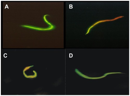
The interaction between microfilariae and nanosilver was further investigated with EB and AO differential staining.Citation22 AO permeates into healthy live cells to stain the nuclear material green, whereas cellular access of EB is restricted, unless there is membrane damage. Mf treated with AgNPs (50 μM) and staurosporine (0.5 μM) were stained orange-yellow with EB (), which reflected loss in surface membrane integrity. This contrasted with the profile of control parasites as well as that of Mf exposed to AuNPs (50 μM), which were stained green with AO ().
Figure 2 Ethidium bromide/acridine orange differential staining of microfilarial forms for the detection of apoptosis. Untreated (A) and gold nanoparticles preincubated (D) nuclei showed green staining due to acridine orange permeation, while organisms treated with silver nanoparticles (B) and staurosporine (C) appeared orange-yellow due to ethidium bromide, suggesting loss of integrity of surface membrane of the parasite shown.
Note: Data are representative of three different experiments.
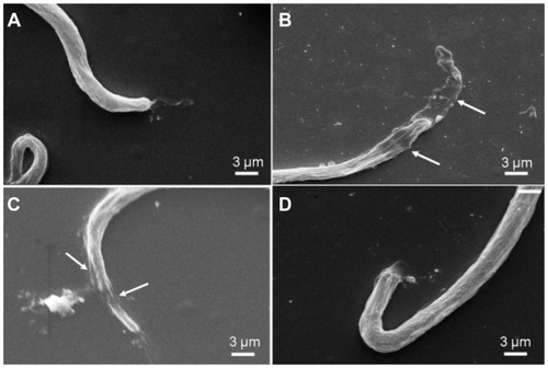
Nanosilver-induced ultrastructural changes in microfilariae were subsequently investigated by electron microscopy. Scanning electron microscopy images reflected marked loss in microfilarial sheath in parasites treated either with staurosporine (0.5 μM) or AgNPs (50 μM), whereas RPMI-treated control microfilariae or parasites exposed to AuNPs (50 μM) exhibited clear undamaged translucent sheaths ().
Figure 3 Scanning electron micrographs of (A) untreated control parasite in Roswell Park Memorial Institute medium; (B) microfilariae treated with staurosporine (0.5 μM); (C) microfilariae treated with silver nanoparticles (50 μM), and (D) microfilariae treated with gold nanoparticles (50 μM).
Note: Data are representative of three different experiments.
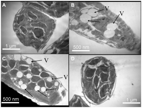
Transmission electron microscopy images of ultrathin sections of microfilariae, pretreated either with staurosporine or AgNPs, revealed multiple large, round vacuoles inside cells (). On the contrary, vacuoles were scanty in parasites treated with AuNPs and absent in RPMI-treated control parasites, which possessed thick homogeneous cuticles and intact cell organelles without vacuoles. The presence of vacuoles in ultrastructural sections of staurosporine-treated as well as AgNP-treated microfilariae further confirmed the apoptotic rationale in the pharmacodynamics of AgNPs. It may be noted that, conventional mechanisms for the study of apoptosis such as terminal deoxynucleotidyl transferase-mediated deoxyuridine triphosphate nick end labeling assay and DNA fragmentation are not technically viable in a parasite model. Citation17 Taken together, the above findings were consistent with apoptotic changes induced in microfilariae in the presence of nanosilver similar to that elicited by staurosporine. Nanogold of comparable size and concentration, on the other hand, had no adverse effect on parasites.
Figure 4 Transmission electron micrographs through sections of (A) untreated control parasite in Roswell Park Memorial Institute medium; (B) microfilariae treated with staurosporine (0.5 μM); (C) microfilariae treated with silver nanoparticles (50 μM); and (D) microfilariae treated with gold nanoparticles (50 μM).
Notes: Arrows point to the vacuoles; data are representative of three different experiments.
Abbreviation: V, vacuoles.
DEC has been shown to induce apoptosis in the microfilariae of W. bancrofti,Citation17 though the drug is not known to be filaricidal in vitro.Citation26 Thus, apoptosis induction in vitro by DEC may not be sufficient to kill the parasite, which could couple with the innate inflammatory response of the host to elicit effective antifilarial activity in vivo. In support of this, the cuticles of adult and microfilarial B. malayi were found to be resistant to hydrogen peroxide, a strong oxidant as well as apoptosis-inducing agent, which is attributable to the presence of α-tocopherol in the lipid fraction of the parasite surface.Citation27 Interestingly, DEC in combination with hydrogen peroxide was found to have marked synergism against filarial parasites.Citation28 The above facts underpinned the relative ineffectiveness of DEC as the sole filaricidal agent and prompts researchers to strategize combinations with apoptosis inducers as a novel lead for antifilarial therapeutics.
In an earlier report, the enhanced antibacterial potency of AgNPs was analyzed.Citation11 Commendable efforts have been made to explore the underlying molecular mechanism of the antimicrobial activity of silver.Citation29 Dissolved silver ions could be responsible for some of the biological actions of AgNPs against microorganismsCitation30,Citation31 through their interaction with and inhibition of the thiol groups of vital enzymes.Citation29,Citation32 It has been recently demonstrated that the rate of ionization of silver from nanoparticles is significantly affected by the presence of salts and biomolecules in the suspension medium.Citation33 Biological media contain large numbers of interfering molecules in abundance, which can thus influence the release of silver ions. It is unlikely that silver ions will rise to a significant level under the present experimental conditions, as reducing agents like glucose present in RPMI would keep these ions in the reduced state, and high concentration of chloride and phosphate ions in the medium would generate insoluble salts of silver (eg, silver chloride and silver phosphate). Besides this, the presence of citric acid during the process of AgNP synthesis would result in surface coating of the nanoparticles with citrate ions which would further limit the release of silver ions, as reported recently.Citation33 Thus, the observed antimicrofilarial activity reported in this study is most likely attributable to AgNPs per se and not to silver ions.
Conclusion
The main embargo on the current drug DEC is its empirical use without rationale. DEC-based mass drug administration policy demands longer durations of therapy, which may lack patient compliance and thus lead to frequent failure.Citation34 AgNPs, on the other hand, are capable of inducing apoptosis in mammalian cells.Citation14–Citation16 The effective microfilaricidal activity of AgNPs at relatively low concentration as reported in this study, with evidence of apoptotic induction in microfilariae, can project nanosilver as a potential drug adjuvant against lymphatic filariasis. Unlike DEC, which is effective against microfilariae only in vivo and not in vitro,Citation35 nanosilver promises greater therapeutic effectiveness. Remarkably, a recent report has demonstrated the therapeutic potential of AgNPs against leishmaniasis, another parasitic disease caused by Leishmania tropica.Citation36 However, the antiparasitic mechanism elucidated in this study was oxidative attack by AgNPs, which was further potentiated by the concomitant ultraviolet exposure. This mechanism is hardly of therapeutic relevance in the present study as filarial parasites are known to be endowed with robust antioxidant defenses. On the contrary, based on evidence presented in the present study, apoptosis is proposed as the mechanistic basis of the antifilarial activity of nanosilver. Interestingly, there is a subtle link reported between oxidative stress and apoptosis induction.Citation37 Filarial parasites are lymphatic system-specific organisms. Given the wide distribution attainable by AgNPs in tissues including lymphatics,Citation38 administered nanosilver is expected to achieve a desired local antifilarial therapeutic concentration in the infected population. Nanosilver may act synergistically with DEC and thus may be effective either individually or as an adjunct (at lower individual doses) to this standard drug, after critical evaluation of safety parameters. Although there have been concerns regarding the toxicity of nanoparticles in general,Citation39 and silver in particular,Citation16,Citation40 nanosilver might be considered to have a safe therapeutic window. The 50% lethal dose of nanosilver (100 μM), which is double the complete inhibitory concentration value (50 μM) as reported in the present study, supports this contention. The report on Leishmania parasites has demonstrated the synergism between nanosilver and ultraviolet exposure in their antiparasitic effect.Citation36 This study did not find any serious toxicity concern with silver. Thus, it can be inferred that AgNPs may be used as an adjuvant to DEC to potentiate synergistic apoptotic impact, so that the dose requirement of nanosilver may be further scaled down. To summarize, the present report provides the first ever conclusive proof in support of apoptosis as a novel stratagem in antifilarial drug designing and nanosilver as a valid lead in the research on antifilarial therapeutics.
Acknowledgments
Grants received by D Dash from the Indian Council of Medical Research and the Department of Biotechnology (DBT), Government of India, and the equipment support from the Department of Science and Technology Unit on Nanoscience and Technology, Banaras Hindu University, are gratefully acknowledged. K Goswami, RD Sharma, and MVR Reddy would like to thank DBT for funding the “Repository for Filarial Parasite and Reagents.” Electron microscopy was carried out at the Sophisticated Analytical Instrument Facility (Department of Science and Technology), All India Institute of Medical Sciences, New Delhi, India. SK Singh is a recipient of a research fellowship from the University Grants Commission, New Delhi, India.
Disclosure
The authors report no conflicts of interest in this work.
References
- OttesenEADukeBOKaramMBehbehaniKStrategies and tools for the control/elimination of lymphatic filariasisBull World Health Organ19977564915039509621
- FanPCDiethylcarbamazine treatment of bancroftian and malayan filariasis with emphasis on side effectsAnn Trop Med Parasitol19928643994051463361
- Tropical Diseases, Special Programme for Research and Training (TDR)Strategic direction for lymphatic filariasis research: disease burden and epidemiological trends [article on the Internet]GenevaTDR2012 Available from: http://www.who.int/tdr/diseases/lymphfil/direction/en/index.htmlAccessed February 3, 2012
- World Health OrganizationDefining the roles of vector control and xenomonitoring in the Global Programme to Eliminate Lymphatic Filariasis. Report of the informal consultationJanuary 29–31, 2002Geneva SwitzerlandGenevaWorld Health Organization2002 Document: WHO/CDS/CPE/PVC/2002.3
- Indian Council of Medical ResearchProspects of elimination of lymphatic filariasis in IndiaICMR Bull2002325–6114
- McGarryHFPlantLDTaylorMJDiethylcarbamazine activity against Brugia malayi microfilariae is dependent on inducible nitric-oxide synthase and the cyclooxygenase pathwayFilaria J2005441215932636
- VerhasseltVGoldmanMWillemsFOxidative stress up-regulates IL-8 and TNF-alpha synthesis by human dendritic cellsEur J Immunol19982811388638909842932
- SahareKNAnandharamanVMeshramVGAnti- microfilarial activity of methanolic extract of Vitex negundo and Aegle marmelos and their phytochemical analysisIndian J Exp Biol200846212813118335811
- BagSTawariNRSharmaRGoswamiKReddyMVDeganiMSIn vitro biological evaluation of biguanides and dihydrotriazines against Brugia malayi and folate reversal studiesActa Trop20101131485119769933
- KimJSKukEYuKNAntimicrobial effects of silver nanoparticlesNanomedicine2007319510117379174
- ShrivastavaSBeraTRoyASinghGRamchandraraoPDashDCharacterization of enhanced antibacterial effect of novel silver nanoparticlesNanotechnology20071822225103225111
- GopinathPGogoiSKChattopadhyayAGhoshSSImplications of silver nanoparticle induced cell apoptosis for in vitro gene therapyNanotechnology200819707510407511321817629
- GopinathPGogoiSKSanpuiPPaulAChattopadhyayAGhoshSSSignaling gene cascade in silver nanoparticle induced apoptosisColloids Surf B Biointerfaces201077224024520197232
- FoldbjergaROlesenPHougaardMDangDAHoffmannHJAutrupHPVP-coated silver nanoparticles and silver ions induce reactive oxygen species, apoptosis and necrosis in THP-1 monocytesToxicol Lett2009190215616219607894
- MiuraNShinoharaYCytotoxic effect and apoptosis induction by silver nanoparticles in HeLa cellsBiochem Biophys Res Commun2009390373373719836347
- AshaRaniPVLowKuhMunGHandeMPValiyaveettilSCytotoxicity and genotoxicity of silver nanoparticles in human cellsACS Nano20093227929019236062
- PeixotoCASantosACAyresCFMolecular evidence for apoptosis in microfilariae of Wuchereria bancrofti induced by diethylcarbamazineParasitol Res2008103371772118497999
- BarcinskiMADosReisGAApoptosis in parasites and parasite-induced apoptosis in the host immune system: a new approach to parasitic diseasesBraz J Med Biol Res199932439540110347800
- ShrivastavaSBeraTSinghSKSinghGRamachandraraoPDashDCharacterization of antiplatelet properties of silver nanoparticlesACS Nano2009361357136419545167
- ShrivastavaSSinghSKMukhopadhyayASinhaASMandalRKDashDNegative regulation of fibrin polymerization and clot formation by nanoparticles of silverColloids Surf B Biointerfaces201182124124620870397
- OkuyaSTanabeKTanizawaYOkaYLeptin increases the viability of isolated rat pancreatic islets by suppressing apoptosisEndocrinology2001142114827483011606450
- RibbleDGoldsteinNBNorrisDAShellmanYGA simple technique for quantifying apoptosis in 96-well platesBMC Biotechnol20055121915885144
- AlvesLCBraynerFASilvaLFPeixotoCAThe ultrastructure of infective larvae (L3) of Wuchereria bancrofti after treatment with diethylcarbamazineMicron2005361677215582480
- MullenPPARP cleavage as a means of assessing apoptosisLangdonSPCancer Cell Culture: Methods and ProtocolsTotowa, NJHumana Press2004171181
- BöhmIThe apoptosis marker enzyme poly-(ADP-ribose) polymerase (PARP) in systemic lupus erythematosusZ Rheumatol2006656541544 German16541210
- MackenzieCDKronMADiethylcarbamazine: a review of its action in onchocerciasis, lymphatic filariasis and inflammationTrop Dis Bull198582R136
- SmithVPSelkirkMEGounarisKBrugia malayi: resistance of cuticular lipids to oxidant-induced damage and detection of α-tocopherol in the neutral lipid fractionExp Parasitol19988821031109538864
- SharmaRDJanardhanaPBGajalakshmiDReddyMVGoswamiKNovel pharmaceutical rationale against human lymphatic filariasis: an oxidative premiseAsian Pac J Trop Med2009213034
- MoronesJRElechiguerraJLCamachoAThe bactericidal effect of silver nanoparticlesNanotechnology200516102346235320818017
- HwangETLeeJHChaeYJAnalysis of the toxic mode of action of silver nanoparticles using stress-specific bioluminescent bacteriaSmall20084674675018528852
- ChoiODengKKKimNJRossLJrSurampalliRYHuZThe inhibitory effects of silver nanoparticles, silver ions, and silver chloride colloids on microbial growthWater Res200842123066307418359055
- MatsumuraYYoshikataKKunisakiSTsuchidoTMode of bactericidal action of silver zeolite and its comparison with that of silver nitrateAppl Environ Microbiol20036974278428112839814
- KittlerSGreulichCDiendorfJKollerMEppleMToxicity of silver nanoparticles increases during storage because of slow dissolution under release of silver ionsChem Mater2010221645484554
- NandhaBSadanandaneCJambulingamPDasPDelivery strategy of mass annual single dose DEC administration to eliminate lymphatic filariasis in the urban areas of Pondicherry, South India: 5 years of experienceFilaria J2007671217718908
- MaizelsRMDenhamDADiethylcarbamazine (DEC): immunopharmacological interactions of an antifilarial drugParasitology1992105SupplS49601308929
- AllahverdiyevAMAbamorESBagirovaMAntileishmanial effect of silver nanoparticles and their enhanced antiparasitic activity under ultraviolet lightInt J Nanomedicine201162705271422114501
- ButtkeTMSandstromPAOxidative stress as a mediator for apoptosisImmunol Today19941517108136014
- LankveldDPOomenAGKrystekPThe kinetics of the tissue distribution of silver nanoparticles of different sizesBiomaterials201031328350836120684985
- El-AnsaryAAl-DaihanSOn the toxicity of therapeutically used nanoparticles: an overviewJ Toxicol2009200975481075481820130771
- El BadawyAMSilvaRGMorrisBScheckelKGSuidanMTTolaymatTMSurface charge-dependent toxicity of silver nanoparticlesEnviron Sci Technol201145128328721133412