Abstract
Hierarchically nanosized hydroxyapatite (HA) with flower-like structure assembled from nanosheets consisting of nanorod building blocks was successfully synthesized by using CaCl2, NaH2PO4, and potassium sodium tartrate via a hydrothermal method at 200°C for 24 hours. The effects of heating time and heating temperature on the products were investigated. As a chelating ligand and template molecule, the potassium sodium tartrate plays a key role in the formation of hierarchically nanostructured HA. On the basis of experimental results, a possible mechanism based on soft-template and self-assembly was proposed for the formation and growth of the hierarchically nanostructured HA. Cytotoxicity experiments indicated that the hierarchically nanostructured HA had good biocompatibility. It was shown by in-vitro experiments that mesenchymal stem cells could attach to the hierarchically nanostructured HA after being cultured for 48 hours.
Objective
The purpose of this study was to develop facile and effective methods for the synthesis of novel hydroxyapatite (HA) with hierarchical nanostructures assembled from independent and discrete nanobuilding blocks.
Methods
A simple hydrothermal approach was applied to synthesize HA by using CaCl2, NaH2PO4, and potassium sodium tartrate at 200°C for 24 hours. The cell cytotoxicity of the hierarchically nanostructured HA was tested by MTT (3-(4,5-dimethylthiazol-2-yl)-2,5-diphenyltetrazolium bromide) assay.
Results
HA displayed the flower-like structure assembled from nanosheets consisting of nanorod building blocks. The potassium sodium tartrate was used as a chelating ligand, inducing the formation and self-assembly of HA nanorods. The heating time and heating temperature influenced the aggregation and morphology of HA. The cell viability did not decrease with the increasing concentration of hierarchically nanostructured HA added.
Conclusion
A novel, simple and reliable hydrothermal route had been developed for the synthesis of hierarchically nanosized HA with flower-like structure assembled from nanosheets consisting of nanorod building blocks. The HA with the hierarchical nanostructure was formed via a soft-template assisted self-assembly mechanism. The hierarchically nanostructured HA has a good biocompatibility and essentially no in-vitro cytotoxicity.
Introduction
Hierarchical nanostructures have attracted a great deal of attention due to their novel optical, magnetic, and catalytic properties, and they have potential applications as important components and interconnects in nanodevices.Citation1–Citation5 It is well known that both the “bottom-up” and “top-down” strategies are two effective approaches for fabricating nanodevices.Citation6–Citation8 The self-assembly of building blocks such as nanoparticles, nanorods, nanobelts, and complex nanocrystals with well defined morphology, structure, size, and size distribution is considered a “bottom-up” approach.Citation9–Citation11 To date, a variety of hierarchical nanostructures of noble metals,Citation12 metal sulfides,Citation13,Citation14 metal oxides,Citation15–Citation18 metal hydroxides,Citation19 metal carbonates,Citation20 and metal silicatesCitation21 have been synthesized via self-assembly.
Hydroxyapatite [HA, Ca10(PO4)6(OH)2] is a typical biomineral that is abundant in organisms. It can be used as bone scaffoldsCitation22 and luminescence materials,Citation23 and it also has many important applications in drug delivery,Citation24,Citation25 and biomedical engineering,Citation26 based on its chemical and biological similarity with the mineral constituents of human bones and teeth.Citation27–Citation29 To date, various derivatives of HA, such as carbonated HA,Citation30–Citation32 strontium HA,Citation33,Citation34 F-substituted HA,Citation35,Citation36 and HA-based nanocomposites,Citation37,Citation38 have been reported. Some successful methods including emulsion, Citation39,Citation40 hydrothermal method,Citation41,Citation42 solvothermal method,Citation43 sonochemical precipitation,Citation44 hard-template method,Citation45 sol-gel method,Citation46 biomimetic method,Citation47,Citation48 and microwave irradiationCitation49 have been employed in the synthesis of HA. Gajjeraman et alCitation50 used organic constituents of bones and teeth to control the nucleation and assembly of hierarchical HA. Wang et alCitation51 reported the fabrication of HA nanorods by tuning the interfaces between surfactants and the central atoms of HA based on the liquid-solid-solution mechanism. Ortega et alCitation52 used functionalized silica as a template for the nucleation and growth of HA coatings under mild conditions, with an aging period of 6 hours. Wang et alCitation53 employed poly(styrene sulfonate) as a modifier in the synthesis of HA microspheres, with controlled size and hierarchical structure by hydrothermal method. Ryu et alCitation54 reported the synthesis of polydopamine-assisted HA inspired by the adhesion mechanism of mussels via a biomineralizing route. Nassif et alCitation55 presented the synthesis of HA nanocrystals via NH3 vapor diffusion into a CaCl2-NaH2PO4 mixed solution and B- or A-type carbonate-apatite phases by the addition of NaHCO3 or (NH4)2CO3. In a previous paper, the authors reported the preparation of hierarchically nanostructured HA hollow spheres assembled from nanorods by using CaCl2, NaH2PO4, and potassium sodium tartrate via a solvothermal method at 200°C for 24 hours in a mixture solvent of water/N, N-dimethylformamide.Citation43 Most natural biomaterials have complex hierarchical microstructures.Citation56 Therefore, the research of hierarchically nanostructured HA is important to the understanding of the biomineralization mechanism and the realization of controllable synthesis of other hierarchical nanostructures.
Reported herein is a simple hydrothermal approach to the synthesis of hierarchically nanostructured HA with flower-like morphology consisting of nanosheets, which are assembled from nanorod building blocks. The potassium sodium tartrate was used as a chelating ligand and template molecule to induce the synthesis of hierarchically nanostructured HA. The formation mechanism of hierarchically nanostructured HA is proposed based on the experimental details. Cytotoxicity experiments of the hierarchically nanostructured HA have been also carried out. The synthetic method presented here is simple, low-cost, and environmentally friendly (ie, does not use any organic solvents or organic structure-directing surfactants. Furthermore, the final products in this work enrich the hierarchically nanostructured HA architecture family.
Materials and methods
All chemicals were of analytical grade and used as received without further purification. All experiments were conducted under ambient atmosphere. In a typical experiment, 0.001 mol of CaCl2 and 0.001 mol of C4H4O6KNa · 4H2O (potassium sodium tartrate) were added into 15 mL of water under vigorous stirring for 30 minutes. Then, 0.0006 mol of NaH2PO4 was added into the above solution under vigorous stirring for another 30 minutes. The obtained solution was hydrothermally treated at a fixed temperature (200°C) for 24 hours without stirring and shaking, and then was allowed to cool to room temperature naturally. Subsequently, the precipitates were separated from the solution by centrifugation, washed by ethanol several times, and dried at 60°C. Finally, a white powder was obtained. This sample was denoted as sample 1.
In order to investigate the formation mechanism of hierarchically nanostructured HA, another sample was synthesized without C4H4O6KNa · 4H2O, keeping the other conditions the same with those in the preparation of sample 1. This sample was denoted as sample 7. Moreover, another sample was synthesized without NaH2PO4 at room temperature for 30 minutes, keeping the other conditions the same. This sample was denoted as sample 8. Another sample was also synthesized without NaH2PO4, keeping the other conditions the same. This sample was denoted as sample 9. The detailed experimental parameters for the synthesis of some typical samples were listed in .
Table 1 Detailed experimental parameters for the synthesis of some typical samples by hydrothermal method
The pH values of the reaction solutions in the preparation of sample 1 (with potassium sodium tartrate) and sample 7 (without potassium sodium tartrate) were 4.92 and 3.97 before hydrothermal treatment, respectively. After the reaction solutions had been hydrothermally treated, the pH values were 3.74 and 2.66 in the preparation of sample 1 and sample 7, respectively.
X-ray powder diffraction (XRD) patterns were recorded with 2θ range from 2° to 70° on a D/Max 2200-PC diffractometer (Rigaku Corporation, Tokyo, Japan) with CuKα radiation (λ = 0.15418 nm) and graphite monochromator at ambient temperature. Field emission scanning electron microscopy (FE-SEM) images were recorded on a JSM-6700F field-emission scanning electron microscope (JEOL, Tokyo, Japan). SEM images were recorded on a Hitachi 3400 N scanning electron microscope. All samples were Au coated prior to the observation by FE-SEM and SEM. Transmission electron microscopy (TEM) imaging, selected area electron diffraction (SAED), high-resolution TEM (HR-TEM) imaging, and energy-dispersive X-ray analysis (EDS) were performed on a JEM-2010 electron microscope (JEOL) with the accelerating voltage of 200 kV. A Fourier transform infrared (FTIR) spectroscopy was carried out with a Nexus spectrometer (Thermo Nicolet, Madison, WI), using the KBr disk method. The Ca/P atomic ratio was also obtained by inductively coupled plasma optical emission spectrometry (ICP-OES), using an Optima 5300DV (PerkinElmer Life and Analytical Sciences, Shelton, CT).
The human gastric carcinoma cells (MGC-803), which were cultured in a RPMI-1640 medium supplemented with 10% fetal bovine serum and 1% penicillin-streptomycin at 37°C for 48 hours, were used for the cell viability test. The cells were seeded in 96-well flat-bottom microassay plates at a concentration of 1 × 10⁴ cells/mL, and cultured for 24 hours. The sterilized HA samples were added into wells at a concentration from 10 to 100 μg/mL, and were co-cultured with cells for 48 hours. The sample free tissue culture plate was used as a control. Cell viability was quantitatively analyzed by MTT (3-(4,5-dimethylthiazol-2-yl)-2,5-diphenyltetrazolium bromide) assay. Data are representative as the mean value of five parallel experiments.
Results and discussion
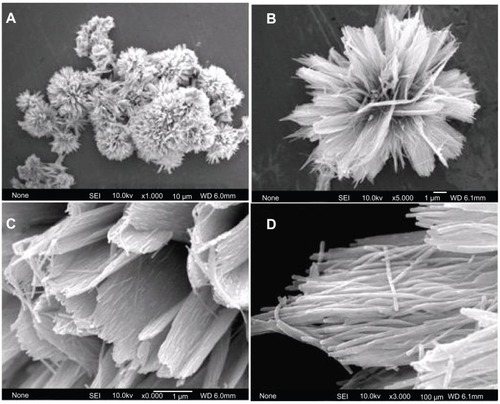
The typical sample (sample 1) was synthesized by using CaCl2, NaH2PO4, and potassium sodium tartrate via a hydrothermal method at 200°C for 24 hours. The morphology of the sample was investigated with FE-SEM. shows HA with flower-like morphology assembled from nanosheets. shows a unit of typical flower-like morphology. shows that the nanosheets are assembled from substructured nanorods as the basic unit. displays an individual nanosheet. The nanorods have diameters of 25–50 nm and lengths of about several micrometers. This is the first report on the synthesis of hierarchically nanostructured HA with flower-like morphology assembled from nanosheets consisting of nanorod units.
Figure 1 Field emission scanning electron microscopy images of hierarchically nanostructured hydroxyapatite (sample 1).
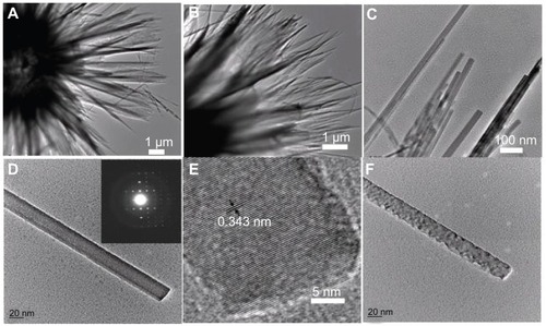
The morphology and microstructure of the sample were further investigated by TEM, SAED, and HR-TEM. and B show TEM images of the hierarchically nanostructured HA, from which one can see the nanosheets with relatively uniform size. displays the edges of the nanosheets built up by nanorods. An individual nanorod with smooth surface is shown in , the inset of which shows the corresponding SAED pattern, indicating the single crystalline structure of the basic nanorod unit. shows the corresponding HR-TEM image of HA nanorod. The periodic fringe spacing of 3.43 Å corresponds to the d-spacing of (002) plane of the hexagonal HA. As seen in , the surface of the nanorod became rough after exposure to electron beam irradiation. Similar cases of one-dimensional structures changing their morphology under electron beam irradiation have been reported in; for example, PbCrO4 rods,Citation57 Ag6Mo10O33 rods,Citation58 and Bi nanotubes,Citation59 etc.
Figure 2 TEM images of hierarchically nanostructured hydroxyapatite (sample 1): (A and B) the edges of the flower-like assembly; (C) the edges of the nanosheets; (D) an individual nanorod; (E) the corresponding high-resolution TEM micrograph of (D); and (F) the nanorod after exposure to electron beam irradiation. The inset of (D) shows the corresponding selected area electron diffraction pattern.
Abbreviation: TEM, transmission electron microscopy.
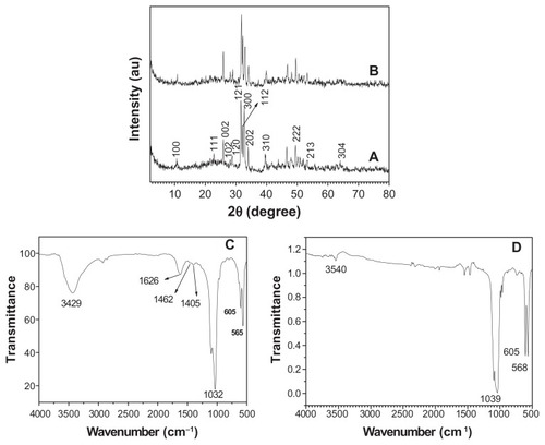
The crystal phase and molecular structure of the typical sample were characterized by XRD and FTIR. shows the XRD pattern of the typical sample, which consisted of a well-crystalline phase with a hexagonal-structured HA (JCPDS 84-1998). No peaks from impurities such as CaHPO4 were observed. The FTIR spectra (shown at (C) and (D) in ) were used to identify the functional groups of the samples. shows the FTIR spectrum of the same sample as in (A). A broad strong peak with a center around 3429 cm−1 can be assigned to the adsorbed water on HA.Citation60 The characteristic stretching mode of the -OH vibration on HA was located around 3570 cm−1,Citation61 which is not clearly observed in because of the overlapping with the strong band of the adsorbed water around 3429 cm−1.Citation60 Furthermore, the weak peaks at about 1405 and 1462 cm−1 should be attributed to the absorption bands of CO3 2−, indicating the presence of carbonate ions, which came from the atmosphere.Citation62 The intense peaks located at 1032, 605, and 565 cm−1 can be attributed to the PO4 3−.Citation63 It should be noted that the similar result that the characteristic stretching mode of the –OH vibration was overlapped with the strong band of the adsorbed water has been observed in a previous study on HA.Citation64 The relative intensity of the peak at about 3540 cm−1 decreased after the calcination of the sample at 800°C for 3 hours (see (D) in ), compared with that in (C). The XRD pattern showed the calcination effect on the crystal structure (see (B) in ); the pattern shows that the sample after calcination at 800°C for 3 hours had a similar XRD pattern with that in (A), indicating that the crystal phase of the sample did not change. However, the relative intensity of (112) and (300) planes in (B) in increased compared with those in (A). The crystal units of HA could be rearranged after high temperature calcination, resulting in better crystallized planes such as (112) and (300) planes. On the basis of the XRD and FTIR results, one can confirm that the sample is HA.
Figure 3 A typical XRD pattern (A) and FTIR spectrum (C) of hydroxyapatite powders prepared by hydrothermal method at 200°C for 24 hours; XRD pattern (B) and FTIR spectrum (D) of the sample prepared by calcination of (A) at 800°C for 3 hours.
Abbreviations: FTIR, Fourier transform infrared; XRD, X-ray powder diffraction.
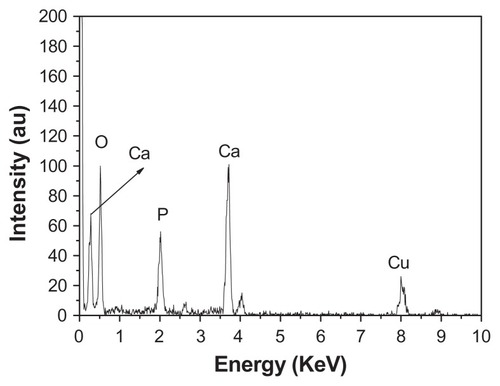
EDS is a technique used to identify the elemental composition of the typical product, as shown in . The figure shows that the sample consisted of Ca, P, and O, with a Ca/P atomic ratio of 1.62, in good agreement with the stoichiometric composition of HA, further implying the formation of pure phase HA. The Cu signal comes from the TEM grid. The Ca/P atomic ratio was also tested by ICP-OES. The obtained value of the Ca/P atomic ratio was 1.73, which is close to the result obtained by EDS analysis.
Figure 4 Energy-dispersive X-ray analysis spectrum of hierarchically nanostructured hydroxyapatite (sample 1).
Investigation has been conducted on the effect of the reaction time on the crystallinity and morphology of HA. All the samples consisted of a single phase of HA (see ). Moreover, the peak intensity of the samples increased with the increasing heating time, indicating that the crystallinity was gradually improved during the reaction. When the hydrothermal time was 12 hours, hierarchically nanostructured HA was observed (). Compared with that shown in , the size of the flower-like particles decreased and the nanorods were not clearly observed (). When the reaction time was 3 hours, spherical structures assembled from nanosheets and flower-like structures consisting of nanorods were obtained (). When the heating time was increased to 4 hours, there were no significant differences in the morphologies of the samples for 3- and 4-hour hydrothermal treatments (). When the heating time was increased to 6 hours, though some irregular morphologies were also observed, the number of flower-like structures consisting of nanosheets increased (). , , and , clearly show the morphology evolution process of hierarchically nanostructured HA during the hydrothermal process.
Figure 5 X-ray powder diffraction patterns of hierarchically nanostructured hydroxyapatite prepared by hydrothermal method at 200°C for different lengths of time: 3 hours (sample 3) (A); 4 hours (sample 4) (B); 6 hours (sample 5) (C); and 12 hours (sample 2) (D).
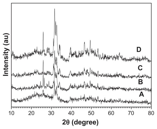
Figure 6 Scanning electron microscopy images of hierarchically nanostructured hydroxyapatite prepared by hydrothermal method at 200°C for 12 hours (sample 2).
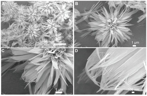
Figure 7 Scanning electron microscopy images of hierarchically nanostructured hydroxyapatite prepared by hydrothermal method at 200°C for different lengths of time: (A) 3 hours (sample 3); (B) 4 hours (sample 4); (C) 6 hours (sample 5).
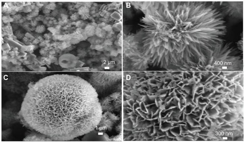
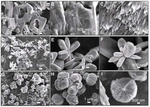
The influence of heating temperature on the morphology of prepared HA was also studied. HA sample with a different morphology () was prepared at 160°C for 24 hours. The flower-like assemblage of nanorods and spherically assembled nanosheets were observed as the major morphologies, while the big blocks were observed as the minor one (). show the typical flower-like structure assemblage of nanorods and spherically assembled nanosheets, respectively. displayed the detailed structure of a single sphere, from which the saw-like structure was observed at the end of the nanosheets.
Figure 8 Scanning electron microscopy images of hierarchically nanostructured hydroxyapatite prepared by hydrothermal method at 160°C for 24 hours (sample 6).
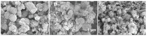
On the basis of the above experimental results, soft-template-assisted self-assembly could be proposed as a formation mechanism of the hierarchically nanostructured HA consisting of nanosheets assembled from nanorods, which is briefly illustrated in . At the initial stage, the complex of Ca2+-tartrate was firstly formed by nucleation. Then, HA with similar morphology was obtained by the addition of NaH2PO4. After that, the individual nanorods aggregated into a sheet-like structure, and the nanosheets aggregated into the hierarchically nanostructured HA by using Ca2+-tartrate as the template. The degree of the assembly and the size of the congeries increased along with the increasing reaction time. Finally, hierarchically nanostructured HA with flower-like morphology assembled from nanosheets consisting of nanorod building blocks was fabricated. It should be noted that the potassium sodium tartrate played a key role in the synthesis of hierarchically nanostructured HA. When the potassium sodium tartrate was not present, the monetite was formed, and the corresponding morphology is shown in . The figure shows the big blocks with irregular shapes, and no hierarchically nanostructured HA with flower-like structure consisting of nanosheets was observed. The potassium sodium tartrate was used as a chelating ligand inducing the synthesis and self-assembly of hierarchically nanostructured HA. In the presence of C4H4O6 2−, the complex of Ca2+-tartrate was formed due to the strong coordination ability of C4H4O6 2−.Citation65 displayed the SEM images of the sample synthesized by using CaCl2 and C4H4O6 KNa · 4H2O at room temperature for 30 minutes before hydrothermal treatment. The figures show that flakes and congregated flakes were obtained. Hierarchical spherical nanostructures consisting of nanosheets were observed after hydrothermal treatment, as shown in . The complex of Ca2+-tartrate reacted with NaH2PO4 to form HA, and then hierarchically nanostructured HA with flower-like units assembled from nanosheets consisting of nanorod building blocks was then gradually formed. Therefore, the complex of Ca2+-tartrate was used as a soft-template to induce the evolution of HA with special structure and morphology. Moreover, the pH values of the reaction solutions in sample 7 (without potassium sodium tartrate) before and after hydrothermal treatment were 3.97 and 2.66, respectively; while the pH values of the reaction solutions with potassium sodium tartrate before and after hydrothermal treatment were increased to 4.92 and 3.74, respectively. These results implied that the addition of potassium sodium tartrate increased the pH value in the system and favored the fabrication of HA, for the most stable phase of HA existed in a basic solution.Citation66 In our previous work, with the assistance of potassium sodium tartrate the hierarchically nanostructured HA, hollow spheres were obtained via a solvothermal process at 200°C for 24 hours in a mixture solvent of water/N,N-dimethylformamide.Citation64 The C4H4O6 2− has been also used in the preparation of CuO spheresCitation67 and Cu2O nanocages.Citation68 Moreover, it is well known that the HA nanorods are formed due to the anisotropy of HA.Citation69–Citation71 The growth and self-assembly of HA nanorods also induced the formation of hierarchically nanostructured HA with increasing reaction time. Finally, the hierarchically nanostructured HA was synthesized. Generally speaking, the morphology evolution of hierarchically nanostructured HA is very complicated. The various reagents and steps including chelating ligand, soft-template, nucleation, growth, and self-assembly were involved in the evolution of hierarchically nanostructured HA, which was also affected by the intrinsic growth habit of HA itself. Of course, the detailed formation mechanism of hierarchically nanostructured HA needs to be further explored.
Figure 9 Schematic representation of the formation mechanism of hierarchically nanostructured HA.
Abbreviation: HA, hydroxyapatite.
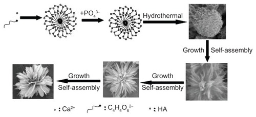
Figure 10 Scanning electron microscopy images of (A–C) the sample synthesized by using CaCl2 and NaH2PO4 without the addition of C4H4O6 KNa · 4H2O at 200°C for 24 hours (sample 7); (D–F) the sample synthesized by using CaCl2 and C4H4O6 KNa · 4H2O at room temperature for 30 minutes (sample 8); and (G–I) the sample synthesized by using CaCl2 and C4H4O6 KNa · 4H2O at 200°C for 24 hours (sample 9).
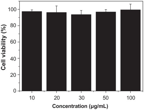
In the synthesis system, CaCl2, NaH2PO4, and potassium sodium tartrate have almost no biological toxicity, and thus are biologically safe. Therefore, it is expected that the hierarchically nanostructured HA is biologically compatible. Mesenchymal stem cells are multipotent stem cells that can differentiate into a variety of cell types and are widely used as seed cells in tissue engineering. The MTT assay was performed as a simple colorimetric assay to measure cell cytotoxicity. In this study, the cytotoxicity of the hierarchically nanostructured HA was tested by comparison with the reference tissue culture plate (). When the hierarchically nanostructured HA concentration in the well was 10 μg/mL, the corresponding cell viability value was 97.2%. Only a slight reduction (2.8%) in cellular viability was observed with the addition of a small amount of hierarchically nanostructured HA. It is interesting to note that the cell viability value was still 99.5%, even though the concentration of hierarchically nanostructured HA increased from 10 to 100 μg/mL after incubation for 48 hours. clearly shows that the cell viability did not decrease with the increasing concentration of hierarchically nanostructured HA added. These results indicate that the hierarchically nanostructured HA has a slight effect on the normal human fibroblast cell viability after 48 hours culture, and little in-vitro cytotoxicity, and can be expected to be used as a biocompatible material.
Figure 11 Viability of normal human fibroblasts incubated with hierarchically nanostructured hydroxyapatite at different concentrations.
Notes: They were determined by counting the survival cells per well in comparison with untreated cells. The error bars denote standard deviations.
Conclusion
In summary, we report a novel, simple, and reliable hydrothermal route for the synthesis of hierarchically nanosized HA with flower-like structure assembled from nanosheets consisting of nanorod building blocks. The potassium sodium tartrate was used as a chelating ligand inducing the formation and self-assembly of HA nanorods. The heating time and heating temperature influenced the aggregation and morphology of HA. Since the HA with the hierarchical nanostructure was formed via a soft-template-assisted self-assembly, prolonged heating time would facilitate the molecular rearrangement and improve the crystallinity of the building blocks such as nanosheets and nanorods. Different reaction temperatures would provide different Gibbs energy in the sealed reaction system, thus leading to different final hierarchical nanostructures. The research of the mechanism of the hierarchical HA does a favor to the synthesis of other nanomaterials with the similar morphology and structure. Cytotoxicity experiments indicate that the hierarchically nanostructured HA has good biocompatibility and essentially no in-vitro cytotoxicity.
Acknowledgments
Financial support from the National Natural Science Foundation of China (31070511), Research Fund for the Doctoral Program of Higher Education of China (20100014120010), China Postdoctoral Science Special Foundation (201003059), and the Program for New Century Excellent Talents in University is gratefully acknowledged.
Disclosure
The author reports no conflicts of interest in this work.
References
- CominiEBarattoCFagliaGFerroniMVomieroASberveglieriGQuasi-one dimensional metal oxide semiconductors: preparation, characterization and application as chemical sensorsProgr Mater Sci200954167
- SnyderMATsapatsisMHierarchical nanomanufacturing: from shaped zeolite nanoparticles to high-performance separation membranesAngew Chem Int Ed Engl2007467560757317694585
- XuAWMaYRColfenHBiomimetic mineralizationJ Mater Chem200717415449
- SanchezCArribartHGuilleMMGBiomimetism and bioinspiration as tools for the design of innovative materials and systemsNat Mater2005427728815875305
- Soler-IlliaGJSanchezCLebeauBPatarinJChemical strategies to design textured materials: from microporous and mesoporous oxides to nanonetworks and hierarchical structuresChem Rev20021024093413812428985
- ChengJYRossCASmithHIThomasELTemplated self-assembly of block copolymers: top-down helps bottom-upAdv Mater20061825052521
- ShenharRRotelloVMNanoparticles: scaffolds and building blocksAcc Chem Res20033654956112859216
- HamleyIWNanotechnology with soft materialsAngew Chem Int Ed Engl2003421692171212707884
- ShimizuTBottom-up synthesis and morphological control of high-axial-ratio nanostructures through molecular self-assemblyPolym J200335122
- ClaridgeSACastlemanAWKhannaSNMurrayCSenAWeissPSCluster-assembled materialsACS Nano2009324425519236057
- ImaiHBiomineralization I: crystallization and self-organization processTop Curr Chem200727043
- LopesWAJaegerHMHierarchical self-assembly of metal nanostructures on diblock copolymer scaffoldsNature200141473573811742395
- MaYRQiLMMaJMChengHMHierarchical, star-shaped PbS crystals formed by a simple solution routeCryst Growth Des20044351354
- ChenXYWangXWangZHYangXGQianYTHierarchical growth and shape evolution of HgS dendritesCryst Growth Des20055347350
- ZhangDFSunLDJiaCJYanZGYouLPYanCHHierarchical assembly of SnO2 nanorod arrays on alpha-Fe2O3 nanotubes: a case of interfacial lattice compatibilityJ Am Chem Soc2005127134921349316190701
- HuXLYuJCGongJMFast production of self-assembled hierarchical alpha-Fe2O3 nanoarchitecturesJ Phys Chem C20071111118011185
- LiuBZengHCRoom temperature solution synthesis of mono-dispersed single-crystalline ZnO nanorods and derived hierarchical nanostructuresLangmuir2004204196420415969417
- LiLLiYGaoSYKoshizakiNOrdered Co3O4 hierarchical nanorod arrays: tunable superhydrophilicity without UV irradiation and transition to superhydrophobicityJ Mater Chem20091983668371
- YangLXZhuYJTongHLiangZHWangWWHierarchical beta-Ni(OH)2 and NiO carnations assembled from nanosheet building blocksCryst Growth Des2007727162719
- LiangXHXiangJHZhangFSXingLSongBChenSWFabrication of hierarchical CaCO3 mesoporous spheres: particle-mediated self-organization induced by biphase interfaces and SAMsLangmuir2010265882588820020762
- WuJZhuYJCaoSWChenFHierachically nanostructured mesoporous spheres of calcium silicate hydrate: surfactant-free sonochemical synthesis and drug-delivery system with ultrahigh drug-loading capacityAdv Mater20102274975320217783
- TampieriASprioSRuffiniACelottiGLesciIGRoveriNFrom wood to bone: multi-step process to convert wood hierarchical structures into biomimetic hydroxyapatite scaffolds for bone tissue engineeringJ Mater Chem20091949734980
- ZhangCMYangJQuanZWHydroxyapatite nano- and microcrystals with multiform morphologies: controllable synthesis and luminescence propertiesCryst Growth Des2009927252733
- HouZYYangPPLianHZMultifunctional hydroxyapatite nanofibers and microbelts as drug carriersChem Eur J2009156973698219496099
- MaMYZhuYJLiLCaoSWNanostructured porous hollow ellipsoidal capsules of hydroxyapatite and calcium silicate: preparation and application in drug deliveryJ Mater Chem20081827222727
- WhiteAABestSMKinlochIAHydroxyapatite-carbon nanotube composites for biomedical applications: a reviewInt J Appl Ceramic Technol20074113
- CaiYRTangRKCalcium phosphate nanoparticles in biomineralization and biomaterialsJ Mater Chem20081837753787
- DuCWangYJProgress in the biomineralization study of bone and enamel and biomimetic synthesis of calcium phosphateJ Inorg Mater200924882888
- PalmerLCNewcombCJKaltzSRSpoerkeEDStuppSIBiomimetic systems for hydroxyapatite mineralization inspired by bone and enamelChem Rev20081084754478319006400
- XiaoJWZhuYCRuanQCBiomacromolecule and surfactant complex matrix for oriented stack of 2-dimensional carbonated hydroxyapatite nanosheets as alignment in calcified tissuesCryst Growth Des20101014921499
- ChengXKHuangZLLiJQSelf-Assembled growth and pore size control of the bubble-template porous carbonated hydroxyapatite microsphereCryst Growth Des20101011801188
- ChengXKHeQJLiJQHuangZLChiRAControl of pore size of the bubble-template porous carbonated hydroxyapatite microsphere by adjustable pressureCryst Growth Des2009927702775
- ZhangCMChengZYYangPPArchitectures of strontium hydroxyapatite microspheres: solvothermal synthesis and luminescence propertiesLangmuir200925135911359819670837
- TanSHChenXGYeYSunJDaiLQDingQHydrothermal removal of Sr2+ in aqueous solution via formation of Sr-substituted hydroxyapatiteJ Hazard Mater201017955956320363558
- HuiJFXiangGLXuXXZhuangJWangXMonodisperse F-substituted hydroxyapatite single-crystal nanotubes with amphiphilic surface propertiesInorg Chem2009485614561619476318
- ZhangHGZhuQSWangYMorphologically controlled synthesis of hydroxyapatite with partial substitution of fluorineChem Mater20051758245830
- LiuJKCaoTJLuYLuoCXFacile preparation of assembly hydroxyapatite spheres to produce nanocompositeMater Technol2009248891
- ZhangYVenugopalJREl-TurkiARamakrishnaSSuBLimCTElectrospun biomimetic nanocomposite nanofibers of hydroxyapatite/chitosan for bone tissue engineeringBiomaterials2008294314432218715637
- ShumHCBandyopadhyayABoseSWeitzDADouble emulsion droplets as microreactors for synthesis of mesoporous hydroxyapatiteChem Mater20092155485555
- BoseSSahaSKSynthesis and characterization of hydroxyapatite nanopowders by emulsion techniqueChem Mater20031544644469
- NeiraISKolen’koYVLebedevOIAn effective morphology control of hydroxyapatite crystals via hydrothermal synthesisCryst Growth Des20099466474
- ChaudhryAAHaqueSKelliciSInstant nano-hydroxyapatite: a continuous and rapid hydrothermal synthesisChem Commun20062122862288
- MaMGZhuJFSolvothermal synthesis and characterization of hierarchically nanostructured hydroxyapatite hollow spheresEur J Inorg Chem200955225526
- JevticMMitricMSkapinSJancarBIgnjatovicNUskokovicDCrystal structure of hydroxyapatite nanorods synthesized by sonochemical homogeneous precipitationCryst Growth Des2008822172222
- EthirajanAZienerULandfesterKSurface-functionalized polymeric nanoparticles as templates for biomimetic mineralization of hydroxyapatiteChem Mater20092122182225
- BigiABoaniniERubiniKHydroxyapatite gels and nano-crystals prepared through a sol–gel processJ Solid State Chem200417730923098
- KithvaPGrondahlLMartinDTrauMBiomimetic synthesis and tensile properties of nanostructured high volume fraction hydroxyapatite and chitosan biocomposite filmsJ Mater Chem201020381389
- ZhangYJLuJJA mild and efficient biomimetic synthesis of rodlike hydroxyapatite particles with a high aspect ratio using polyvinylpyrrolidone as capping agentCryst Growth Des2008821012107
- López-MacipeAGómez-MoralesJRodríguez-ClementeRNanosized hydroxyapatite precipitation from homogeneous calcium/citrate/ phosphate solutions using microwave and conventional heatingAdv Mater1998104953
- GajjeramanSNarayananKHaoJJQinCLGeorgeAMatrix macromolecules in hard tissues control the nucleation and hierarchical assembly of hydroxyapatiteJ Biol Chem20072821193120417052984
- WangXZhuangJPengQLiYDLiquid-solid-solution synthesis of biomedical hydroxyapatite nanorodsAdv Mater20061820312034
- OrtegaIJobbágyMFerrerMLdel MonteFUrease functionalized silica: a biohybrid substrate to drive self-mineralizationChem Mater20082073687370
- WangYSHassanMSGunawanPLauRWangXXuRPolyelectrolyte mediated formation of hydroxyapatite microspheres of controlled size and hierarchical structureJ Colloid Interface Sci2009339697719660764
- RyuJKuSHLeeHParkCBMussel-inspired polydopamine coating as a universal route to hydroxyapatite crystallizationAdv Funct Mater20102021322139
- NassifNMartineauFSyzgantsevaOIn vivo inspired conditions to synthesize biomimetic hydroxyapatiteChem Mater20102236533663
- MeyersMAChenPYLinAYMSekiYBiological materials: structure and mechanical propertiesProgr Mater Sci2008531206
- WangWWZhuYJSynthesis of PbCrO4 and Pb2CrO5 rods via a microwave-assisted ionic liquid methodCryst Growth Des20055505507
- CuiXJYuSHLiLLSelective synthesis and characterization of single-crystal silver molybdate/tungstate nanowires by a hydrothermal processChem Eur J20041021822314695566
- LiYDWangJWDengZXBismuth nanotubes: a rational low-temperature synthetic routeJ Am Chem Soc20011239904990511583558
- PandaRNHsiehMFChungRJChinTSFTIR, XRD, SEM and solid state NMR investigations of carbonate-containing hydroxyapatite nano-particles synthesized by hydroxide-gel techniqueJ Phys Chem Solids200364193199
- JokanoviæVIzvonarDDramiæaninMHydrothermal synthesis and nanostructure of carbonated calcium hydroxyapatiteJ Mater Sci Mater Med20061753954616691352
- KumarRPrakashKHCheangPKhorKATemperature driven morphological changes of chemically precipitated hydroxyapatite nanoparticlesLangmuir2004205196520015986652
- KuriakoseTAKalkuraSNPalanichamyMSynthesis of stoichiometric nano crystalline hydroxyapatite by ethanol-based sol-gel technique at low temperatureJ Cryst Growth2004263517523
- MaMGZhuYJChangJMonetite formed in mixed solvents of water and ethylene glycol and its transformation to hydroxyapatiteJ Phys Chem B2006110142261423016854124
- ShlyakhovaEVYudanovNFShubinYVYudanovaLIBulushevaLGOkotrubAVCatalytic synthesis of carbon nanotubes using Ni- and Co-doped calcium tartratesCarbon20094717011707
- ChowLCDevelopment of self-setting calcium phosphate cementsJ Ceramic Soc Japan199199954964
- XuYYChenDRJiaoXLFabrication of CuO pricky microspheres with tunable size by a simple solution routeJ Phys Chem B2005109135611356616852697
- LuCHQiLMYangJHOne-pot synthesis of octahedral Cu2O nanocages via a catalytic solution routeAdv Mater20051725622567
- KoutsopoulosSKinetic study on the crystal growth of hydroxyapatiteLangmuir20011780928097
- WangXZhuangJPengQLiYDLiquid-solid-solution synthesis of biomedical hydroxyapatite nanorodsAdv Mater20061820312034
- WangJWShawLLMorphology-enhanced low-temperature sintering of nanocrystalline hydroxyapatiteAdv Mater20071923642369