 ?Mathematical formulae have been encoded as MathML and are displayed in this HTML version using MathJax in order to improve their display. Uncheck the box to turn MathJax off. This feature requires Javascript. Click on a formula to zoom.
Abstract
The aim of the present study was to prepare solid lipid nanoparticles (SLNs) for the oral delivery of frankincense and myrrh essential oils (FMO). Aqueous dispersions of SLNs were successfully prepared by a high-pressure homogenization method using Compritol 888 ATO as the solid lipid and soybean lecithin and Tween 80 as the surfactants. The properties of the SLNs such as particle size, zeta potential (ZP), and drug encapsulation efficiency (EE) were investigated. The morphology of SLNs was observed by transmission electron microscopy (TEM). The crystallinity of the formulation was analyzed by differential scanning calorimetry (DSC) and X-ray diffraction (XRD). In addition, drug evaporation release and antitumor activity were also studied. Round SLNs with a mean size of 113.3 ± 3.6 nm, a ZP of −16.8 ± 0.4 mV, and an EE of 80.60% ± 1.11% were obtained. DSC and XRD measurements revealed that less ordered structures were formed in the inner cores of the SLN particles. Evaporation loss of the active components in FMO could be reduced in the SLNs. Furthermore, the SLN formulation increased the antitumor efficacy of FMO in H22-bearing Kunming mice. Hence, the presented SLNs can be used as drug carriers for hydrophobic oil drugs extracted from traditional Chinese medicines.
Introduction
Frankincense and myrrh are gum resins obtained from the genera Boswellia and Commiphora, respectively. Both genera belong to the family Burseraceae, which is native to Northeast Africa and the Middle East.Citation1,Citation2 Frankincense and myrrh have been used for medical purposes in China and India for thousands of years.Citation3 In traditional Chinese medicine, frankincense and myrrh have synergistic effects when used in combination (generally in a 1:1 ratio), and the book Compendium of Materia Medica introduced the therapeutic characteristics of the combination of frankincense and myrrh during the Ming Dynasty. Modern pharmacological research has revealed that essential oils are the primary effective components in frankincense and myrrh oil (FMO) that exhibit a broad spectrum of biological activities such as antimicrobial, anti-inflammatory, and antitumor activities.Citation4–Citation6
However, despite these pharmacological functions, insufficient attention has been focused on the side effects of FMO. As with other essential oils, the instability and poor water solubility of FMO result in poor oral bioavailability, which limits its clinical application.Citation7 The components of FMO are sensitive to light, air, and high temperature, and FMO stimulates the gastrointestinal tract, making it unsuitable for oral administration. Therefore, a formulation that can overcome the aforementioned problems is highly desired. The conventional method of generating the FMO formulation is beta-cyclodextrin (β-CD) inclusion. Although this method is inexpensive, it has not advanced beyond the empirical stage because the process is cumbersome, and the yielded product is unstable and may contain organic solvent residue. The rapid development of nanodrug delivery systems may support the production of essential oil formulations. Several studies have revealed that nanostructured lipid carriers and self-microemulsifying drug delivery systems enhance the stability and water solubility of essential oils.Citation8,Citation9
Solid lipid nanoparticles (SLNs), a new nanoparticle-based drug-delivery system with particles that range in diameter from 10 to 1000 nm, have attracted considerable attention. The advantages of SLNs compared to conventional drug-delivery systems include improved efficacy, reduced toxicity, protection of active compounds, and enhanced biocompatibility.Citation10 Moreover, SLNs can be produced on a large scale for oral drug delivery.Citation11 Given these features, it was hypothesized that SLNs would be an ideal delivery system for FMO, protecting it from environmental degradation and enhancing its antitumor efficacy. As well as this, a freeze-drying procedure following entrapment in SLNs could make FMO more stable and convenient for oral administration.
Therefore, the aim of the present study was to prepare SLNs for the oral delivery of FMO (FMO-SLNs) that are capable of improving the stability and antitumor efficacy of FMO. It must be emphasized that the method for investigating the drug-encapsulation efficiency (EE) of FMO-SLN focuses on specific molecules, and as essential oils such as FMO are complex mixtures of numerous molecules, the use of one or two indexed components is insufficient for comprehensively assessing the EE of FMO-SLNs. Thus, in the present study, the fingerprint similarity (FS) between the total FMO in SLNs and the entrapped FMO in SLNs (stopped by a filter membrane) was determined to evaluate the EE from a macroscopic perspective.
Materials and methods
Materials
Compritol 888 ATO® (glyceryl dibehenate/behenate) was donated by Gattefossé (Saint-Priest, France). Tween 80® was purchased from Sigma Aldritch (St Louis, MO). Soybean lecithin (Lipoid S 100) was purchased from Lipoid GmbH (Ludwigshafen, Germany). Frankincense and myrrh were purchased from Shanghai Kangqiao Medicine Co, Ltd (Shanghai, China). Octyl acetate (OA; purity > 98%) and β-elemene (β-E; purity > 98%) were purchased from the National Institutes for Food and Drug Control (Beijing, China). 5-Fluorouracil (5-FU) was obtained from Shanghai Xudong Haipu Pharmaceutical Co, Ltd (Shanghai, China). Double-distilled water was obtained through the use of a Millipore® Simplicity System (Millipore, Billerica, MA), and all organic solvents were of analytical reagent grade.
Animal models
Male Kunming mice of clean grade, weighing 18–22 g, were provided by the Laboratory Animal Center of the Shanghai University of Traditional Chinese Medicine. All experimental procedures were performed in accordance with the approval of the Animal Ethical Committee, Shanghai University of Traditional Chinese Medicine. The animals were kept in an agreeable environment for at least 1 week before the start of the study.
Extraction of FMO
FMO was extracted using a distillation method.Citation12 Eighteen kg of powdered frankincense and myrrh (9 kg each) were distilled in a steam apparatus with an aqueous-phase recycling system for 7 hours. The obtained FMO was dried over sodium sulfate and used as the basic material. FMO was stored at 4°C until use.
Gas chromatography spectrometry analysis (GC)
FMO was analyzed on an Agilent 7890A GC system (Agilent Technologies, Santa Clara, CA) coupled with a flame ionization detector and a split–splitless injector. An Agilent 19091-413 HP-5 capillary column (30 m × 0.32 mm × 0.25 μm) was used. The temperature of the injector and detector was 250°C. The speed of the carrier gas (nitrogen) was 2 mL/min. The temperature was set to increase from 60°C to 160°C at a rate of 5°C/minute and from 160°C to 300°C at a rate of 10°C/minute; the analyte was then isothermally held for 2 minutes at 300°C. One microliter was injected in the splitless model.
Preparation of FMO-SLNs
FMO-SLNs were prepared by high-pressure homogenization. Citation13 In brief, the lipid phase, consisting of Compritol 888 ATO (3% w/w) and FMO (2% w/w), was heated to 5°C–10°C above the melting point of Compritol 888 ATO. The aqueous phase containing a mixture of surfactants (soybean lecithin, 2% w/w and Tween 80, 2% w/w) in double-distilled water was simultaneously prepared at the same temperature. A pre-emulsion was obtained by adding the aqueous phase to the lipid phase through a constant flow pump under stirring at 800 rpm for 30 minutes at a temperature above the melting temperature. The hot pre-emulsion was further passed through a high-pressure homogenizer (NS1001L; GEA, Parma, Italy) at 800 bar for six cycles, and the FMO-SLNs were formed after cooling the mixture to room temperature in an ice bath.Citation14
Freeze-drying of FMO-SLNs
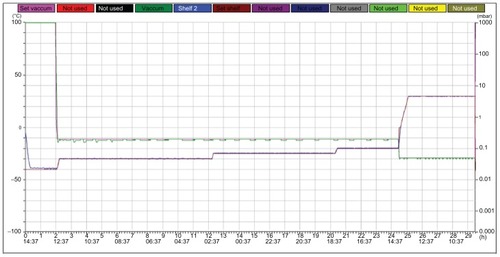
Mannitol (10%) was used as the cryoprotecting agent, and FMO-SLNs in suspension were frozen in a deep freezer (Christ Epsilon 2–4 LSC, Martin Christ Gefriertrocknungsanlagen GmbH, Osterode am Harz, Germany) according to the freeze curve shown in . The FMO-SLN powder was then obtained and stored at 4°C for DSC and XRD analysis.
Figure 1 Freeze-drying curves of FMO-SLNs.
Abbreviation: FMO-SLNs, frankincense and myrrh oil–solid lipid nanoparticles.
Characterization of FMO-SLNs
Particle size and ZP
The size and ZP of FMO-SLNs were measured using a Nano ZS90 Zetasizer (Malvern Instruments Ltd, Worcestershire, UK).
EE and LC
Free FMO (nonentrapped in the FMO-SLNs) was separated by an ultrafiltration method.Citation15 OA (21.06% in FMO) and β-E (2.58% in FMO) were selected as the indexed components. Centrifugal filter tubes (molecular weight cut-off = 10 kDa, Nanosep®; Pall Corporation, Port Washington, NY) were used to determine the EE. After a suitable dilution, FMO-SLNs (0.5 mL) were placed in the upper chamber of a centrifugal filter tube and then centrifuged at 5000 rpm for 15 minutes. The separated part collected at the bottom of the tube was subjected to GC analysis to determine the OA and β-E content. The total drug content in FMO-SLNs was determined after extraction with dehydrated alcohol in an ultrasonic bath. Drug loading capacity was presented as percent entrapped drug to lipid ratio. Encapsulation efficiency could be calculated by the following equations:
where WTotal OA and WTotal β-E are the weights of OA and β-E in SLNs, respectively, and WFree OA and WFree β-E are the mean weights of untrapped OA and β-E, respectively.
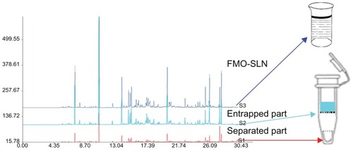
FMO is composed of hundreds, or even thousands, of components with different molecular structures. The composition of FMO is so complex that the use of only two indexed components did not facilitate comprehensive assessment of the EE of FMO-SLNs. Thus, the FS between the total FMO in SLNs and the entrapped FMO in SLNs (stopped by the filter membrane) was determined to evaluate the EE from a macroscopic point of view. The fingerprint of FMO was obtained using the established GC method mentioned in the section discussing gas chromatography spectrometry analysis, and the method of operation is shown in .
Figure 2 Schematic diagram for evaluation of the EE of FMO-SLNs.
Abbreviations: EE, encapsulation efficiency; FMO-SLNs, frankincense and myrrh oil–solid lipid nanoparticles.
TEM
The morphology of FMO-SLNs was observed using a transmission scanning microscope (JEM-1230; JEOL, Tokyo, Japan). After a suitable dilution was created, samples were placed on a film-coated copper grid. Thereafter, a drop of 2% phosphotungstic acid was added to the film and allowed to dry for 10 minutes before observation.
DSC
DSC was performed using a differential scanning calorimeter (Shimadzu DSC-60; Shimadzu Corporation, Kyoto, Japan). A heating rate of 10°C/minute was used, and the temperature range was 20°C–100°C. An empty aluminum pan was used for reference.
XRD
Crystalline structures of the FMO-SLN powder were assessed with an X-ray diffractometer (Rigaku Corporation, Tokyo, Japan). A Cu-Kα radiation source was used, and samples were scanned over a 2θ range of 2°–50° with a scanning rate of 5°/minute.
In vitro evaporation release
To assess the chemical stability of components from unformulated and formulated FMO, samples were placed in open vials and stored at 35°C for 6 days. During this time, essential oil was extracted from the samples by immersing them in dehydrated alcohol for 30 minutes in an ultrasonic bath and the selected components were analyzed using the established GC method. Three groups were used: unformulated FMO, FMO-β-CD, and FMO-SLN. FMO-β-CD comprised the β-CD inclusion compounds of FMO. FMO-β-CD was prepared using a coprecipitation method at a β-CD/FMO molar ratio of 3:1.Citation16
In vivo antitumor activity
H22-bearing Kunming mice were used as a model to assess the in vivo antitumor activity of FMO.Citation17,Citation18 H22 cells were injected and incubated in the abdominal cavity of mice for 6–8 days before they were collected and resuspended in PBS. The collected H22 cells (2 × 10⁶/mL) were then inoculated subcutaneously into the right forelimb of each mouse at a concentration of 0.2 mL. After injection, the H22-bearing mice were randomly divided into six groups (ten mice/group) that were administered one of the following treatments: saline (control group), 5-FU (reference drug), FMO suspension (prepared with 5% Tween 80), FMO-β-CD, blank SLN, or FMO-SLN. 5-FU was administered via intraperitoneal injection once every 2 days at a dose of 25 mg/kg body weight. All other drugs were administered orally for 10 consecutive days (saline, 0.2 mL/mouse; FMO suspension, FMO-β-CD, blank SLN, and FMO-SLN, 100 mg/kg body weight). After 10 days, the mice were killed, and tumors, spleens, and thymi were excised. The tumor growth inhibition rate was calculated using the following formula: tumor inhibitory rate (%) = (C − T)/C × 100%, where “C” and “T” are the average tumor weights in the control and test groups, respectively. The spleen and thymus indices were calculated as follows: spleen/thymus index = WS/T/WM, where WS/T is the weight of the spleen or thymus and WM is the weight of the mouse.
Statistical analysis
Data are expressed as the mean ± standard deviation and analyzed using one-way ANOVA. P-values less than 0.05 were considered significant.
Results and discussion
Extraction and GC analysis
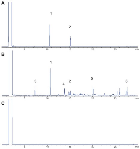
A green essential oil (FMO) was obtained at a good yield of 0.83% by distillation. The established gas chromatograms are shown in , which indicates that the selected indexed components (from peaks 1 to 6) could be separated under the GC condition, and that the pharmaceutical adjuvants (including Compritol 888 ATO, Tween 80, and soybean lecithin) did not influence the analysis. The gas chromatograms of FMO revealed that FMO is a complex mixture of organic compounds. Among the marked peaks, peaks 1 and 2 correspond to OA and β-E, respectively. OA is present at the highest concentration (21.06%) in FMO, and β-E (2.58%) exerts a favorable antitumor activity;Citation19–Citation21 therefore, these compounds were chosen as the indexed components. Peaks 1 to 6 were used to assess the in vitro evaporation release of unformulated and formulated FMO.
Figure 3 GC of a standard sample (A), FMO-SLN (B), and blank SLN (C); peaks 1 and 2 correspond to OA and β-E, respectively.
Abbreviations: FMO-SLNs, frankincense and myrrh oil–solid lipid nanoparticles; GC, gas chromatography; OA, octyl acetate; β-E, β-elemene.
Preparation of FMO-SLNs
Production techniques for SLNs include high-pressure homogenization,Citation22 microemulsion,Citation23 an ultrasound-based method,Citation24 and solvent emulsification/evaporation.Citation25,Citation26 In the present study, SLNs were prepared by high-pressure homogenization. High-pressure homogenization is a reliable and powerful technique for the preparation of SLNs, and it has been utilized on a large scale in the pharmaceutical industry. In our preliminary study, FMO was found to have high solubility in different lipids, such as Compritol 888 ATO, Precirol ATO 5, glycerin monostearate, and stearin. However, a high EE was obtained using Compritol 888 ATO as the lipid. A mixture of soybean lecithin and Tween 80 was used as the surfactant. Other details of the SLN formulations are given in .
Table 1 Sample composition (%) of FMO-SLNs (w/w)
Characterization of FMO-SLNs
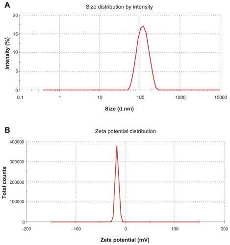
shows the typical particle size distribution of FMO-SLNs. As revealed in this figure, FMO-SLNs were found to be nanometric and unimodal with a relatively narrow size distribution that ranged from 43.8 to 219.5 nm. The average diameter was 113.3 ± 3.6 nm, and the polydispersity index value was 0.25. ZP is also an important surface characteristic of SLNs, and the measurement of ZP allows predictions of the storage stability of SLNs.Citation27 As revealed in , the FMO-SLNs possessed a high ZP with the value of −16.8 ± 0.4 mV, indicating that the surface of the FMO-SLNs was negatively charged.
Figure 4 Particle size (A) and ZP (B) of FMO-SLNs.
Abbreviations: FMO-SLNs, frankincense and myrrh oil–solid lipid nanoparticles; ZP, zeta potential.
EE is a very important characteristic for judging the quality of SLNs. Generally, the methods of assessing EE include ultracentrifugation,Citation28 Sephadex minicolumn centrifugation,Citation29 dialysis,Citation30 and the ultrafiltration method used in the present study. As mentioned in the section on extraction and GC analysis, FMO is a complex mixture of abundant compounds. Therefore, two components were selected as indexed components to evaluate the EE from a microscopic point of view, and the FS between the total FMO in SLNs and the entrapped FMO in SLNs was determined to evaluate the EE from a macroscopic point of view. As is shown in , a high incorporation capacity of SLNs was observed because of the high lipophilicity of FMO and its good compatibility with Compritol 888 ATO. The FS was 98.68% ± 0.57%, indicating that the most abundant components were entrapped by the SLNs. The results of the present study confirm those of previous work, indicating that hydrophobic drugs could be incorporated well into lipid carriers with high EE.Citation31
Table 2 EE and LC of SLNs (n = 3)
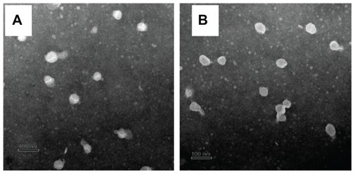
The morphologies of the blank SLNs and FMO-SLNs were observed by TEM. As shown in , most of the particles were round and uniform in size. No significant difference was found between the blank SLNs and FMO-SLNs.
Figure 5 TEM images of the blank SLNs (A) and FMO-SLNs (B).
Abbreviations: FMO-SLNs, frankincense and myrrh oil–solid lipid nanoparticles; TEM, transmission electron microscopy.
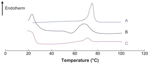
As has been reported previously,Citation32,Citation33 DSC can be used to determine thermodynamic variations related to morphological changes, because different lipid modifications possess different melting points and melting enthalpies. In the present study, DSC was performed to investigate the melting and crystallization behavior of the lipid matrices to detect whether these characteristics were changed by SLN preparation. As shown in , the bulk lipid (Compritol 888 ATO) displayed a melting point of 75°C. The physical mixture and SLN heating curves differed distinctly from those of the bulk lipid, and shifts of the melting point of the physical mixture and SLNs to 68.3°C and 70.9°C, respectively, were observed. The reduction of the melting point of the physical mixture was likely caused by the low-boiling-point components in FMO. presents the melting points, enthalpy, and crystallinity of the samples. The enthalpy and crystallinity of FMO-SLNs were lower than those of the initial lipid, indicating an increased number of lattice defects in FMO-SLNs.Citation34 The results indicate that the perfect crystal structure of Compritol 888 ATO was changed by SLN preparation, and a new less-ordered phase was formed.
Table 3 Melting points, enthalpy, and crystallinity of each sample
Figure 6 DSC curves of (A) Compritol 888 ATO, (B) physical mixture, and (C) FMO-SLNs.
Abbreviations: DSC, differential scanning calorimetry; FMO-SLNs, frankincense and myrrh oil–solid lipid nanoparticles.
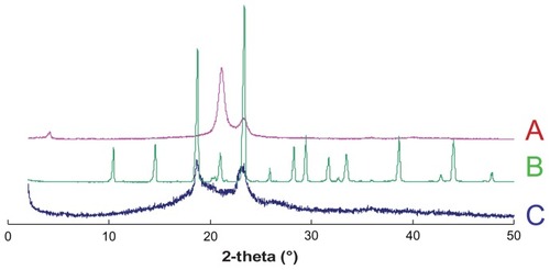
As a further microstructure investigation of FMO-SLNs, XRD was performed to confirm the findings obtained by DSC. The X-ray patterns of Compritol 888 ATO, mannitol, and FMO-SLNs are shown in . As indicated in , the bulk Compritol 888 ATO displayed three peaks at positions 2θ = 4.16°, 21.16°, and 23.30°, while shows that the peaks at 2θ = 4.16° and 21.16° disappeared. In two blunt peaks appeared that were likely influenced by the cryoprotectant (mannitol), which has a high degree of crystallinity (). From these results, it was confirmed that Compritol 888 ATO exhibited a less-ordered structure in SLNs.
Figure 7 X-ray spectra of (A) Compritol 888 ATO, (B) mannitol, and (C) FMO-SLNs.
Abbreviation: FMO-SLNs, frankincense and myrrh oil–solid lipid nanoparticles.
In vitro evaporation release
Generally, essential oils are unstable when exposed to air, light, and high temperature, which may result in the rapid evaporation and degradation of some active components and further lead to a dramatic decrease in activity.Citation35 Several studies have demonstrated that the incorporation of essential oils into SLNs helps to increase their stability.Citation36,Citation37 To investigate the capability of SLNs to prevent the rapid evaporation of FMO, six components that possess different boiling points according to the established GC spectrum were chosen as the indexed components. β-CD inclusion is the most common technique to incorporate essential oils, and thus, in the present study, the evaporation release behavior of FMO-β-CD was determined as a control. The data for the in vitro evaporation release of unformulated FMO, FMO-β-CD, and FMO-SLNs are presented in . clearly shows the rapid evaporation loss of all six selected components from the initial FMO, with evaporation beginning on the second day with large losses of more than 50% seen after 6 days of storage (excluding component 4). Compared with the initial FMO, FMO-β-CD () and FMO-SLN () exhibited a considerable decrease in evaporation loss, and no component exhibited an evaporation loss exceeding 50% after 6 days of storage, indicating that both β-CD inclusion and SLN incorporation can prevent the evaporation loss of components in FMO.
Table 4 Evaporation release of components
In vivo antitumor activity
H22 tumor-bearing mice were used as an animal model to assess whether FMO-SLNs exhibit antitumor activity. As shown in , neither saline nor blank SLN had any measurable effect on tumor growth, indicating that the SLN vehicle had no influence on antitumor activity. Compared with the findings for the saline group, significant antitumor effects (P < 0.01) were observed for the FMO suspension, FMO-β-CD, and FMO-SLNs. The tumors in these three groups were considerably smaller than those in the saline group. The inhibition rates of the FMO suspension, FMO- β-CD, and FMO-SLNs (at a concentration of 100 mg/kg) were 31.23%, 34.81%, and 43.66%, respectively. These results indicate that the inhibition rate of FMO-β-CD against tumor growth was similar to that of the FMO suspension (P > 0.05), and that the slight enhancement of the inhibition rate may be attributable to the increased dissolution of FMO by β-CD inclusion. These findings also suggest that the antitumor efficacy of the FMO-SLNs was significantly higher (P < 0.05) than that of the FMO suspension and FMO-β-CD at the same dosage. These results confirmed that SLNs represent a better delivery system for antitumor therapy.
Table 5 In vivo antitumor effects in H22-bearing mice (n = 10)
Changes in the body weight of H22-bearing mice over the course of the study are also presented in . The weight of the mice in the saline and blank SLN groups increased rapidly, which may have been caused by tumor growth. The rates of the weight increase in the 5-FU and FMO-SLN groups were lower than those in the saline group.
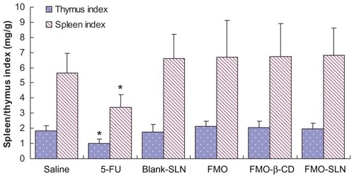
The spleen and thymus are the two primary immune organs related to antitumor activity.Citation38 As shown in , the 5-FU group exhibited significant reductions (P < 0.05) in both the spleen and thymus indices as compared to the saline group. Slight enhancements of these indices were observed in the FMO suspension, FMO-β-CD, and FMO-SLN groups, indicating that FMO could enhance the immune response in tumor-bearing mice. The results confirmed the previous findings that traditional Chinese medicines can exert antitumor effects on the immune system.Citation39
Figure 8 Evaluation of the thymus and spleen indices in tumor-bearing mice.
Note: *P < 0.05 compared with the saline group.
Numerous investigations have shown that SLNs can increase the antitumor efficacy of drugs while reducing their systemic side effects.Citation40 This may be due to the controlled release behavior and targetable distribution of SLNs. A future study will focus on the organ distribution of FMO-SLNs.
Conclusion
In this study, FMO-SLNs were successfully prepared by high-pressure homogenization. Compritol 888 ATO showed reasonable FMO solubilization capacity. The poorly water-soluble drug FMO was efficiently encapsulated into the nanoparticles. Particles prepared under proper formulation conditions were spherical with diameters of <220 nm. A high EE was obtained using the described method. Physicochemical characterization revealed that efficiently prepared SLNs encapsulated the drug. The drug evaporation release study showed that SLN incorporation could prevent the evaporation loss of FMO components to a desirable degree. Furthermore, FMO-SLNs possess significantly higher antitumor efficacy. Hence, the developed SLNs can be used to increase the stability and/or to improve the in vivo antitumor efficacy of FMO. However, further studies should focus on the absorption and distribution of the FMO-SLNs.
Acknowledgments
This work was financially supported by the Subject Chief Scientist Program (10XD14303900) from the Science and Technology Commission of Shanghai Municipality, and the Program (NCET08-0898) for New Century Excellent Talents and Program (No IRT1071) for Changjiang Scholars and Innovative Research Team in Universities from the State Education Ministry, PR China.
The authors gratefully acknowledge Dr Zhenghai Zhang of Jiangsu Provincial Academy of Chinese Medicine for his helpful suggestions.
Disclosure
The authors report no conflicts of interest in this work.
References
- FrankAUngerMAnalysis of frankincense from various Boswellia species with inhibitory activity on human drug metabolising cytochrome P450 enzymes using liquid chromatography mass spectrometry after automated on-line extractionJ Chromatogr A200611121–225526216364338
- MassoudAEl-SisiSSalamaOMassoudAPreliminary study of therapeutic efficacy of a new fasciolocidal drug derived from Commiphora molmol (myrrh)Am J Trop Med Hyg2001652969911508399
- DöhlingCBoswellia serrata (Frankincense)-from traditional Indian medicine (Ayurveda) to evidence-based medicinePhytomedicine2008156540
- Van VuurenSFKamatouGPPViljoenAMVolatile composition and antimicrobial activity of twenty commercial frankincense essential oil samplesS Afr J Bot2010764686691
- TiptonDALyleBBabichHDabbousMKhIn vitro cytotoxic and anti-inflammatory effects of myrrh oil on human gingival fibroblasts and epithelial cellsToxicol in Vitro200317330131012781209
- AshryKMEl-SayedYSKhamissRMEl-AshmawyIMOxidative stress and immunotoxic effects of lead and their amelioration with myrrh (Commiphora molmol) emulsionFood Chem Toxicol201048123624119818824
- PillmoorJBWrightKTerryASNatural products as a source of agrochemicals and leads for chemical synthesisPestic Sci1993392131140
- ZhaoXLYangCRYangKLLiKXHuHYChenDWPreparation and characterization of nanostructured lipid carriers loaded traditional Chinese medicine, zedoary turmeric oilDrug Dev Ind Pharm201036777378020136496
- YaoGLiYPreparation, characterization and evaluation of self-microemulsifying drug delivery systems (SMEDDSs) of Ligusticum chuanxiong oilBiomed Pharmacother2011113642
- SoutoEBMüllerRHLipid nanoparticles: effect on bioavailability and pharmacokinetic changesHandb Exp Pharmacol201019711514120217528
- MüllerRHMäderKGohlaSSolid lipid nanoparticles (SLN) for controlled drug delivery – a review of the state of the artEur J Pharm Biopharm200050116117710840199
- SharmaMAgrawalSKSharmaPRCytotoxic and apoptotic activity of essential oil from Ocimum viride towards COLO 205 cellsFood Chem Toxicol201048133634419852999
- SilvaACGonzález-MiraEGarcíaMLPreparation, characterization and biocompatibility studies on risperidone-loaded solid lipid nanoparticles (SLN): high pressure homogenization versus ultrasoundColloids Surf B Biointerfaces201186115816521530187
- RidolfiDMMarcatoPDJustoGZCordiLMachadoDDuránNChitosan-solid lipid nanoparticles as carriers for topical delivery of tretinoinColloids Surf B Biointerfaces201293364022244299
- LiuYWangPFSunCWheat germ agglutinin-grafted lipid nanoparticles: Preparation and in vitro evaluation of the association with Caco-2 monolayersInt J Pharm20103971–215516320599600
- LiXHJinZYWangJComplexation of allyl isothiocyanate by α- and β-cyclodextrin and its controlled release characteristicsFood Chem20071032461466
- ZhangJWangXLuHAmifostine increases cure rate of cisplatin on ascites hepatoma 22 via selectively protecting renal thioredoxin reductaseCancer Lett20082601–212713618039557
- LouHGaoLWeiXOridonin nanosuspension enhances antitumor efficacy in SMMC-7721 cells and H22 tumor bearing miceColloids Surf B Biointerfaces201187231932521676599
- YaoYQDingXJiaYCHuangCXWangYZXuYHAnti-tumor effect of beta-elemene in glioblastoma cells depends on p38 MAPK activationCancer Lett2008264112713418442668
- SunYHLiuGFZhangYQZhuHRenYShenYMSynthesis and in vitro anti-proliferative activity of β-elemene monosubstituted derivatives in HeLa cells mediated through arrest of cell cycle at the G1 phaseBioorg Med Chem20091731118112419128976
- XuLYTaoSJWangXMThe synthesis and anti-proliferative effects of beta-elemene derivatives with mTOR inhibition activityBioorg Med Chem200614155351535616617020
- MüllerRHMehnertWLucksJSSolid lipid nanoparticles (SLN) – an alternative colloidal carrier system for controlled drug deliveryEur J Pharm Biopharm19954116269
- GascoMRSolid lipid nanospheres from warm micro-emulsionsPharm Technol Eur19979115258
- SpeiserPLipidnanopellets als tragersystem fur Arzneimittel zur peroralen anwendungEuropean Patent EP 016782511141989
- SjöstörmBBergenståhlBPreparation of submicron drug particles in lecithin-stabilized o/w emulsions. I. Model studies of the precipitation of cholesteryl acetateInt J Pharm1992842107116
- SiekmannBWestesenKInvestigations on solid lipid nanoparticles prepared by precipitation in o/w emulsionsEur J Pharm Biopharm1996422104109
- MüllerRHZetapotential und Partikelladung in der LaborpraxisStuttgart, GermanyWissenschaftliche Verlagsgesellschaft1996
- BhalekarMRPokharkarVMadgulkarAPatilNPatilNPreparation and evaluation of miconazole nitrate-loaded solid lipid nanoparticles for topical deliveryAAPS Pharm Sci Tech2009101289296
- HouDXieCHuangKZhuCThe production and characteristics of solid lipid nanoparticles (SLNs)Biomaterials200324101781178512593960
- ElsieOTiwariSBUdupaNNiosome entrapped β-cyclodextrin methotrexate complex as a drug delivery systemIndian J Pharmacol1999314279284
- LiHZhaoXMaYZhaiGLiLLouHEnhancement of gastrointestinal absorption of quercetin by solid lipid nanoparticlesJ Control Release2009133323824418951932
- CastelliFPugliaCSarpietroMGRizzaLBoninaFCharacterization of indomethacin-loaded lipid nanoparticles by differential scanning calorimetryInt J Pharm20053041–223123816188405
- HuFQJiangSPDuYZYuanHYeYQZengSPreparation and characteristics of monostearin nanostructured lipid carriersInt J Pharm20063141838916563671
- HouDZXieCSHuangKJZhuCThe production and characteristics of solid lipid nanoparticles (SLNs)Biomaterials200324101781178512593960
- MogulMGAkinHHasirciNControlled release of biologically active agents for purposes of agricultural crop managementResources, Conservation and Recycling1996161–4289320
- WissingSAMäderKMüllerRHProlonged efficacy of the insect repellent lemon oil by incorporation into solid lipid nanoparticles (SLN)Third World Meeting APGI/APVMainz, GermanyAPV2000439440
- WissingSAMäderKMüllerRHSolid lipid nanoparticles (SLN) as a novel carrier system offering prolonged release of the perfume Allure (Chanel)Proceedings of the International Symposium on Controlled Release of Bioactive MaterialsParis, France2000311312
- GaoZZhangDGuoCPaclitaxel efficacy is increased by parthenolide via nuclear factor-kappaB pathways in in vitro and in vivo human non-small cell lung cancer modelsCurr Cancer Drug Targets201010770571520578985
- FengLJiaXBShiFChenYIdentification of two polysaccharides from Prunella vulgaris L. and evaluation on their anti-lung adenocarcinoma activityMolecules20101585093510320714287
- ZhangPChenLZhangZLinLLiYPharmacokinetics in rats and efficacy in murine ovarian cancer model for solid lipid nanoparticles loading docetaxelJ Nanosci Nanotechnol201010117541754421137978