
Loutfy SA, Elberry MH, Farroh KY, et al. Int J Nanomedicine. 2020;15:2699–2715.
The authors have advised on page 2712 is incorrect. The correct is shown below.
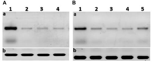
Figure 8 (A) The scanned densitometry Western blot of viral replication in Huh7 (a) versus β-actin (b); lane 1, protein levels of infected untreated cells: lane 2, infected cells treated with curcumin: lane 3, infected cells treated with CsNPs: lane 4, infected cells treated with curcumin chitosan nanocomposite. (B) The scanned densitometry western blot of viral entry (a) versus β-actin (b) protein levels in positive; lane 1, untreated infected cells: lane 2, cells treated with curcumin: lane 3, cells treated with CsNPs: lane 4, cells treated with curcumin chitosan nanocomposite: lane 5, cells treated with sofosbuvir. HCV core protein at size of 22 KD.
The authors apologize for this error and advise it does not affect the results of the paper