This article refers to:
Duan X, Lu L, Wang Y, et al. Int J Nanomedicine. 2017;12:6705–6719.
The authors have advised that , and on pages 6713, 6715 and 6716, respectively, of the published paper are incorrect. The errors were introduced by the Editorial staff during the publication process. The correct figures are as follows.
Figure 6 Apoptosis and survival of GFP-MSCs at implantation site.
Notes: TUNEL analysis shows that apoptotic cells peaked at 1 week, and then decreased over time in animals grafted with labeled GFP-MSCs (A), unlabeled GFP-MSCs (B), and PBS (C). Graph show the percentages of TUNEL-positive cells in animals grafted with labeled and unlabeled GFP-MSCs (D). Graph shows the number of apoptotic GFP-MSCs in animals grafted with labeled and unlabeled GFP-MSCs (E). Graph shows the percentage of viable GFP-positive cells in animals grafted with labeled and unlabeled GFP-MSCs (F). *p<0.05 between labeled GFP-MSCs and PBS; #p<0.05 between unlabeled GFP-MSCs and PBS; Bar =15 μm.
Abbreviations: MSC, mesenchymal stem cell; PBS, phosphate-buffered saline.
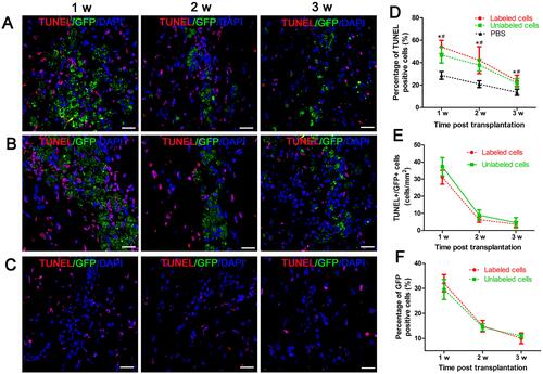
Figure 8 Histology of the grafted cells in the injection site.
Notes: Fluorescence immunostaining micrographs show that GFP-MSCs remained in the injection site in animals grafted with polymersome-labeled GFP-MSCs and unlabeled GFP-MSCs. Only a few surviving GFP-MSCs differentiated into GFAP-positive astrocytes (arrows), but not into NeuN-positive neurons. A large number of cells were phagocytized by macrophages (arrows) (A). Graphs show the numbers of GFAP+GFP+ (B), CD11b+GFP+ (C), and the percentage of GFP-MSCs (D) in animals grafted with polymersome-labeled GFP-MSCs and unlabeled GFP-MSCs. Bar =15 μm.
Abbreviation: MSC, mesenchymal stem cell.
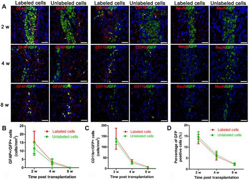
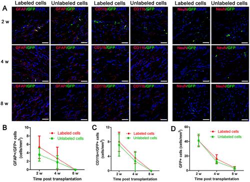
Figure 9 Histology of migrating cells in the corpus callosum.
Notes: Fluorescence immunostaining micrographs show that a small amount of GFP-MSCs migrated to the corpus callosum in animals treated with polymersome-labeled cells and unlabeled cells. Only a few surviving GFP-MSCs were differentiated into GFAP-positive astrocytes (arrows) and were phagocytized by macrophages (arrows), but no cells differentiated into NeuN-positive neurons (A). Graphs show the numbers of GFAP+GFP+ (B), CD11b+GFP+ (C), and GFP-MSCs (D) in animals grafted with polymersome-labeled GFP-MSCs and unlabeled GFP-MSCs. Bar =15 μm.
Abbreviation: MSC, mesenchymal stem cell.