 ?Mathematical formulae have been encoded as MathML and are displayed in this HTML version using MathJax in order to improve their display. Uncheck the box to turn MathJax off. This feature requires Javascript. Click on a formula to zoom.
Abstract
Background
Resveratrol, like other natural polyphenols, is an extremely photosensitive compound with low chemical stability, which limits the therapeutic application of its beneficial effects. The development of innovative formulation strategies, able to overcome physicochemical and pharmacokinetic limitations of this compound, may be achieved via suitable carriers able to associate controlled release and protection. In this context, nanotechnology is proving to be a powerful strategy. In this study, we developed novel cationic chitosan (CS)- and anionic alginate (Alg)-coated poly(d,l-lactide-co-glycolide) nanoparticles (NPs) loaded with the bioactive polyphenolic trans-(E)-resveratrol (RSV) for biomedical applications.
Methods
NPs were prepared by the nanoprecipitation method and characterized in terms of morphology, size and zeta potential, encapsulation efficiency, Raman spectroscopy, swelling properties, differential scanning calorimetry, and in vitro release studies. The protective effect of the nanosystems under the light-stressed RSV and long-term stability were investigated.
Results
NPs turned out to be spherical in shape, with size ranging from 135 to about 580 nm, depending on the composition and the amount of polyelectrolytes, while the encapsulation efficiencies increased from 8% of uncoated poly(d,l-lactide-co-glycolide) (PLGA) to 23% and 32% of Alg- and CS-coated PLGA NPs, respectively. All nanocarriers are characterized by a biphasic release pattern, and more effective controlled release rates are obtained for NPs formulated with higher polyelectrolyte concentrations. Stability studies revealed that encapsulation provides significant protection against light-exposure degradation, by reducing the trans–cis photoisomerization reaction. Moreover, the nanosystems are able to prevent the degradation of trans isoform and the leakage of RSV from the carrier for a period of 6 months.
Conclusion
Our findings indicated that the newly developed CS- and Alg-coated PLGA NPs are suitable to be used for the delivery of bioactive RSV. The encapsulation of RSV into optimized polymeric NPs provides improved drug loading, effective controlled release, and protection against light-exposure degradation, thus opening new perspectives for the delivery of bioactive related phytochemicals to be used for (nano)chemoprevention/chemotherapy.
Introduction
The research and application of natural polyphenols have recently attracted great interest in functional food, nutraceutical, and pharmaceutical companies, due to their potential health benefits.Citation1–Citation3 For example, chemoprevention by using natural products has emerged as an important strategy, with potential to prevent the occurrence of cancer by inhibition, reversing, or retarding the carcinogenesis process.Citation4–Citation6 Among the polyphenolic compounds, the bioactive phytoalexin trans-(E)-resveratrol (RSV), found in red wine, legumes, berries, peanuts, and pistachios,Citation7 has gained considerable attention as a preventive agent of several important pathologies, such as neurodegenerative processes, viral infections, vascular diseases, and cancers.Citation8–Citation12 RSV has demonstrated potential cancer chemopreventive properties by suppressing the proliferation process at the initiation, promotion, and progression stages of several cancer cell lines, such as lymphocytes, breast, glia, and skin and prostate tumor cells.Citation12–Citation14
Despite promising results in preclinical settings, the extensive use of RSV has met only limited success, largely due to its instability,Citation15 poor solubility,Citation16 inefficient systemic delivery, and low bioavailability.Citation17 RSV is more biologically active compared to its cis (Z)-isoform, and therefore requires the formulation of a finished protecting product, able to retain the structural integrity of the bioactive molecules until consumption or administration.Citation18
In this context, the encapsulation of RSV into polymeric or lipid-based matrices is a major challenge, and nanotechnology represents a powerful strategy.Citation19 This approach provides prototypes as carriers/vehicles able to offer a greater surface area and the potential to increase solubility, stability, bioavailability, and controlled release as well as targeted delivery of the encapsulated active agents.Citation20–Citation24 Moreover, nanoparticles (NPs) tend to extravasate passively through the leaky vasculature that is characteristic of solid tumors (angiogenic blood vessels in tumor tissues have gaps as large as about 400–800 nm), and preferentially accumulate through the enhanced permeability and retention (EPR) effect.Citation25,Citation26 The increased drug concentration near the cell membrane generates a concentration gradient that promotes drug influx into the cell. Additionally, NPs are taken up by cells through an endocytosis pathway, thus resulting in a higher cellular uptake of the entrapped drug, thereby enabling them to escape from the effect of P-glycoprotein pumps.Citation27
More recently, Siddiqui and Mukhtar introduced a novel concept of “nanochemoprevention” where nanotechnology was exploited to augment the outcome of chemoprevention.Citation23,Citation27,Citation28 This proof-of-principle study demonstrated the usefulness of nanoparticulate technology to enhance the efficacy of natural agents, such as (−)-epigallocatechin 3-gallate (EGCG), loaded in polylactic acid–polyethylene glycol (PLA-PEG) NPs in the preclinical setting.Citation23
Encapsulation of EGCG as well as related natural polyphenols (ie, curcumin) into NPs increased their inhibitory effect on cancer cell proliferation, also improving their pharmacokinetic properties and bioavailability.Citation29–Citation31
Several studies have also been reported on the use of nanotechnology to vehicle RSV. In particular, RSV incorporation into mPEG-poly(ɛ-caprolactone)–based NPs resulted in significantly higher cytotoxicity against malignant glioma cells compared to an equivalent dose of free biomolecule.Citation32
Solid-lipid NPs loaded with RSV also contributed to effectiveness of RSV on decreasing cell proliferation and demonstrated potential benefits for prevention of skin cancer.Citation33 Moreover, other authors investigated the effect of RSV incorporated in liposome on the proliferation and ultraviolet B (UVB) protection of cells, in order to enhance the efficacy in the prevention and treatment of human skin disorders caused by excessive exposure to UV radiation.Citation34 Among the polymeric materials, poly(d,l-lactide-co-glycolide) (PLGA) is a widely used copolymer approved by the FDA for various medical and pharmaceutical applications, such as drug delivery.Citation35–Citation37 The combination of PLGA as a hydrophobic polymer and many natural hydrophilic biopolymers like gelatinCitation38 or sodium alginate (Alg) provides advantages for both the hydrophilic and the hydrophobic nanoparticulate systems.Citation36,Citation39 Chitosan (CS) and Alg are two major naturally occurring polysaccharides with hydrophilic characteristics that have been gaining increasing interest in the biomedical field, and particularly in the drug-delivery area.Citation39 CS is a polycationic polymer that has one amino group and two hydroxylic functionalities in the repeating glycosidic residue.Citation40
On the other hand, Alg is an anionic polymer extracted from brown algae, and bears carboxylic acid groups that may introduce negative charge to the polymer at appropriate pH values.Citation41 More recently, these polymers have been used as coating materials to make polyelectrolyte multilayer–charged PLGA NPs,Citation42 and to achieve cohesive colloidal gels as scaffold in tissue engineering,Citation43 such as to construct composite microspheres.Citation44
It is a consistent opinion that the future for efficient RSV delivery lies in the development of innovative formulation strategies able to overcome each of the physicochemical, pharmacokinetic, and metabolic limitations that characterize this compound.Citation19
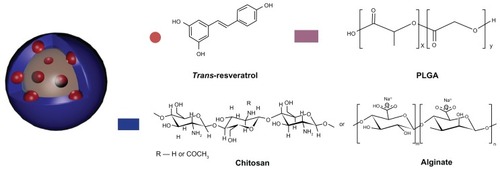
In this study, we examined the feasibility of encapsulating RSV into PLGA NPs coated on the surface with cationic and anionic polymers (), in order to obtain suitable drug carriers able to combine protection and controlled release. The NPs were prepared by a solvent-displacement method using different amounts of CS and Alg. The influence of coating composition on morphology, size and zeta potential, encapsulation efficiency, swelling capacity and in vitro release was also evaluated. Moreover, the protective effect of the NPs under the light stress of encapsulated RSV in comparison with free compound and long-term stability of nanosystems were investigated and are herein detailed.
Figure 1 Chemical structure of RSV, PLGA, CS, and Alg, and schematic representation of the nanoprototypes.
Abbreviations: RSV, trans-(E)-resveratrol; PLGA, poly(d,l-lactide-co-glycolide); CS, chitosan; Alg, alginate.
Materials and methods
Chemicals and reagents
Poly(d,l-lactide-co-glycolide) (lactide:glycolide = 75:25 molar ratio, inherent viscosity 0.14–0.22 dL/g, acid terminated, molecular weight 4000–15,000), CS (low molecular weight, 75%–85% deacetylated chitin, viscosity 200–800 cP, 1% in 1% acetic acid), alginic acid sodium salt (Alg, medium viscosity ≥ 200 cP, 2%), and RSV were purchased from Sigma- Aldrich (Steinheim, Germany). All other chemicals were analytical grade and were used without further purification.
Formulation of NPs
RSV-loaded NPs were prepared by a nanoprecipitation method as previously described,Citation42 with minor modification. Briefly, the PLGA polymer and RSV dissolved in 10 mL acetone were added dropwise into a CS solution (0.1%, 0.25%, and 0.5% w/v acetic acid solution in 0.1% v/v, 5 mL) or Alg (0.1, 0.25, and 0.5% w/v in distilled water, 5 mL) under gentle stirring. The set of NPs and their composition is reported in .
Table 1 Theoretical composition of RSV-loaded NPs
The resulting milky colloidal suspension was evaporated at room temperature to remove the organic solvent. NPs were collected by centrifugation at 13,000 rpm for 10 minutes and washed with water to remove the unencapsulated RSV and residual CS or Alg. PLGA NPs were prepared by adding the polymer and RSV organic solution to water. The obtained NPs were lyophilized for further characterization and utilization.
Morphology, particle-size analysis and zeta potential
The morphology of NPs was characterized by scanning electron microscopy (SEM) (model DSM 962; Carl Zeiss, Jena, Germany). A drop of NP suspension was placed on an aluminum stub and dried under vacuum for 12 hours. The samples were then analyzed at 20 kV acceleration voltage after gold sputtering, under an argon atmosphere.
Particle size and size distribution of NPs were determined by photon correlation spectroscopy using a Malvern Zetasizer Nano ZS (Malvern Instruments, Malvern, UK), at a temperature of 25°C and a scattering angle of 90°.
Zeta potential of the NPs was measured at 25°C with a Zeta Plus zeta potential analyzer (Brookhaven Instruments, Holtsville, NY). The samples were diluted with distilled water and sonicated for several minutes before measurement. Data were obtained by the averaging of three measurements.
Raman spectroscopy
The chemical composition of the RSV, CS, and Alg (raw materials), and PLGA, PLGA-CS, and PLGA-Alg dried NPs was analyzed by Raman spectroscopy (Senterra Raman microscope; Bruker Optics, Ettlingen, Germany) using an excitation wavelength of 532 nm at 5 mW. Raman spectra were recorded by averaging five acquisitions of 5 seconds with a 50× objective, in the range 180–3200 cm−1.
Differential scanning calorimetry
Differential scanning calorimetry (DSC) scans of empty and drug-loaded nanoparticles were performed on a DSC Q100 V 9.0 calorimeter (TA Instruments, New Castle, DE). Indium was used to calibrate the instrument. The thermograms of samples were obtained at a scanning rate of 10°C/minute in a 30°C–500°C temperature range and performed under an Ar purge (50 mL/minute). The thermal measurements were carried out on pure RSV, PLGA, CS, Alg, on drug-loaded PLGA-, PLGA-CS–, and PLGA-Alg–coated NPs, and on the physical mixture of PLGA/RSV, PLGA/CS/RSV, and PLGA/Alg/RSV.
Drug-loading content, encapsulation efficiency, and yield of production
The amount of RSV encapsulated was determined by dissolving an aliquot of NPs (1.0 mg) in 0.1 mL of methylene chloride. The organic solvent was evaporated and the residue dissolved in 0.1 mL of mobile phase A:B (21:79 v/v), where solvent A was trifluoroacetic acid (TFA) in water (0.1/99.9 v/v) and solvent B was acetonitrile/TFA/water (95/0.07/4.93 v/v). The obtained solution was analyzed by modification of a previously reported high-performance liquid chromatography (HPLC) method.Citation45 Chromatographic analyses were performed on an HPLC 1200 system (Agilent Technologies, Santa Clara, CA), equipped with a quaternary pump, an autosampler, and a diode array detector. Analyses were carried out using a Jupiter C18 column (250 × 2.0 mm, 5-μm pore size; Phenomenex, Torrance, CA). The injection volume was 25 μL and the analyte was eluted at a flow rate of 0.2 mL/minute for an isocratic elution period of 25 minutes. The column was thermostated at 25°C and detection was carried out by monitoring the absorbance signals at 306 nm. HPLC was calibrated with standard solutions of 5–50 μg/mL of RSV (correlation coefficient, R2 = 0.9999).
The drug-loading content (DLC%), drug-entrapment efficiency (EE%), and yield of NPs (YP%) were presented by the following equations, respectively:
Swelling index (SI)
The swelling properties of the NPs in phosphate-buffered saline (PBS, pH 7.4) were determined by swelling them to their equilibrium. Accurately weighted amounts of NPs (about 3 mg) were suspended in PBS solution at 37°C for 6 hours. At predetermined time intervals (1, 2, 4, and 6 hours), the swollen NPs were collected by centrifugation at 13,000 rpm for 5 minutes, and the excess surface water was removed carefully using the filter paper. NPs were then immediately weighted on an electronic microbalance. The SI of the specimens was calculated by the following equation:Citation46
In vitro drug release
For the in vitro release studies, about 1.0 mg of RSV-loaded NPs were suspended in 1.0 mL of PBS with different pH (ie, 6.8 and 7.4) and then incubated at 37°C (Thermomixer; HLC BioTech, Bovenden, Germany). At predetermined time intervals, samples were centrifuged at 21,000 rpm for 5 minutes and the isolated NPs were fully dissolved in 1 mL of mobile phase. The concentration of RSV released from the NPs was determined by HPLC analysis, as described above. Each experiment was performed in triplicate.
RSV protection
The protective effect of the NPs under the light stress of RSV was investigated and compared with free RSV. Uncoated PLGA, CS- and Alg PLGA–coated NPs (1.0 mg), and free RSV (reference sample) in 5% ethanol solution were added into a glass flask and exposed for 120 minutes to the UV light set at 254 nm at a distance of 10 cm. After 120 minutes, NP samples were collected by centrifugation, lyophilized, and the residue was dissolved in 1 mL of eluent mixture. RSV content was analyzed using the HPLC method previously reported,Citation47 with some modifications.
The gradient elution program was: 0–4 minutes 20% B linear increase to 30% B; 4–8 minutes linear increase to 40% B; 8–12 minutes linear increase to 65%; 12–16 minutes linear increase to 80% B; 16–20 minutes linear increase to 95% B; 20–24 minutes linear increase to 100% B; 24–26 min 100% B; and 26–28 min linear decrease to 20% B, with a final hold at 20% B until 36 min, where solvent A was TFA in water (0.1/99.9 v/v) and solvent B was acetonitrile/TFA/water (95/0.07/4.93 v/v). The effect of encapsulation on the stability of RSV was evaluated by the retention percentage of trans isomer (ie, RSV), namely the ratio of the content of compound retained in the original one in the samples.
Long-term chemical stability test
Long-term stability of RSV-loaded NPs was evaluated by monitoring RSV retention and chemical stability during storage. Aqueous NP suspensions were incubated under static conditions at 4°C, 75% relative humidity, for 6 months. At determined time intervals, the suspensions were centrifuged at 14,000 rpm for 10 minutes, supernatant was removed, and NPs were dissolved in dichloromethane. The amount of RSV was detected by HPLC analysis, as described above.
Statistical analysis
All data were subjected to one-way analysis of variance (GraphPad Prism, version 5.03; GraphPad, San Diego, CA). Individual differences were evaluated using a nonparametric post hoc test (Tukey’s test) and considered statistically significant at P < 0.05.
Results and discussion
Formulation of NPs
In this study, we developed cationic and anionic PLGA NPs using CS and Alg as surface modifiers in order to allow the encapsulation of RSV, and we investigated the influence of polyelectrolyte coatings on the characteristics of NPs. Nanosystems were obtained by the solvent-displacement method, which involves the precipitation of polymer from an organic solution and the diffusion of the organic solvent in the aqueous medium in the presence or absence of a surfactant.Citation48 The coating was performed by precipitation of PLGA NPs in the polyelectrolyte solution, CS or Alg, which introduced on the surface positive and negative charges, respectively.
Particle-size analysis and zeta potential
The morphology of nanosystems was investigated by SEM analyses. SEM images of all batches are displayed in .
Figure 2 SEM images of PLGA (A), PLGA-CS1 (B), PLGA-CS2 (C), PLGA-CS3 (D), PLGA-Alg1 (E), PLGA-Alg2 (F), and PLGA-Alg3 (G) RSV-loaded NPs.
Abbreviations: RSV, trans-(E)-resveratrol; PLGA, poly(d,l-lactide-co-glycolide; NPs, nanoparticles.
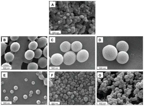
The NPs showed smooth surface and spherical shape, with differences in size depending on the type and amount of polyelectrolytes used on nanoformulation. The uncoated PLGA NPs are characterized by an average diameter of 129.4 ± 22.6 nm. The coating of PLGA surface with different CS concentrations (ie, 5.0, 12.5, 25.0 mg) determined a significant increase of particles size, that was found to be 420.82 ± 22.58, 465.25 ± 39.15, and 552.56 ± 30.62 nm for PLGA-CS1, PLGA-CS2, and PLGA-CS3 NPs, respectively ().
Figure 3 Mean diameter (A) and zeta potential (B) of uncoated PLGA NPs (PLGA) and NPs coated with different amounts (5.0, 12.5, and 25.0 mg) of CS and Alg.
Notes: *Significantly different from the PLGA; #significantly different from other CS-coated NPs.
Abbreviations: PLGA, poly(d,l-lactide-co-glycolide; NPs, nanoparticles.
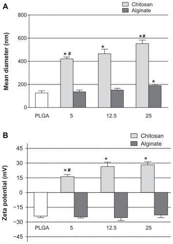
PLGA-CS3 NPs obtained using the highest concentration of CS resulted in significantly larger NPs in regard to NPs produced with lower CS amount (eg, PLGA-CS1, 5.0 mg, P < 0.05), suggesting that particle size is related to the concentration of CS used in the particle preparation.
On the other hand, the precipitation of PLGA in Alg solution at different concentrations did not influence particle size with respect to the uncoated PLGA nanosystems. The mean diameter of Alg-coated NPs was only significantly increased for PLGA-Alg3 (190.01 ± 8.71 nm), prepared with 25.0 mg Alg, compared to uncoated PLGA NPs (P < 0.05). Besides, all formulations were characterized by polydispersity index ranging from 0.27 to 0.38 and exhibited a unimodal particle-size distribution, typical of monodispersed systems.
The differences in size observed in the coated NPs can be mainly related to the different physicochemical properties of CS and Alg polymers, which influence the absorption mechanism on the surface of the polymeric nanoprototypes. In fact, it is well known that the PLGA particle coating with surfactant or polymers is a complex interfacial phenomenon that involves physical adsorption and/or electrostatic interactions between the polymer chains and the coating material. During nanoprecipitation into polyelectrolyte solutions, the electrostatic attraction was proposed as the predominant mechanism, especially in the formation of the first monomolecular adsorption layer.Citation49
More specifically, since the protonated amino groups of CS provide opportunities of intermolecular hydrogen bonding with carboxylic end groups of PLGA, the adsorption of CS on the surface of PLGA continues even though a positively charged surface has been achieved, in which hydrogen bonds or Van der Waals forces can be involved.Citation50 The subsequent layers of CS may perhaps be adsorbed on the first layer by the physical mechanism. On the contrary, the electrostatic interactions were reduced with anionic Alg, and in this case the coating mechanism could be mainly attributed to physical adsorption on the surface of PLGA. As coating with 5 and 12.5 mg of Alg did not determine a significant increase in mean diameter of NPs, a lower amount of Alg was expected to coat on the surface of a particle compared to CS, due to the large surface-to-volume ratio of the particles. Additionally, it is noted that polyelectrolytes can change their conformation depending on the solution concentration.Citation51,Citation52 In fact, in diluted solution the polymer chains are expanded, while in concentrated solution the interchain interactions of polyelectrolyte dominate over intrachain ones. On the basis of these considerations, we can hypothesize the formation of a more compact structure of coating layer in the PLGA-Alg3 NPs with respect to PLGA-Alg1 and PLGA-Alg2 NPs.
The efficiency of NP surface modification can be measured by estimating the zeta potential of the aqueous suspension containing NPs. The surface charge values may be positive or negative depending upon the nature of the polymer or the material used for surface modification.Citation53
As shown in , the zeta potential of the uncoated PLGA NPs was negative (−24 ± 1.5 mV), as expected, because of the carboxyl end groups on PLGA. Otherwise, all CS-coated NPs were electropositive, confirming the presence of such polymer on the PLGA surface. Zeta potential significantly increased (P < 0.05) with CS concentration from 16.2 ± 2.1 for PLGA-CS1 to 26.5 ± 4.4 mV for PLGA-CS2. The further addition of the polymer did not significantly affect the zeta potential, thus suggesting that a steady surface charge was reached.
On the other hand, the zeta potential of Alg-coated NPs was negative and ranged from −22.9 ± 2.7 to −26.2 ± 2.2 mV, with no significant differences between different batches. This finding can be probably related to the low amount of Alg that coats the surface of NPs. Moreover, due to a constant apparent surface charge per unit area (for the exposed carboxyl groups), the layers beyond the first few do not increase the zeta potential.Citation49
Drug loading, encapsulation efficiency, and yield of production
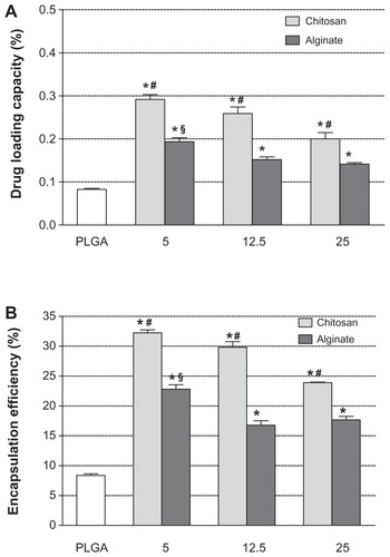
shows the dependence of drug-loading content and encapsulation efficiency of NPs on the composition and the amount of polyelectrolytes used. In general, the RSV encapsulation efficiency in the uncoated PLGA NPs (8.36% ± 0.27%) was significantly lower (P < 0.05) with respect to coated formulations. This suggests that during the precipitation process, only a low amount of RSV can be entrapped into PLGA matrix.
Figure 4 RSV loading content (A) and encapsulation efficiencies (B) of uncoated PLGA NPs and nanosystems coated with different amounts (5.0, 12.5, and 25.0 mg) of CS and Alg.
Notes: *Significantly different from the PLGA; #significantly different from other CS-coated NPs; §significantly different from other Alg coated-NPs.
Abbreviations: RSV, trans-(E)-resveratrol; PLGA, poly(d,l-lactide-co-glycolide); CS, chitosan; Alg, alginate; NPs, nanoparticles.
Previous studies on PLGA NPs revealed that the hydrophobic polymer does not promote an efficient loading of hydrophilic molecules, as well as several peptidic compounds.Citation54 To improve the drug-encapsulation properties of PLGA NPs, a possible strategy consists in the modification of the PLGA surface by coating with stabilizing hydrophilic bioadhesive polymers or surfactants.Citation55 In the present study, we highlighted that cationic CS and anionic Alg coating on PLGA NPs determined a significant improvement in RSV entrapment efficiency.
As can be seen in , the EE% values ranged from about 32% for PLGA-CS1 to about 24% for PLGA-CS3 NPs containing the lowest and the highest CS concentrations, respectively. A similar behavior was observed with batches containing Alg, in which the EE% was found to be 23% and 18% for PLGA-Alg1 and PLGA-Alg3, respectively. The higher loading efficiency of CS- and Alg-coated PLGA NPs could be explained by taking into account the chemical properties of RSV and its affinity for hydrophilic polymers. In fact, the RSV is an amphiphilic molecule, with hydrophobic aromatic rings and hydrophilic phenolic hydroxyl groups.Citation56 Because of the presence of multiple hydroxyl groups, RSV can establish extensive hydrogen bonding and thus promote complexation with CS and Alg polyelectrolytes capable of making H-bonds.Citation57 As a result, during the solvent-evaporation process, the RSV remains embedded between polymeric chains of CS or Alg, and therefore on the coating layer on the PLGA surface. Moreover, the increased encapsulation values of RSV observed in CS-coated NPs with respect to batches containing Alg could be related to the presence of different functional groups (OH and NH2) in CS that might improve the ionic interactions with RSV, and thus determine a larger percentage of entrapped RSV.
The amount of RSV loaded was found to increase in the presence of lower CS or Alg concentrations. This is due to the increase in the initial RSV-to-polyelectrolyte ratio. Upon further addition of polyelectrolytes, the RSV interacts with a larger number of molecules without leading to an extensive enlargement of the coating layer. Thus, it remains in the aqueous solution and is subsequently washed out during the centrifugation procedure. Furthermore, the yields of production obtained ranged from 50% to 65%, with the highest values obtained for uncoated PLGA NPs.
Raman spectroscopy
The qualitative composition of NPs was investigated by Raman spectroscopy. The characteristic peaks obtained from RSV, PLGA, CS, and Alg raw materials () were compared with the peaks for uncoated PLGA, PLGA-CS3, and PLGA-Alg3 RSV–loaded NPs, chosen as examples ().
Figure 5 (A) Raman spectra of RSV, PLGA, CS, and Alg (raw materials); (B) PLGA, PLGA-CS3, and PLGA-Alg3 RSV loaded NPs.
Abbreviations: RSV, trans-(E)-resveratrol; PLGA, poly(d,l-lactide-co-glycolide); CS, chitosan; Alg, alginate; NPs, nanoparticles.
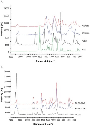
The spectrum of RSV is characterized by the olefinic band at 995 cm−1, C–O stretching vibrations at 1160 cm−1, the intense C–C olefinic stretching at 1600 cm−1, and C–C aromatic double-bond stretching at 1630 cm−1.
The PLGA spectrum displayed bands at 2882, 2946, and 3000 cm−1 corresponding to C–H stretching vibrations, a peak at 1766 cm−1 due to C=O bond stretching vibration, peaks at 1130 cm−1 and 1044 cm−1, due to C–O stretching. The C–H bend was observed at 1453 cm−1. In the spectrum of CS, the peak at 2886 cm−1 was assigned to C–H stretching, the absorption band of the carbonyl (C=O) stretching of amide I at 1630 cm−1, and the bending vibrations of the N–H (N-acetylated residues, amide II band) at 1600 cm−1.Citation58 The band at 1376 cm−1 belongs to the N–H stretching of the amide. The peaks observed at 1090 and 1117 cm−1 were characteristic of C–O stretch of –CH–OH in cyclic alcohols and of –CH2–OH in primary alcohols, respectively.Citation59
As far as the characteristics of Alg are concerned, Raman spectroscopy showed a band at 2932 cm−1 due to C–H stretching and bands around 1095 cm−1 due to C–O–C stretching attributed to its saccharide structure. In addition, the bands at 1617 and 1412 cm−1 were assigned to asymmetric and symmetric stretching peaks of carboxylate salt groups.Citation60
The characteristic absorption bands of C–C olefinic stretching at 1600 cm−1 and C–C aromatic double-bond stretching at 1630 cm−1 of RSV appeared in the loaded uncoated PLGA and CS- and Alg-coated NPs, which probably indicates that RSV molecules were fitted into the polymeric network.Citation61
Differential scanning calorimetry
As shown in , the DSC thermograms of CS and Alg raw materials exhibited the exothermic peaks centered at 320°C and 249°C, respectively, coinciding with the exothermic behavior of the polymer decomposition.Citation62,Citation63 In , the DSC scan of pure PLGA showed an endothermic event (57°C) and an endothermic decomposition peak centered at 372°C. The DSC curve of RSV showed the characteristic endothermic peaks corresponding to the melting point at 270°C and to the glass transition point at 363°C.Citation64
Figure 6 DSC curves of (A) pure CS and Alg, (B) pure RSV and PLGA, (C) uncoated PLGA, PLGA-CS3, and PLGA-Alg3 NPs, and (D) physical mixture of PLGA/RSV, PLGA/CS/RSV and PLGA/Alg/RSV.
Abbreviations: RSV, trans-(E)-resveratrol; PLGA, poly(d,l-lactide-co-glycolide); CS, chitosan; Alg, alginate; NPs, nanoparticles.
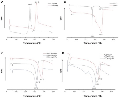
The disappearance of the RSV melting peak in the DSC thermograms of RSV-loaded NPs () indicates the absence of the drug crystalline state, suggesting that entrapped RSV is in an amorphous state of a solid molecular dispersion into the polymer matrix.Citation65
Furthermore, in the thermogram of RSV-loaded PLGA NPs, the endothermic peak of PLGA appeared at about 375°C, as observed in the pure PLGA. The scans of PLGACS3 and PLGA-Alg3 NPs, chosen as examples, exhibited a considerable shifting of the endothermic decomposition peak of PLGA from about 375° to 317° and 327°C, respectively. According to Nie et al,Citation66 this finding suggests that both CS and Alg were physically blended with PLGA in the NPs.
Additionally, the DSC scans of the physical mixtures of PLGA/CS/RSV and PLGA/Alg/RSV () confirmed the presence of a shifting of the PLGA decomposition peak with respect to PLGA/RSV, suggesting an intermolecular interaction formed between the components of the systems.Citation67
Swelling properties of NPs
The swelling properties of polymeric NPs can strongly influence their drug-release profiles.Citation37 The SI of different batches of NPs was measured as the function of time (until 6 hours), and results are shown in .
Figure 7 (A and B) Swelling index of different NPs as a function of time in PBS solution (pH 7.4, 37°C).
Abbreviations: PLGA, poly(d,l-lactide-co-glycolide); NPs, nanoparticles; PBS, phosphate-buiffered saline.
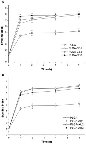
It was observed that all samples swelled relatively quickly in the first 60 minutes and reached a stable equilibrium within about 2 hours. The plots showed that the swelling capacity of uncoated PLGA NPs (SI about 5) was significantly lower (P < 0.05) than that of coated NPs (SI about 8). Since the concentration of polymer used limitedly regulated SI of NPs in both formulations, no significant differences of SI for batches containing CS or Alg were observed. The large SI and high swelling rate of CS- and Alg-coated NPs can be related to their hydrophilic properties with respect to hydrophobic PLGA polymer.
In vitro drug release
The in vitro release experiments were performed in PBS at pH 6.8 and 7.4, chosen to simulate both the slightly acidic microenvironment of extracellular fluid in most tumors and the physiological conditions, respectively.Citation68,Citation69
All tested formulations showed an overlapping of release profiles in different dissolution media (data not shown), suggesting that the pH of the medium did not significantly affect the release of RSV from NPs.
shows the release profiles of RSV from NPs in PBS solution (pH 7.4) at 37°C. Formulations are characterized by similar release profiles but different release rates, depending on the properties and the amount of the polyelectrolyte used. On the whole, NPs showed a biphasic release pattern, where the initial burst of RSV release can be strongly related to the swelling results. When the NPs are exposed to medium, the extended liquid penetration into the systems promotes a fast dissolution of the drug adsorbed, incorporated or weakly bonded onto the surface of NPs with a large surface area.
Figure 8 (A and B) In vitro release profiles (PBS, pH 7.4, 37°C) of RSV from NPs.
Abbreviations: RSV, trans-(E)-resveratrol; PLGA, poly(d,l-lactide-co-glycolide); PBS, phosphate-buffered saline.
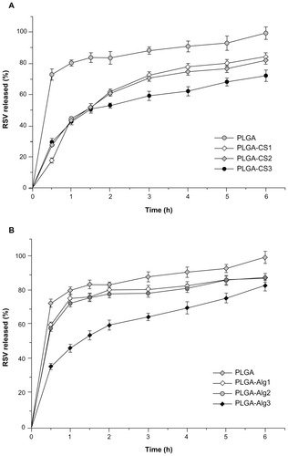
In the slower-release phase, the RSV was progressively released under the dual influence of diffusion through the polymer matrix and polymer degradation.
PLGA NPs showed a first burst release of RSV (about 70%) in the first 30 minutes, followed by a slower and complete release within 6 hours. According to literature data, this finding may be related to small NPs with a relatively large surface, thus resulting in a larger RSV fraction exposed to the leaching medium. Smaller NP size also leads to a shorter diffusion pathway of the matrix-entrapped RSV molecules.Citation70
Regarding CS-containing NPs, these showed a similar drug-release profile within the first 1.5 hours, corresponding to about 50% of RSV released. After 2 hours, PLGA-CS1 and PLGA-CS2 containing 12.5 mg and 25 mg of CS, respectively, showed superimposed release profiles, and released about 80% of RSV during 6 hours. The increase of CS content to 25 mg (PLGA-CS3) led to a significant decrease in the RSV release rate (30% of RSV retained in the 6-hour period). The presence of Alg, 5 and 12.5 mg in PLGA-Alg1 and PLGA-Alg2 NPs, respectively, reduces the release rate compared to PLGA NPs, but results in a significantly higher RSV release with respect to PLGA-CS analogous batches. On the other hand, the release rate of PLGA-Alg3 systems was slightly delayed and comparable to that of PLGA-CS3 NPs during the first 2 hours, followed by a significant improvement of RSV release, corresponding to about 80% during the 6 hours. It has been claimed that the initial burst release is significantly reduced when using polyelectrolyte layers.Citation71 Accordingly, the release rate of RSV from coated NPs was found to decrease with increasing polyelectrolyte concentration, suggesting that the RSV molecules have to pass through an additional layer of diffusional resistance created by the coating material.
In order to investigate the kinetics and mechanism of drug release from NPs, and to confirm that the mechanism governing RSV release from NPs is predominantly diffusion-mediated, the release data were fitted to several mathematical models (zero-order [Q = k0t], first order [ln {100 − Q} = ln Q0 − k1t], Higuchi [Q = kH t1/2], and Korsmeyer–Peppas models).Citation72
shows the correlation values (R2) used as an indicator of the best fitting of the models considered for different RSV-NP batches. The R2 values for first-order kinetics of different polymeric NPs (ranging between 0.7643 and 0.9791) were greater than that of zero order (ranging between 0.7596 and 0.9368) and corresponded with that of Higuchi’s square root of time (ranging between 0.7991 and 0.9827). Also, to understand the drug-release mechanism, the data were fitted to the Korsmeyer–Peppas exponential model Mt/M∞ = ktn, where Mt/M∞ is the fraction of drug released after time ‘t’, ‘k’ is a kinetic constant, and ‘n’ is the release exponent that characterizes the different drug-release mechanism. Values of the exponent n ≤ 0.5 indicate a Fickian or quasi-Fickian diffusion mechanism, whereas for values 0.5 < n < 1, an anomalous mechanism for drug release occurs.Citation73
Table 2 Coefficient of determination (R2) and release exponent (n) of kinetic data analysis of RSV release from different polymeric nanoparticles
In all formulations, n values were found to be in the range of 0.11–0.42, confirming that the drug release from NPs followed a quasi-Fickian diffusion mechanism.
RSV protection
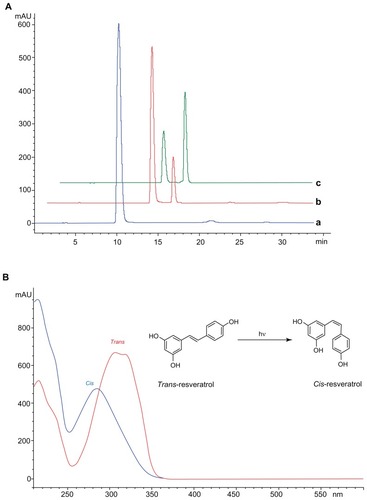
RSV naturally occurs in two isoforms, cis and trans, and to date most studies indicate that the trans isomer is biologically more active and more stable than its cis isoform.Citation18 RSV (ie, trans-RSV) is known to be a highly photosensitive compound and easily degraded by light and heat, which limits its efficacy and application.Citation74 In spite of this, we focused our attention on investigating the protective effect of the NPs on degradation of the trans stereoisomeric form of RSV under UV irradiation. As depicted in , before the UV light exposure of RSV, only the peak of trans isoform was detected at 10 minutes (blue line). After 30 minutes of irradiation (red line), a second peak appeared in the chromatogram at about 13 minutes, representing the cis isoform. With further exposure time (120 minutes), the intensity of trans isomer peak decreased, while the peak of cis increased. The collected UV-visible spectra of two peaks () was found to be similar to literature data.Citation47
As shown in , the encapsulation of RSV into NPs largely prevented its degradation with respect to free RSV. After 120 minutes of exposure, only 20% of trans isoform was detected in the reference sample (unencapsulated RSV), which was significantly reduced if compared with that obtained in the formulations.
Figure 9 The retention percentage of unencapsulated RSV and uncoated PLGA, PLGA-CS1, PLGA-CS3, PLGA-Alg1, and PLGA-Alg3 NPs exposed to UV lamp for 120 minutes.
Notes: *Significantly different from the free RSV; #significantly different from PLGA; §significantly different from other CS-coated NPs; °significantly different from other Alg-coated NPs.
Abbreviations: RSV, trans-(E)-resveratrol; PLGA, poly(d,l-lactide-co-glycolide); CS, chitosan; Alg, alginate, NPs, nanoparticles.
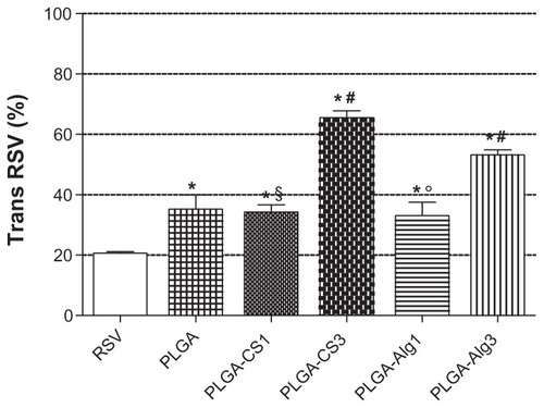
In the uncoated PLGA NPs, the retention percentage of RSV was about 35%. The coating of PLGA NPs with lower polyelectrolyte concentration (PLGA-CS1 and PLGA-Alg1) did not produce a significant increase in protection, probably due to the preferential localization of RSV between polymeric chains of the coating layer, as previously mentioned.
Also, for PLGA-Alg3 and PLGA-CS3, the amount of RSV detected was 53% and 66%, respectively, suggesting that the larger polyelectrolyte content, with respect to PLGA-Alg1 and PLGA-CS1, reduced the RSV fraction exposed to UV light irradiation, thus improving the protection against degradation.
Long-term chemical stability of entrapped RSV
The chemical integrity of drugs encapsulated in nanoparticles is a fundamental aspect of the overall stability evaluation of formulations, in particular to evaluate the eventual leaking of the drug from the carrier during storage.Citation75 The stability of colloidal polymeric carriers depends on their storage conditions (temperature and pH), the type and the molecular weight of the polymer used,Citation76 and the chemical stability of entrapped drugs.Citation75 Consequently, for each specific formulation, the corresponding stability experiments should be performed to assess the quality of the product. In this study, the amount and chemical stability of RSV in NPs stored at 4°C was investigated by HPLC for 6 months. As reported in , for all formulations the amount of RSV loaded did not change significantly with respect to 0 day (), and no peak was detected due to absorption of cis resveratrol. This finding suggests that NPs showed a negligible release of the encapsulated RSV and are able to prevent the degradation of trans isoform for the long term.
Figure 10 RSV loading content of uncoated PLGA NPs and nanosystems coated with different amounts (5.0, 12.5, and 25.0 mg) of CS and Alg after 6 months of storage at 4°C.
Notes: *Significantly different from PLGA; #significantly different from other CS-coated NPs; §significantly different from other Alg-coated NPs.
Abbreviations: RSV, trans-(E)-resveratrol; PLGA, poly(d,l-lactide-co-glycolide); CS, chitosan; Alg, alginate; NPs, nanoparticles.
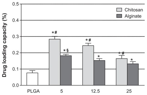
Figure 11 (A) HPLC chromatograms of RSV (trans-resveratrol) before (a) and after exposure to UV for 30 minutes (b) and 120 minutes (c). (B) UV-Vis spectra of trans- and cis-resveratrol conversion induced by UV exposure.
Abbreviations: HPLC, high-performance liquid chromatography; UV-Vis, UV visible.
Conclusion
Our results demonstrate that the newly developed CS- and Alg-coated PLGA NPs are suitable to be used for the encapsulation of bioactive RSV. The amount of cationic or anionic polyelectrolytes employed as surface modifiers resulted in differences in size, surface charge, encapsulation capacity, and in vitro release behavior of RSV-loaded nanoprototypes. Stability studies revealed that encapsulation provides significant protection against light-exposure degradation, thereby contributing to increase the protection of the encapsulated RSV. The translational potential of these novel nanovehicles warrants biological evaluation in several experimental models, eg, focusing on prostate and skin cancer as target diseases. We believe that the insights obtained from this study can contribute to validate the nanoformulation of natural dietary agents, ie, “nanochemoprevention,” as a strategy to improve their potential ability to prevent and suppress cancers by intravenous or topical application, as well as to reduce the risk of cancer development in a larger context.
Acknowledgments
The research activities presented were done within a project agreement with Sardinian Technology Park, Porto Conte Ricerche, Italy. Mario Sechi gratefully acknowledges the Regione Autonoma della Sardegna for financial support of grant CRP-25920, “Development of novel targeted nanodevices for the prevention, diagnosis and treatment of prostate cancer,” within the frame of “Legge regionale n 7/2007, promozione della ricerca scientifica e dell’innovazione tecnologica in Sardegna – Annualità 2010.” The authors also thank Professor Sergio Uzzau and Dr Tonina Roggio for their organizational support and Dr Paolo Sanna for graphic design.
Disclosures
The authors report no conflicts of interest in this work.
References
- ManachCScalbertAMorandCRémésyCJiménezLPolyphenols: food sources and bioavailabilityAm J Clin Nutr200479572774715113710
- ScalbertAManachCMorandCRémésyCJiménezLDietary polyphenols and the prevention of diseasesCrit Rev Food Sci Nutr200545428730616047496
- ScalbertAJohnsonITSaltmarshMPolyphenols: antioxidants and beyondAm J Clin Nutr200581Suppl 1215S217S15640483
- SurhYJCancer chemoprevention with dietary phytochemicalsNat Rev Cancer200331076878014570043
- KhanNAfaqFMukhtarHCancer chemoprevention through dietary antioxidants: progress and promiseAntioxid Redox Signal200810347551018154485
- YangCSLandauJMHuangMTNewmarkHLInhibition of carcinogenesis by dietary polyphenolic compoundsAnnu Rev Nutr20012138140611375442
- FrémontLBiological effects of resveratrolLife Sci200066866367310680575
- SunAYWangQSimonyiASunGYBotanical phenolics and neurodegenerationBenzieIFFWachtel-GalorSHerbal Medicine: Biomolecular and Clinical Aspects2nd edBoca RatonCRC Press2011
- BaurJAPearsonKJPriceNLResveratrol improves health and survival of mice on a high-calorie dietNature2006444711733734217086191
- CampagnaMRivasCAntiviral activity of resveratrolBiochem Soc Trans201038Pt 1505320074034
- YanYGaoYYLiuBQNiuXFZhuangYWangHQResveratrol-induced cytotoxicity in human Burkitt’s lymphoma cells is coupled to the unfolded protein responseBMC Cancer20101044520723265
- AggarwalBBBhardwajAAggarwalRSSeeramNPShishodiaSTakadaYRole of resveratrol in prevention and therapy of cancer: preclinical and clinical studiesAnticancer Res2004245A2783284015517885
- GoswamiSKDasDKResveratrol and chemopreventionCancer Lett200928411619261378
- AhmadNAdhamiVMAfaqFFeyesDKMukhtarHResveratrol causes WAF-1/p21-mediated G(1)-phase arrest of cell cycle and induction of apoptosis in human epidermoid carcinoma A431 cellsClin Cancer Res2001751466147311350919
- SignorelliPGhidoniRResveratrol as an anticancer nutrient: molecular basis, open questions and promisesJ Nutr Biochem200516844946616043028
- LuZChengBHuYLZhangYHZouGLComplexation of resveratrol with cyclodextrins: solubility and antioxidant activityFood Chem200911311720
- JuanMEBuenafuenteJCasalsIPlanasJMPlasmatic levels of trans-resveratrol in ratsFood Res Int2002352195199
- MuninAEdwards-LévyFEncapsulation of natural polyphenolic compounds; a reviewPharmaceutics201134793829
- AmriAChaumeilJCSfarSCharrueauCAdministration of resveratrol: what formulation solutions to bioavailability limitations?J Control Release2012158218219321978644
- ZhangLGuFXChanJMWangAZLangerRSFarokhzadOCNanoparticles in medicine: therapeutic applications and developmentsClin Pharmacol Ther200883576176917957183
- ByrneJDBetancourtTBrannon-PeppasLActive targeting schemes for nanoparticle systems in cancer therapeuticsAdv Drug Deliv Rev200860151615162618840489
- RanganathanRMadanmohanSKesavanANanomedicine: towards development of patient-friendly drug-delivery systems for oncological applicationsInt J Nanomedicine201271043106022403487
- SiddiquiIAAdhamiVMBharaliDJIntroducing nanochemoprevention as a novel approach for cancer control: proof of principle with green tea polyphenol epigallocatechin-3-gallateCancer Res20096951712171619223530
- SannaVPintusGRoggioAMTargeted biocompatible nanoparticles for the delivery of (−)-epigallocatechin 3-gallate to prostate cancer cellsJ Med Chem20115451321133221306166
- HobbsSKMonskyWLYuanFRegulation of transport pathways in tumor vessels: role of tumor type and microenvironmentProc Natl Acad Sci U S A1998958460746129539785
- SannaVSechiMNanoparticle therapeutics for prostate cancer treatmentMaturitas2012731273222341739
- SannaVRoggioAMPosadinoAMNovel docetaxel-loaded nanoparticles based on poly(lactide-co-caprolactone) and poly(lactide-co-glycolide- co-caprolactone) for prostate cancer treatment: formulation, characterization and cytotoxicity studiesNanoscale Res Lett20116126021711774
- SiddiquiIAMukhtarHNanochemoprevention by bioactive food components: a perspectivePharm Res20102761054106020221894
- SiddiquiIAAdhamiVMChamcheuJCMukhtarHImpact of nanotecnology in cancer: emphasis on nanochemopreventionInt J Nanomedicine2012759160522346353
- SannaVSechiMNanoparticle therapeutics for prostate cancer treatmentNanomedicine20128Suppl 1S31S3622640911
- SiddiquiAIShuklaYMukhtarHNanoencapsulation of natural products for chemopreventionJ Nanomed Nanotechnol20112104e
- ShaoJLiXLuXEnhanced growth inhibition effect of resveratrol incorporated into biodegradable nanoparticles against glioma cells is mediated by the induction of intracellular reactive oxygen species levelsColloids Surf B Biointerfaces2009721404719395246
- TeskacKKristlJThe evidence for solid lipid nanoparticles mediated cell uptake of resveratrolInt J Pharm20103901616919833178
- CaddeoCTeskacKSinicoCKristlJEffect of resveratrol incorporated in liposomes on proliferation and UV-B protection of cellsInt J Pharm20083631–218319118718515
- ShiveMSAndersonJMBiodegradation and biocompatibility of PLA and PLGA microspheresAdv Drug Deliv Rev199728152410837562
- MakadiaHKSiegelSJPoly lactic-co-glycolic acid (PLGA) as biodegradable controlled drug delivery carrierPolymers2011331377139722577513
- DavaranSRashidiMRPourabbasBDadashzadehMHaghshenasNMAdriamycin release from poly(lactide-co-glycolide)-polyethylene glycol nanoparticles: synthesis, and in vitro characterizationInt J Nanomedicine20061453553917722284
- LiSHCaiSXLiuBMaKWWangZPLiXKIn vitro characteristics of poly(lactic-co-glycolic acid) microspheres incorporating gelatin particles loading basic fibroblast growth factorActa Pharmacol Sin200627675475916723096
- GeorgeMAbrahamTEPolyionic hydrocolloids for the intestinal delivery of protein drugs: alginate and chitosan – a reviewJ Control Release2006114111416828914
- AgrawalPStrijkersGJNicolayKChitosan-based systems for molecular imagingAdv Drug Deliv Rev2010621425819861142
- TakkaSGürelAEvaluation of chitosan/alginate beads using experimental design: formulation and in vitro characterizationAAPS Pharm Sci Tech2010111460466
- ZhouJRomeroGRojasEMaLMoyaSGaoCLayer by layer chitosan/alginate coatings on poly(lactide-co-glycolide) nanoparticles for antifouling protection and folic acid binding to achieve selective cell targetingJ Colloid Interface Sci2010345224124720227712
- WangQJamalSDetamoreMSBerklandCPLGA-chitosan/PLGA-alginate nanoparticle blends as biodegradable colloidal gels for seeding human umbilical cord mesenchymal stem cellsJ Biomed Mater Res A201196352052721254383
- ChoiDHParkCHKimIHChunHJParkKHanDKFabrication of core-shell microcapsules using PLGA and alginate for dual growth factor delivery systemJ Control Release2010147219320120647022
- SunBRibesAMLeandroMCBelchiorAPSprangerMIStilbenes: quantitative extraction from grape skins, contribution of grape solids to wine and variation during wine maturationAnal Chim Acta20065631–2382390
- ElgindyNElkhodairyKMolokhiaAElZoghbyABiopolymeric nanoparticles for oral protein delivery: design and in vitro evaluationJ Nanomed Nanotechnol201123110
- López-HernándezJPaseiro-LosadaPSanches-SilvaATLage-YustyMAStudy of the changes of trans-resveratrol caused by ultraviolet light and determination of trans- and cis-resveratrol in Spanish white winesEur Food Res Technol2007225789796
- Quintanar-GuerreroDAllemannEFessiHDoelkerEPreparation techniques and mechanism of formation of biodegradable nanoparticles from preformed polymersDrug Dev Ind Pharm19982412111311289876569
- GuoCGemeinhartRAUnderstanding the adsorption mechanism of chitosan onto poly(lactide-co-glycolide) particlesEur J Pharm Biopharm200870259760418602994
- MahmoodiMKhosroshahiMEAtyabiFLaser thrombolysis and in vitro study of tPA release encapsulated by chitosan coated PLGA nanoparticles for AMIInt J Biol Biomed Eng2010423542
- MichlerGHBaltá CallejaFJPart III. Nano- and microcomposites: interphaseKarger-KocsisJFakirovSNano- and Micromechanics of Polymers Blends and CompositesCincinnatiHanser2009
- SomasundaranPMehtaSCYuXKrishnakumarSCap 6: colloid systems and interfaces stability of dispersions through polymer and surfactant adsorptionBirdiKSHandbook of Surface and Colloid Chemistry3rd edBoca RatonCRC Press2008
- SoppimathKSAminabhaviTMKulkarniARRudzinskiWEBiodegradable polymeric nanoparticles as drug delivery devicesJ Control Release2001701–212011166403
- ZhengCHGaoJQZhangYPLiangWQA protein delivery system: biodegradable alginate-chitosan-poly(lactic-co-glycolic acid) composite microspheresBiochem Biophys Res Commun200432341321132715451441
- TrimailleTPichotCDelairTSurface functionalization of poly(D,L-lactic acid) nanoparticles with poly(ethylenimine) and plasmid DNA by the layer-by-layer approachColloids Surf A Physicochem Eng Asp200322113948
- RiouVVernhetADocoTMoutounetMAggregation of grape seed tannins in model wine – effect of wine polysaccharidesFood Hydrocoll20021611723
- HandiqueJGBaruahJBPolyphenolic compounds: an overviewReact Funct Polym200252163188
- SankaliaMGMashruRCSankaliaJMSutariyaVBReversed chitosan-alginate polyelectrolyte complex for stability improvement of alpha-amylase: optimization and physicochemical characterizationEur J Pharm Biopharm200765221523216982178
- ChenSCWuYCMiFLLinYHYuLCSungHWA novel pH-sensitive hydrogel composed of N,O-carboxymethyl chitosan and alginate cross-linked by genipin for protein drug deliveryJ Control Release200496228530015081219
- SartoriCFinchDSRalphBDetermination of the cation content of alginate thin films by FTIR spectroscopyPolymer19973814351
- SocratesGInfrared and Raman Characteristic Group Frequencies: Tables and Charts3rd edChichesterJ Wiley & Sons2001
- WanYLuXDalaiSZhangJThermophysical properties of polycaprolactone/chitosan blend membranesThermochim Acta20094871–23338
- Fernández-HervásMJHolgadoMAFiniAFellJTIn vitro evaluation of alginate beads of a diclofenac saltInt J Pharm19981631–22334
- LeeCWYenFLHuangHWResveratrol nanoparticle system improves dissolution properties and enhances the hepatoprotective effect of resveratrol through antioxidant and anti-inflammatory pathwaysJ Agric Food Chem201260184662467122480310
- TzengCWYenFLWuTHEnhancement of dissolution and antioxidant activity of kaempferol using a nanoparticle engineering processJ Agric Food Chem20115995073508021417334
- NieHLeeLYTongHWangCHPLGA/chitosan composites from a combination of spray drying and supercritical fluid foaming techniques: new carriers for DNA deliveryJ Control Release2008129320721418539352
- MakraduliLCrcarevskaMSGeskovskiNDodovMGGoracinovaKFactorial design analysis and optimisation of alginate-Ca-chitosan microspheresJ Microencapsul Epub July 3, 2012
- DanhierFFeronOPréatVTo exploit the tumor microenvironment: passive and active tumor targeting of nanocarriers for anti-cancer drug deliveryJ Control Release2010148213514620797419
- LeeESOhKTKimDYounYSBaeYHTumor pH-responsive flower-like micelles of poly(L-lactic acid)-b-poly-(ethylene glycol)- b-poly(L-histidine)J Control Release20071231192617826863
- BriggerIDubernetCCouvreurPNanoparticles in cancer therapy and diagnosisAdv Drug Deliv Rev200254563165112204596
- TanJPWangQTamKCControl of burst release from nanogels via layer by layer assemblyJ Control Release2008128324825418457895
- MehtaAKYadavKSSawantKKNimodipine loaded PLGA nanoparticles: formulation optimization using factorial design, characterization and in vitro evaluationCurr Drug Deliv20074318519317627492
- KorsmeyerRWGurneyRDoelkerEBuriPPeppasNAMechanism of solute release from porous hydrophilic polymerJ Pharm Sci19831512535
- OralloFComparative studies of the antioxidant effects of cis- and trans-resveratrolCurr Med Chem2006131879816457641
- AbdelwahedWDegobertGStainmesseSFessiHFreeze-drying of nanoparticles: formulation, process and storage considerationsAdv Drug Deliv Rev200658151688171317118485
- LemoineDFrancoisCKedzierewiczFPreatVHoffmanMMaincentPStability study of nanoparticles of poly(ɛ-caprolactone), poly(D,L-lactide) and poly(D,L-lactide-co-glycolide)Biomaterials19961722219121978922605