
Yang Y, Ren S, Zhang X, et al. Int J Nanomedicine. 2018;13:3751-3762.
The authors have advised that there were some editing errors for on page 3756 and on page 3759.
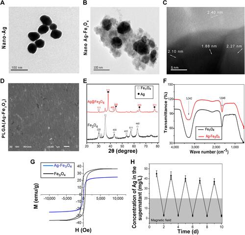
did not clearly depict the boundary of Ag and Fe3O4. Thus, this corrigendum replaces the image 1C with a better resolution, as described in the “Results” section “PLGA(Ag-Fe3O4) nanocomposites preparation and characterization)” on pages 3755-3756.
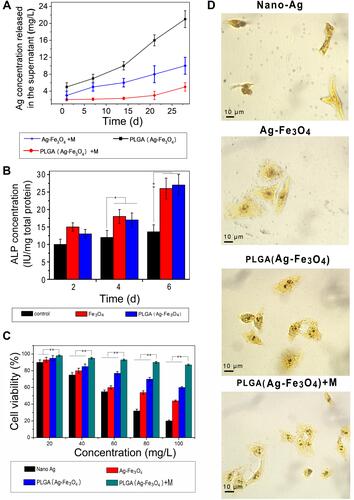
Image 3D has reported to have repeated region in the PLGA(Ag-Fe3O4) on the published proof. After a thorough check to the original images and raw data, the authors found out that there was no repeated region and hereby present the updated image for .
Page 3756, , the correct figure is as follows.
Figure 1 Characterization of PLGA(Ag-Fe3O4).
Page 3759, , the correct figure is as follows.
Figure 3 (A) Storage stability test result of PLGA(Ag-Fe3O4) nanoparticles. (B) ALP activity of osteoblasts in PBS, Fe3O4, or PLGA(Ag-Fe3O4) nanoparticles after 2, 4, and 6 days of differentiation culture; *P<0.05. **P<0.01. (C) Viability of osteoblasts incubated with different concentrations of samples for 24 hours. n=6; **P<0.01. (D) AgNOR staining in nucleoli of osteoblasts cultured with different nanoparticles. Original magnification 1,000×.
The authors apologize for these errors and advise they do not affect the results of the paper.