
Abstract
Objective
To investigate the effects of proanthocyanidins (PA), myricetin, resveratrol, and kaempferol on the modification of dentin collagen and the inhibition of matrix metalloproteinase (MMP) activity, and to evaluate their contributions to the biomimetic remineralization and resin-dentin bonding performance.
Methods
Attenuated total reflection Fourier transform infrared spectroscopy (ATR-FTIR) and in situ zymography were applied to verify the collagen modification and MMP activity inhibition induced by these four polyphenols. Scanning electron microscopy/energy dispersive spectrometer (SEM/EDS) analysis, X-ray diffraction (XRD), ATR-FTIR, Vickers hardness numbers (VHN), and micro-computed tomography (micro-CT) were performed to characterize the remineralized dentin. Microtensile bond strength (μTBS) and nanoleakage were investigated to evaluate the effects of the four polyphenols on resin-dentin bonding durability.
Results
ATR-FTIR and in situ zymography confirmed that these four polyphenols could modify dentin collagen and inhibit MMP activity, respectively. Chemoanalytic characterization exhibited the efficacies of the four polyphenols in promoting dentin biomimetic remineralization. The surface hardness of PA-pretreated dentin was the greatest. Micro-CT results demonstrated that the PAs group possessed the highest amount of dentin surface minerals and the lowest amount of deep-layer minerals. The surface and deep-layer mineral contents of the Myr group were higher than Res and Kae groups. Treatment with these four polyphenols significantly increased the initial μTBS compared with the control group without primer conditioning. μTBS decreased significantly during aging, and the decrease was more severe in the PAs and Kae groups than in the Myr and Res groups. With or without aging, the polyphenol groups exhibited relatively less fluorescence. However, the Myr and Res groups showed less serious nanoleakage after aging.
Conclusion
PA, myricetin, resveratrol, and kaempferol can modify dentin collagen, inhibit MMP activity, promote biomimetic remineralization, and improve resin-dentin bond durability. Compared with PA and kaempferol, myricetin and resveratrol are more effective in improving resin-dentin bonding.
Graphical Abstract
Introduction
The dynamic process of demineralization-remineralization occurs throughout life in the human body even after bone development is complete. When demineralization is dominant, mineral crystals dissolve and inorganic ions such as Ca2+ and PO43- are removed from the organic matrix, exposing type I collagen fibers; thus, the mechanical strength of the bone is reduced, potentially leading to the occurrence of fractures and bone deformities.Citation1 In humans, dentin and bone tissues have similar compositions and inorganic contents; hence, the dynamic balance of demineralization-remineralization also exists in dentin.Citation2,Citation3
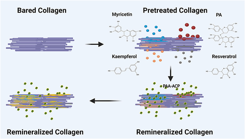
The demineralization of dentin occurs during the bonding procedure, regardless of the self-etch or etch-and-rinse mode. The presence of residual water and acidic ions in the dental adhesive systems as well as the activation of matrix metalloproteinases (MMPs) lead to exposed type I collagen fiber degradation in the deep layers that are not completely encapsulated by the resin matrix. This is the source of the persistent problem of dentin bond durability within the range of available bonding techniques based on the hybrid layer (HL) theory.Citation4,Citation5 Reducing the degradation of collagen fibers in the HL and inducing biomimetic remineralization of demineralized dentin areas are considered as two viable approaches that can be applied individually or combinedly to improve dentin bond durability.Citation5,Citation6 Biomimetic remineralization is based on the fact that mineral backfilling of collagen fibril interstices can replace water molecules to encapsulate collagen fibrils, and thus protect collagen from degrading over time.Citation7 The currently accepted theory of dentin biomimetic remineralization is based on the hypothesis of the polymer-induced liquid precursor (PILP) process, in which liquid-like amorphous calcium phosphate (ACP) nanoparticles are delivered into type I collagen fiber matrices and gradually attract Ca2+, leading to hydroxyapatite (HA) formation.Citation8–10 However, the biomimetic remineralization process through the PILP alone is slow, and only a few HA crystals are formed within 4 weeks; this provides little resistance to the rapid degradation effect of water and MMPs on bare collagen fibers.Citation11,Citation12 Therefore, a compound that resists enzymatic degradation while accelerating the formation of minerals around exposed collagen fibers to further protect collagen is extremely attractive.
Natural polyphenols, such as proanthocyanidins (PA),Citation13 quercetin,Citation14 catechins,Citation15 myricetin,Citation16 resveratrol,Citation17 and kaempferolCitation18 contain multiple phenolic -OH groups, which can form hydrogen bonds with collagen amide carbonyl groups to modify the collagen and inhibit MMPs. These -OH groups at the free end can also chelate with metal cations (e g, Ca2+, Mg2+, and Au2+), thereby potentially accelerating ACP nanoparticle deposition and HA formation.Citation19–22
PA has been shown to have positive effects on dentin remineralization.Citation23–25 In the past 3 years, the clinical value of myricetin, resveratrol, and kaempferol has gradually increased, and these polyphenols have been proven to improve the resin-dentin bonding durability by modifying collagen and inhibiting MMP activity;Citation16–18 however, their contribution to biomimetic remineralization remains unclear.
In this study, we evaluated and compared the effects of four polyphenols (PA, myricetin, resveratrol, and resveratrol) on the promotion of biomimetic remineralization and the bonding performance of resin-dentin. Attenuated total reflection Fourier transform infrared spectroscopy (ATR-FTIR) and in situ zymography were employed to confirm dentin collagen modification and MMP activity inhibition, respectively. Biomimetic remineralization was observed via scanning electron microscopy/energy dispersive spectrometry (SEM/EDS), X-ray diffraction (XRD), ATR-FTIR, and Vickers hardness numbers (VHN), and quantitatively analyzed by micro-computed tomography (micro-CT). Furthermore, the resin-dentin bonding durability was examined by microtensile bond strength (μTBS) and nanoleakage analyses. The null hypotheses were as follows: These four polyphenols (i) cannot modify collagen, (ii) do not contribute to MMP activity inhibition; (iii) have no effect on promoting biomimetic remineralization of demineralized dentin; and (iv) contribute to improving the dentin bond durability little.
Materials and Methods
Preparation of Dentin Slices
This study was conducted in accordance with the Declaration of Helsinki. One hundred and seventeen no-carious human third molars were obtained for in vitro research with the patients’ informed consents according to the protocol approved by the Ethics Committee of Nanjing Medical University, China (No. [2019] 277). The dentin specimens were sectioned into dentin slices with different thicknesses (0.5 ± 0.1 mm for analyzing the interaction between polyphenols and dentin collagen as well as the quantitative analysis of dentin remineralization; 1 ± 0.1 mm for the characterization of dentin remineralization and the analysis of MMP activity; and 3 ± 0.1 mm for μTBS and nanoleakage tests), using a slow speed saw under water irrigation (Isomet 1000, Buehler Ltd., Lake Bluff, IL, USA).Citation26 These teeth were obtained and stored in 0.01% sodium azide solution at 4°C less than 1 month before use.
Interaction Between Polyphenols and Dentin Collagen
In this stage, 55 mg/mL PA/ethanol, 0.2 mg/mL myricetin/ethanol, 10 mg/mL resveratrol/ethanol, and 10 mg/mL kaempferol/ethanol primers were prepared and stored at 4°C before use.Citation16,Citation17,Citation25,Citation27 All the chemical reagents were purchased from Macklin, Shanghai, China. Dentin slices were immersed in 1 M/L hydrochloric acid (HCl) for 12 h for complete demineralization (examined using an intraoral dental X-ray device [Focus 50540-IMG, KAVO, California, USA]),Citation28 rinsed completely with double distilled water, and blotted dry. Thereafter, the slices were divided into five groups and subjected to pretreatment for 30 min (n=1): (1) Ctr1, polyphenol-free/ethanol solution pretreatment as a blank control group; (2) PAs, PA/ethanol solution; (3) Myr, myricetin/ethanol solution; (4) Res, resveratrol/ethanol solution; and (5) Kae, kaempferol/ethanol solution.
The pretreated slices of each group were also thoroughly rinsed and dried, and then examined by ATR-FTIR (Thermo Fisher Scientific, USA) with 32 scans in the range of 500–4000 cm−1 with a resolution of 4.0 cm−1.
Analysis of MMP Activity
The dentin surface was acid-etched with 35% phosphate gel for 15s, rinsed for 30s, and then grouped and pretreated separately as described above (n=5). A layer of adhesive (Single Bond 2, 3M ESPE, St. Paul, MN, USA) was immediately applied according to the manufacturer’s instructions. Subsequently, two 1-mm-thick layers of composite resin (Filtek Z250; 3M ESPE, St. Paul, MN, USA) were placed over the bonded surface with 20s light-curing for each layer using an LED unit (EliparTM S10, 3M ESPE, São Paulo, MN, USA).
Three bonded slabs from each group were wet-polished with #600, #1200, and #2000 silicon-carbide papers to thicknesses of approximately 500 μm, ultrasonically cleaned for 10 min, and then glued to glass slides using cyanoacrylate glue. 50 μL of freshly quenched fluorescein-conjugated gelatin mixture (E-12055; Molecular Probes, Eugene, OR, USA) was applied on the top of each resin-dentin slab and covered with a coverslip.Citation29 After the microscope slides were incubated in the dark at 37 ◦C for 24 h, they were examined with a confocal microscope (Zeiss LSM880 with NLO & Airyscan, Germany).
Characterization of Dentin Remineralization
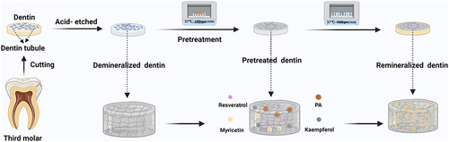
The dentin slices were subjected to acid etching and grouping pretreatment. Thereafter, the slices were immersed in a 10-mL remineralization solution (12 mM Na2HPO4, 20 mM CaCl2·2H2O, 18 mg/mL NaCl, 14 mg/mL Tris, 60 mM HCl, 700 μg/mL polyacrylic acid, pH =7.4±0.1)Citation30 for 7 days and incubated on a shaker (Minquan MQT-60R, Shanghai, China) at 37°C and 100 rpm/min. shows the preparation process of dentin remineralization.
Figure 1 The process of preparing remineralization samples.
Color changes of the dentin surfaces before and after remineralization were observed visually and photographed with a digital camera (D7500, Nikon, Tokyo, Japan) (n=1).
After remineralization, the samples of each group were successively dehydrated for 30 min in solutions with an increasing concentration of ethanol (25 wt%, 50 wt%, 75 wt%, 90 wt%, 95 wt%, and 100 wt%). The untreated sample was used as the control group (labeled as Ctr0). All groups were submitted to SEM/EDS (TESCAN MAIA3, Kohoutovice, Czech Republic), XRD (D8 ADVANCE, Bruker AXS, Germany), ATR-FTIR (Thermo Fisher Scientific, USA), and VHN (TUKON 1102, Wilson, Norwood, USA) testing to observe remineralization.
Three dentin slices were coated with Pt and observed using SEM in the secondary electronic mode with an accelerating voltage of 20 kV and a working distance of 5 mm.
XRD was performed using Ni-filtered Cu K (λ = 1.5418 Å) radiation at ambient temperature, with a 2θ step size, angular range, and scan rate of 0.02°, 20°–80°, and 2° /min, respectively (n=1).
The ATR-FTIR spectra were acquired with 32 scans in the range of 800–4000 cm−1 with a resolution of 4.0 cm−1 (n=1).
The VHN test of dentin surfaces was performed under a load of 100 g for 15s (n=5). One-way analysis of variance (ANOVA) and Tukey’s post hoc least significant difference (LSD) tests were performed using the SPSS 21.0 statistical software package (IBM SPSS, Inc., Chicago, IL, USA) to evaluate the hardness of the dentin surfaces (α = 0.05).
Quantitative Analysis of Dentin Remineralization
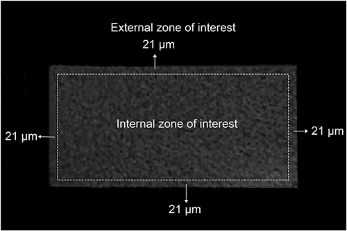
Dentin slices were immersed in 1 M/L HCL for 12 h for complete demineralization; further, the slices were divided into six groups to be treated separately. The zones of interest in the dentin slices were divided (from superficial to deep) into two zones: the external (0–21 μm) and internal (remaining central area) zones of interest. The mineral content of dentin after remineralization for 7 days was detected using a micro-CT (viva CT 80, SCANCO Medical AG, Switzerland) system with a current of 72 mA and a voltage of 55 kV. SkyScan CTAn software (version 1.15.4.0+) was used for data analysis.Citation31
After recording the mineral volume percentage (V%, equivalent to bone volume percentage in bone micro-CT measurements) and mineral separation (Tb. Sp, equivalent to trabecular separation in bone micro-CT measurements), the two-way ANOVA followed by Tukey’s post-hoc LSD tests were performed to evaluate the effects of the primer and zone of interest factors on mineral content (α = 0.05).
μTBS Testing and Nanoleakage
Remineralized dentin specimens were divided into six groups and prepared as described above. A layer of adhesive (Single Bond 2) was applied to the bonding surfaces according to the manufacturer’s instructions. Further, a 4-mm composite resin was placed in layers, with 20s light-curing for each layer that was less than 2 mm. After 24 h of water storage or 3 months of aging plus 10,000 thermocycles (TC-501F, Suzhou, China) for 30s at 5°C and 30s at 55 °C, with a transition time of 3s, the bonded specimens were further sectioned into pieces with dimensions of 1 mm × 1 mm × 7 mm. Each beam was subjected to μTBS testing using a universal testing machine (Instron 3365 ElectroPuls, Boston, MA, USA) at a crosshead speed of 1 mm/min until fracture occurred (n=15). The μTBS values were calculated by dividing the load at failure by the cross-sectional bonding area. The same statistical analysis as that for the micro-CT test was performed to evaluate the effects of the primer and aging factors on μTBS (α = 0.05).
Three dentin-bonded slabs (with or without aging) were selected from each group and observed for nanoleakage within the HL using SEM.Citation29
Results
Interaction Between Polyphenols and Dentin Collagen
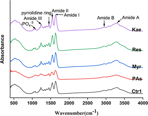
Representative ATR-FTIR spectra of dentin collagen treated with PA, myricetin, resveratrol, or kaempferol are shown in . Typical amide I, II, and III bands as well as the scissoring mode of CH2 groups, which represent dentin collagen, were observed in all groups. There were no obvious changes in the positions of the amide I band in the PAs, Myr, Res, and Kae groups. The AIII/A1454 value for the PAs group was 0.5, whereas those for the Ctr1, Myr, Res, and Kae groups were all approximately equal to 1.0 ().
Table 1 AIII/A1451 Value of Each Group with Different Pretreatment Conditions
Figure 2 ATR-FTIR spectrum of each group with different pretreatment conditions.
Activity Analysis of MMPs
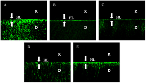
The results of in situ zymography for each group are presented in . A strong and wide green fluorescence band was observed in the HL of the Ctr1 group (), indicating that fluorescein-conjugated gelatin was strongly hydrolyzed. Compared with the Ctr1 group, the fluorescence intensities of the PAs, Myr, Res, and Kae groups () were significantly weakened, and the fluorescence band width was narrowed, indicating inhibited enzymatic activity; the weakest and strongest inhibitions were observed in the Myr () and Kae () groups, respectively.
Figure 3 Typical images of quenched fluorescein-conjugated gelatin substrates of each group. (A) No pretreatment; (B) Pretreatment with PA/ethanol solution; (C) Pretreatment with myricetin/ethanol solution; (D) Pretreatment with resveratrol/ethanol solution; (E) Pretreatment with kaempferol/ethanol solution. R = resin, D = dentin, HL = hybrid layer (between arrowheads).
Characteristics of Dentin Remineralization
Color Change of Dentin Caused by the Four Polyphenols
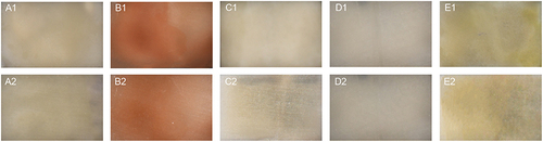
Before remineralization, compared with the Ctr1 group (), the dentin surfaces of the PAs, Kae, Myr, and Res groups showed a brownish red color (), a dark yellow color (), no obvious staining (), and no obvious staining (), respectively. After remineralization, except for the Ctr1 group (), the color lightened in all polyphenol pretreated groups (), although the dentin surface color remained most visible in the PAs group (). Typical images are shown in .
Figure 4 Color changes of dentin surface before (A1–E1) and after (A2–E2) remineralization caused by four polyphenols primers. (A1 and A2) No pretreatment; (B1 and B2) Pretreatment with PA/ethanol solution; (C1 and C2) Pretreatment with myricetin/ethanol solution; (D1 and D2) Pretreatment with resveratrol/ethanol solution; (E1 and E2) Pretreatment with kaempferol/ethanol solution.
SEM/EDS
shows typical SEM images of the remineralized dentin surfaces. The dentin tubules in the Ctr0 group remained completely open ( and ), and the collagen fiber morphology on the tubular wall and between dentin tubules was visible. The dentin tubules in the Ctr1 group remained open, but a small number of crystals formed between the collagen fibers and within the dentin tubules; moreover, the collagen morphology became blurred ( and ). The PAs, Myr, Res, and Kae groups showed a large increase in crystal formation, by which the collagen morphology was completely covered; even the dentin tubule diameter appeared to be significantly reduced ( and ).
Figure 5 Representative SEM images (a) of dentin surfaces after different treatments. (A1 and A2) No treatment; (B1 and B2) Polyphenol-free/ethanol solution pretreatment + remineralization; (C1 and C2) Proanthocyanidins/ethanol solution pretreatment + remineralization; (D1 and D2) Myricetin/ethanol solution pretreatment + remineralization; (E1 and E2) Resveratrol/ethanol solution pretreatment + remineralization; (F1 and F2) Kaempferol/ethanol solution pretreatment + remineralization. Magnification: ×10,000 (A1–F1); ×50,000 (A2–F2). (b) The results of the mapping of each group (At%: Atomic percentage). (c) The results of the EDS of each group (Wt%: Weight percentage).
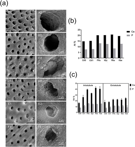
shows the results of the mapping of Ca and P contents on each group. The Ca and P contents were lowest in the Ctr0 group, followed by the Ctr1 group; compared with the Ctr1 group, the Ca and P contents were significantly increased in the PAs, Myr, Res, and Kae groups.
shows the results of the EDS of Ca and P contents on each group. The Ca and P contents within the dentin tubules had the same trend as shown in . However, the Ca and P contents between dentin tubules almost remained unchanged.
XRD and ATR-FTIR
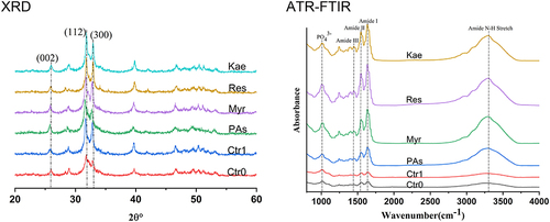
shows XRD and ATR-FTIR spectra of dentin in each group. Typical diffraction peaks representing HA were detected in all groups at 2θ = 25.9 (002), 2θ = 31.8 (112), and 2θ = 32.9 (300). After remineralization, most of the characteristic HA peaks increased. In the ATR-FTIR spectra, the PO43-, amide I, and amide II groups were detected from 885 to 1190 cm−1, 1600 to 1700 cm−1, and 1510 to 1580 cm−1, respectively; the amide III and N-H groups were detected from 1220 to 1340 cm−1. The intensities of typical diffraction peaks in the Ctr1 group were nearly the same as those in the Ctr0 group, but lower than those in the four polyphenol groups. Combining with XRD and ATR-FTIR results, the formation of HA was obtained.
Figure 6 XRD and ATR-FTIR spectra of dentin in each group after different treatments.
VHN
The VHN of the dentin surfaces for each group is shown in . The type of primer had a significant effect on the VHN (p<0.001). The Ctr0 group had the lowest VHN, and the Ctr1 group, which was treated only with remineralization, achieved significantly higher VHN. The VHN of the PAs, Myr, Res, and Kae groups were further significantly increased after treatment with polyphenol/ethanol solution primer compared with those of the Ctr1 group, with the largest VHN in the PAs group (p < 0.001), followed by the Myr group (p < 0.001); the smallest VHN were found in the Res and Kae groups with no significant difference (p = 0.178).
Table 2 Vickers Hardness Numbers (VHN, Mean and Standard Deviation) of Dentin After Different Experimental Primer Treatments
Micro-CT
The reconstructed images of dentin are shown in , and the V% results are shown in . Both primer type and zone of interest had a significant effect on the V% (p < 0.001), and there is a significant interaction between these two factors (p < 0.001). The V% values of the external and internal zones of interest were lowest in the Ctr0 group, followed by the Ctr1 group (p < 0.001). In the external zone of interest, the V% value was greater in the PAs group than in the Myr group (p < 0.001), whereas the V% values were smaller in the Res and Kae groups, with no statistically significant difference (p =0.647). In the internal zone of interest, the V% values were largest and smallest in the Myr and PAs groups, respectively.
Table 3 Mineral Volume Percentage (V%, Mean and Standard Deviation) of Dentin After Different Experimental Treatments
Figure 7 Reconstructed image of dentin and different zones of interest.
The Tb. Sp values of the external zone of interest were 2.00 in all groups, whereas the Tb. Sp values of the internal zone of interest were different. The Ctr0 group showed the highest Tb. Sp value (6.54 μm), followed by the Ctr1 group (3.85 μm). A significant decrease in the Tb. Sp value was observed in the PAs (2.76 μm), Myr (2.56 μm), Kae (2.60 μm), and Res (2.50 μm) groups.
μTBS Testing
summarizes the μTBS results obtained after 24 h of water storage or after aging. The μTBS was significantly affected by the primer solution type and aging (p < 0.001); additionally, a significant interaction was observed between these two factors (p = 0.024).
Table 4 Mean Values (with Standard Deviations in Parentheses) of Micro-Tensile Bond Strength (MPa) for the Six Groups After 24 h of Water Storage and After Aging for 3 Months in Water at 37°C Plus 10,000 Thermocycles
After 24 h of water storage, the μTBS value was lowest in the Ctr0 group and increased in the Ctr1 group, although there was no statistical difference between the values in these two groups (p = 0.286). The μTBS values in the PAs, Myr, Res, and Kae groups were significantly higher than that in the Ctr1 group (p < 0.001), although there was no significant difference between the values in these four groups (p > 0.05). After aging, the μTBS decreased in all six groups; however, the PAs and Kae groups showed a higher decrease in μTBS values than the Myr and Res groups.
Nanoleakage
The representative SEM images of each group after 24 h of water storage or after aging are presented in . After 24 h of water storage, the Ctr0 group showed a large number of dense and continuous depositions with high-electron-density particles within the HL ( and ), and the Ctr1 group showed reduced silver deposition with the HL having continuous but sparse depositions ( and ). The PAs, Myr, and Kae groups showed only a few discontinuous particles scattered at the bottom of the HL (; and ), whereas the Res groups revealed almost no deposition of high-electron-density particles ( and ).
Figure 8 Representative SEM images of the bonded interfaces evaluated after 24 h of water storage (A1–F1 and A2–F2) and after aging for 3 months in water at 37°C plus 10,000 thermocycles (A3–F3 and A4–F4). (A1–A4) No treatment; (B1–B4) No pretreatment + remineralization; (C1–C4) PA/ethanol solution pretreatment + remineralization; (D1–D4) Myricetin/ethanol solution pretreatment + remineralization; (E1–E4) Resveratrol/ethanol solution pretreatment + remineralization; (F1–F4) Kaempferol/ethanol solution pretreatment + remineralization. Magnification: ×1000 (A1–F1 and A3–F3); ×2000 (A2–F2, A4–F4).
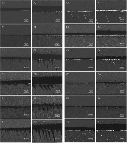
Aging aggravated nanoleakage for all the groups. The Ctr0 group demonstrated the greatest nanoleakage ( and ). Less silver deposition was observed in the Ctr1 group ( and ) than in the Ctr0 group. Among the four polyphenol/ethanol solution pretreatment groups, the amount of silver deposited was larger in the PAs ( and ) and Kae ( and ) groups than in the Myr ( and ) and Res ( and ) groups.
Discussion
The concentrations of the primers investigated in this study were based on previous studies;Citation16,Citation17,Citation25 these primers almost had no cytotoxic effects on cells, and could promote collagen cross-linking. Based on the observation of the color of dentin treated with the four polyphenols, only PA caused a significantly darker staining, which is contrary to the concept of esthetic restoration.
The ATR-FTIR results showed that the amide I group was mainly attributed to the stretching vibration of the C-O group; the amide II group was coupled by the N-H bending vibration and the C-N stretching vibration; and the amide III group resulted from the C-N stretching vibration and N-H bending vibration of the amide bond as well as the swinging vibration of the CH2 group on the glycine backbone and proline side chain, which was attributed to the organic collagen fibrils.Citation32,Citation33 The position of the amide I group was almost unchanged before and after the interaction of PA, myricetin, resveratrol, and kaempferol with dentin collagen, indicating that none of these four polyphenols disrupt the triple helix structure of collagen fibers.Citation34 The AIII/A1454 value, another indicator to evaluate the binding of polyphenols to collagen, is about 1.0 for pure collagen fibers. There was a small increase in the AIII/A1454 value (from 1.07 to 1.16) after the interaction of myricetin, resveratrol, and kaempferol with collagen fibers, indicating that these three polyphenols have no effect on the triple helix structure of collagen fibers.Citation35,Citation36 An AIII/A1454 value of 0.5 after PA interaction is due to the absence of AIII in the ATR-FTIR spectrum of pure PA, which has little effect on its AIII band; nevertheless, the effect is considerable on the band at 1451 cm−1.Citation37 Thus, the current experimental results suggest that the effective interaction of PA, myricetin, resveratrol, and kaempferol with dentin collagen is the result of hydrogen bond formation between the -OH and amide carbonyl groups on the amino acids of collagen. Hence, the null hypothesis 1 is rejected. The hydrogen bond exerts the main force, while covalent, ionic, and hydrophobic interactions play a secondary role.Citation38–40
It has been extensively demonstrated that the -OH groups of polyphenols can act as ligands to bind Ca2+, and in particular, the catechol groups seem to have a stronger chelating effect on Ca2+, which more effectively promotes the deposition of minerals to the collagen matrix.Citation41 In this experiment, the PILP method was adopted to guide the bottom–up assembly of apatite crystallites. Moreover, PA, myricetin, resveratrol, and kaempferol were used as primers to observe the formation of minerals. The typical diffraction peaks of HA and PO43- detected by XRD and ATR-FTIR, respectively, suggested that HA was regenerated in the dentin matrix. Importantly, the intensities of PO43 –and amide peaks increased, which is attributed to the positive effects of polyphenols on mineral formation and the protection of collagen fibers.
Dentin biomimetic remineralization can improve the mechanical properties of demineralized dentin,Citation10 and the differences in dentin remineralization triggered by the four polyphenols resulted in differences in dentin hardness. Concerning the microhardness findings in this experiment, the microhardness value of fresh dentin was 53.82 ± 1.76 MPa; partially demineralized dentin surface hardness caused by 35% phosphate gel was reduced to 38% of the original value, and increased to only 42% after simple remineralization. However, pretreatment with polyphenols for 30 min led to a significant increase in dentin hardness values, resulting in the recovery of 49–68% of fresh dentin, and thus partially compensating for the loss of dentin surface hardness due to demineralization; in this case, the biomimetic remineralization effect was in the following order: PA > myricetin > resveratrol and kaempferol. To analyze the reasons for the differences in the restoration of demineralized dentin hardness using the four polyphenols, we performed SEM/EDS and Micro-CT.
SEM/EDS analysis revealed that a large number of granular substances appeared in the dentin treated with PA, resulting in a more uneven and rough surface. This is because the oligomeric structure of PA molecules limits their permeability, and the large amount of PA molecules bound on the superficial collagen fibers of the HL rapidly transform ACP nanoparticles into HA, which hinders the formation of minerals in deeper collagen fibers to a large extent.Citation42 Therefore, the higher amount of superficial mineral deposition considerably contributed to the highest surface hardness achieved in the PA-treated dentin. The chemical structure and molar mass analysis of the four polyphenols indicated that myricetin, resveratrol, and kaempferol, which are unimolecular, low-molecular-weight compounds with high permeability, can quickly and freely penetrate all interstices to reach the deeper layers and effectively bind to the collagen fiber surface using the -OH group to exert a bottom-up remineralization effect. In cases where the mineralization time was not long enough, the dentin surface had a relatively lower crystalline material mass and Ca and P contents; therefore, it was relatively flat and smooth, as observed by SEM. Interestingly, the EDS results also revealed that remineralization preferentially occurs within than between the dentin tubules over a certain period. This suggests the potential of the four polyphenols in the treatment of dentin sensitivity, although it is beyond the scope of this study.
The quantitative analysis of dentin remineralization performed by micro-CT showed that only PA-treated dentin mineral deposition was highest in the superficial layer and lower in the deeper layers. This result could not be reversed even after the immersion time was extended to 30 min to reduce the effects of incomplete permeability. This finding is consistent with that of a previous study wherein greater mineral deposition was observed in the superficial layer when PA was applied for root surface dentin remineralization.Citation23 However, the other three polyphenols promoted dentin remineralization progressively from superficial to deep layers, indicating that these three polyphenols preferentially penetrated the deep layer to promote remineralization. This is more conducive to the rewrapping of the exposed collagen fibers at the bottom of the HL by HA, thereby avoiding the defect of weak mechanical properties of exposed collagen fibers in the HL. The experimental results are consistent with the present SEM and VHN results. Consequently, null hypothesis 3 is rejected.
It is well established that dentin biomimetic remineralization of the HL facilitates the improvement of the resin-dentin bonding performance.Citation9,Citation43,Citation44 The current μTBS findings revealed that the remineralization treatment alone increased the initial μTBS values by only approximately 5%, whereas the initial μTBS values increased by 30–36% after the pretreatment of the dentin surface with PA, myricetin, resveratrol, and kaempferol. This is because polyphenol-promoted biomimetic remineralization enhances the protection of minerals on collagen fibers.Citation9 Similar to the nanoleakage experiment findings, the effects of PA, myricetin, resveratrol, and kaempferol pretreatments followed by biomimetic remineralization were extremely significant in reducing silver deposition in initial samples.
Biomimetic remineralization, as a progressive dehydration mechanism of water-rich, resin-sparse collagen matrices, enables resin-dentin adhesive joint to resist degradation for a long period of time.Citation45 These four polyphenols are reportedly hydrophobic, which contributes to repelling the invasive effect of water molecules on the HL in a long-term aqueous environment, thereby inhibiting the dissolution of the resin matrix.Citation46 Another function of polyphenols is to protect collagen fibers from enzymatic degradation by inactivating MMPs around them through three mechanisms: down-regulation of endogenous protease expression, protease inactivation/silencing, and protection of cleavage sites within collagen.Citation47 The current in situ enzyme experiment also confirmed the MMP inhibitory properties of these four polyphenols. Despite the strong MMP inhibitory activity of PA, the present micro-CT and SEM/EDS analyses confirmed the formation of a large amount of shallow agglomerated crystals in the superficial layer of dentin after PA treatment, which, to some extent, hindered the formation of resin-dentin tags (the main functional structure of resin-dentin bonding). The low level of deep permeability reduces the protective effect of remineralization on collagen in the deeper layers. Although kaempferol provided superior remineralization in the deep layer compared with PA, the inhibitory activity of MMP was worse. Therefore, the resin-dentin specimens pretreated with PA and kaempferol after aging showed a greater increase in nanoleakage and a greater decrease in the μTBS value, which was not significantly different from the remineralization-only group findings, indicating that a valuable durability improvement could not be achieved by relying solely on PA and kaempferol to promote remineralization. Myricetin and resveratrol elicited a strong ability to penetrate deeper layers to provide biomimetic remineralization and inhibit the activity of MMP, a smaller increase in nanoleakage, and a smaller decrease in μTBS values after aging, thereby suggesting a potential clinical value for improving the durability of resin-dentin bonding. Therefore, null hypothesis 2 is rejected, and null hypothesis 4 is partially rejected.
Conclusion
Based on the current experimental results and within the limits of this study, all four polyphenols are effective in inhibiting MMP activity, accelerating the deposition of ACP nanoparticles, and promoting biomimetic remineralization of demineralized dentin when they are used as bonding interface primers; nevertheless, not all four polyphenols can improve the durability of resin-dentin bonding. Furthermore, the following conclusions can be drawn:
PA, myricetin, resveratrol, and kaempferol can modify collagen by forming hydrogen bonds, without denaturing its three-dimensional structure.
PA, myricetin, resveratrol, and kaempferol can inhibit MMP activity to protect collagen fibers from enzymatic hydrolysis.
PA, myricetin, resveratrol, and kaempferol can promote the deposition of ACP nanoparticles and biomimetic remineralization of demineralized dentin; the effect of PA was mainly exerted on the dentin surface, whereas the effects of myricetin, resveratrol, and kaempferol were more pronounced in deeper layers.
Myricetin and resveratrol can improve the bond durability of resin-dentin by inhibiting MMP activity and promoting biomimetic remineralization at the bottom of the HL with no visible color changes.
In summary, myricetin and resveratrol may avoid the aesthetic and shallow remineralization problems of PA, and represent potential approaches to achieve desirable bonding stability and reduce the frequent replacement of composite restorations in clinic.
Disclosure
The authors report no conflicts of interest in this work.
Acknowledgments
The authors thank Mr Yan Fang (NIGPAS, China) for providing technical support in SEM observation. This work was supported by the National Natural Science Foundation of China [grant number 81970927], the Key R & D Program of Jiangsu Province (Social Development) Project [grant BE2022797], the Medical Research Projects of the Health Department of Jiangsu Province [grant number M2020066, LKM2022025], Jiangsu Province Capability Improvement Project through Science, Technology and Education-Jiangsu Provincial Research Hospital Cultivation Unit (YJXYYJSDW4), and Jiangsu Provincial Medical Innovation Center (CXZX202227).
References
- Abou Neel EA, Aljabo A, Strange A, et al. Demineralization-remineralization dynamics in teeth and bone. Int J Nanomedicine. 2016;11:4743–4763. doi:10.2147/IJN.S107624
- Sharma V, Srinivasan A, Nikolajeff F, Kumar S. Biomineralization process in hard tissues: the interaction complexity within protein and inorganic counterparts. Acta Biomater. 2021;120:20–37. doi:10.1016/j.actbio.2020.04.049
- Cao CY, Mei ML, Li QL, Lo EC, Chu CH. Methods for biomimetic remineralization of human dentine: a systematic review. Int J Mol Sci. 2015;16(3):4615–4627. doi:10.3390/ijms16034615
- Liu Y, Tjaderhane L, Breschi L, et al. Limitations in bonding to dentin and experimental strategies to prevent bond degradation. J Dent Res. 2011;90(8):953–968. doi:10.1177/0022034510391799
- Frassetto A, Breschi L, Turco G, et al. Mechanisms of degradation of the hybrid layer in adhesive dentistry and therapeutic agents to improve bond durability--A literature review. Dent Mater. 2016;32(2):e41–53. doi:10.1016/j.dental.2015.11.007
- Lin HP, Lin J, Li J, Xu JH, Mehl C. In vitro remineralization of hybrid layers using biomimetic analogs. J Zhejiang Univ Sci B. 2016;17(11):864–873. doi:10.1631/jzus.B1600151
- Chesnick IE, Mason JT, Giuseppetti AA, Eidelman N, Potter K. Magnetic resonance microscopy of collagen mineralization. Biophys J. 2008;95(4):2017–2026. doi:10.1529/biophysj.107.120923
- Saeki K, Chien YC, Nonomura G, et al. Recovery after PILP remineralization of dentin lesions created with two cariogenic acids. Arch Oral Biol. 2017;82:194–202. doi:10.1016/j.archoralbio.2017.06.006
- Kim J, Arola DD, Gu L, et al. Functional biomimetic analogs help remineralize apatite-depleted demineralized resin-infiltrated dentin via a bottom-up approach. Acta Biomater. 2010;6(7):2740–2750. doi:10.1016/j.actbio.2009.12.052
- Gu LS, Huffman BP, Arola DD, et al. Changes in stiffness of resin-infiltrated demineralized dentin after remineralization by a bottom-up biomimetic approach. Acta Biomater. 2010;6(4):1453–1461. doi:10.1016/j.actbio.2009.10.052
- Jin W, Jin Y, Duan P, et al. Promotion of collagen mineralization and dentin repair by succinates. J Mater Chem B. 2022;10(30):5826–5834. doi:10.1039/D2TB01005D
- Liang K, Xiao S, Liu H, et al. 8DSS peptide induced effective dentinal tubule occlusion in vitro. Dent Mater. 2018;34(4):629–640. doi:10.1016/j.dental.2018.01.006
- Han B, Jaurequi J, Tang BW, Nimni ME. Proanthocyanidin: a natural crosslinking reagent for stabilizing collagen matrices. J Biomed Mater Res A. 2003;65(1):118–124. doi:10.1002/jbm.a.10460
- Jiang NW, Hong DW, Attin T, Cheng H, Yu H. Quercetin reduces erosive dentin wear: evidence from laboratory and clinical studies. Dent Mater. 2020;36(11):1430–1436. doi:10.1016/j.dental.2020.08.013
- Prakki A, Xiong Y, Bortolatto J, et al. Functionalized epigallocatechin gallate copolymer inhibit dentin matrices degradation: mechanical, solubilized telopeptide and proteomic assays. Dent Mater. 2018;34(11):1625–1633. doi:10.1016/j.dental.2018.08.297
- Baldion PA, Cortes CC, Castellanos JE, Betancourt DE. Effect of myricetin on odontoblast-like cells and its potential to preserve resin-dentin Bonds. J Mech Behav Biomed Mater. 2021;117:104392. doi:10.1016/j.jmbbm.2021.104392
- Peng W, Yi L, Wang Z, Yang H, Huang C. Effects of resveratrol/ethanol pretreatment on dentin bonding durability. Mater Sci Eng C Mater Biol Appl. 2020;114:111000. doi:10.1016/j.msec.2020.111000
- Cho J, Kim H, Yoo K-H, et al. The effect of kaempferol on the dentin bonding stability through matrix metalloproteinases inhibition and collagen crosslink in dentin biomodification. J Dent Sci. 2022. doi:10.1016/j.jds.2022.12.002
- Quideau S, Deffieux D, Douat-Casassus C, Pouysegu L. Plant polyphenols: chemical properties, biological activities, and synthesis. Angew Chem Int Ed Engl. 2011;50(3):586–621. doi:10.1002/anie.201000044
- Kharouf N, Haikel Y, Ball V. Polyphenols in dental applications. Bioengineering. 2020;7:3.
- Shavandi A, Bekhit AEA, Saeedi P, Izadifar Z, Bekhit AA, Khademhosseini A. Polyphenol uses in biomaterials engineering. Biomaterials. 2018;167:91–106. doi:10.1016/j.biomaterials.2018.03.018
- Fine AM. Oligomeric proanthocyanidin complexes: history, structure, and phytopharmaceutical applications. Altern Med Rev. 2000;5(2):144–151.
- Epasinghe DJ, Yiu C, Burrow MF. Synergistic effect of proanthocyanidin and CPP-ACFP on remineralization of artificial root caries. Aust Dent J. 2015;60(4):463–470. doi:10.1111/adj.12249
- Epasinghe DJ, Burrow MF, Yiu CKY. Effect of proanthocyanidin on ultrastructure and mineralization of dentine collagen. Arch Oral Biol. 2017;84:29–36. doi:10.1016/j.archoralbio.2017.09.012
- Abdelshafi MA, Fathy SM, Elkhooly TA, Reicha FM, Osman MF. Bond strength of demineralized dentin after synthesized collagen/hydroxyapatite nanocomposite application. J Mech Behav Biomed Mater. 2021;121:104590. doi:10.1016/j.jmbbm.2021.104590
- Tay FR, Pashley DH. Guided tissue remineralisation of partially demineralised human dentine. Biomaterials. 2008;29(8):1127–1137. doi:10.1016/j.biomaterials.2007.11.001
- Hu WH, Wang HY, Xia YT, et al. Kaempferol, a major flavonoid in ginkgo folium, potentiates angiogenic functions in cultured endothelial cells by binding to vascular endothelial growth factor. Front Pharmacol. 2020;11:526. doi:10.3389/fphar.2020.00526
- Murata M, Sato D, Hino J, et al. Acid-insoluble human dentin as carrier material for recombinant human BMP-2. J Biomed Mater Res A. 2012;100(3):571–577. doi:10.1002/jbm.a.33236
- Han F, Jin X, Yuan X, Bai Z, Wang Q, Xie H. Interactions of two phosphate ester monomers with hydroxyapatite and collagen fibers and their contributions to dentine bond performance. J Dent. 2022;122:104159. doi:10.1016/j.jdent.2022.104159
- Chen C, Mao C, Sun J, et al. Glutaraldehyde-induced remineralization improves the mechanical properties and biostability of dentin collagen. Mater Sci Eng C Mater Biol Appl. 2016;67:657–665. doi:10.1016/j.msec.2016.05.076
- Besinis A, van Noort R, Martin N. Remineralization potential of fully demineralized dentin infiltrated with silica and hydroxyapatite nanoparticles. Dent Mater. 2014;30(3):249–262. doi:10.1016/j.dental.2013.11.014
- Nashy EH, Osman O, Mahmoud AA, Ibrahim M. Molecular spectroscopic study for suggested mechanism of chrome tanned leather. Spectrochim Acta A Mol Biomol Spectrosc. 2012;88:171–176. doi:10.1016/j.saa.2011.12.024
- He L, Mu C, Shi J, Zhang Q, Shi B, Lin W. Modification of collagen with a natural cross-linker, procyanidin. Int J Biol Macromol. 2011;48(2):354–359. doi:10.1016/j.ijbiomac.2010.12.012
- Li K, Yao C, Sun Y, et al. Enhancing resin-dentin bond durability using a novel mussel-inspired monomer. Mater Today Bio. 2021;12:100174. doi:10.1016/j.mtbio.2021.100174
- Albu MG, Ghica MV, Leca M, et al. Doxycycline delivery from collagen matrices crosslinked with tannic acid. Mol Cryst Liq Cryst. 2010;523(1):669–677. doi:10.1080/15421401003724159
- Chen W, Jin H, Zhang H, et al. Synergistic effects of graphene quantum dots and carbodiimide in promoting resin-dentin bond durability. Dent Mater. 2021;37(10):1498–1510. doi:10.1016/j.dental.2021.07.004
- Liu Y, Wang Y. Proanthocyanidins’ efficacy in stabilizing dentin collagen against enzymatic degradation: MALDI-TOF and FTIR analyses. J Dent. 2013;41(6):535–542. doi:10.1016/j.jdent.2013.03.007
- Pierpoint WS. o-Quinones formed in plant extracts. Their reactions with amino acids and peptides. Biochemical J. 1969;112(5):609–616. doi:10.1042/bj1120609
- McRae JM, Falconer RJ, Kennedy JA. Thermodynamics of grape and wine tannin interaction with polyproline: implications for red wine astringency. J Agric Food Chem. 2010;58(23):12510–12518. doi:10.1021/jf1030967
- Shahidi F. Encyclopedia of Food Chemistry: Protein–Phenol Interactions. Elsevier; 2018.
- Zhang X, Li Z, Yang P, et al. Polyphenol scaffolds in tissue engineering. Mater Horiz. 2021;8(1):145–167. doi:10.1039/D0MH01317J
- Xie Q, Bedran-Russo AK, Wu CD. In vitro remineralization effects of grape seed extract on artificial root caries. J Dent. 2008;36(11):900–906. doi:10.1016/j.jdent.2008.07.011
- Tay FR, Pashley DH. Biomimetic remineralization of resin-bonded acid-etched dentin. J Dent Res. 2009;88(8):719–724. doi:10.1177/0022034509341826
- Gungormus M, Tulumbaci F. Peptide-assisted pre-bonding remineralization of dentin to improve bonding. J Mech Behav Biomed Mater. 2021;113:104119. doi:10.1016/j.jmbbm.2020.104119
- Kim YK, Mai S, Mazzoni A, et al. Biomimetic remineralization as a progressive dehydration mechanism of collagen matrices--implications in the aging of resin-dentin bonds. Acta Biomater. 2010;6(9):3729–3739. doi:10.1016/j.actbio.2010.03.021
- Tang HR, Covington AD, Hancock RA. Structure–activity relationships in the hydrophobic interactions of polyphenols with cellulose and collagen. Biopolymers. 2003;70:403–413. doi:10.1002/bip.10499
- Bedran-Russo AK, Pauli GF, Chen SN, et al. Dentin biomodification: strategies, renewable resources and clinical applications. Dent Mater. 2014;30(1):62–76. doi:10.1016/j.dental.2013.10.012