 ?Mathematical formulae have been encoded as MathML and are displayed in this HTML version using MathJax in order to improve their display. Uncheck the box to turn MathJax off. This feature requires Javascript. Click on a formula to zoom.
Abstract
We prepared solid dispersions (SDs) of tanshinone IIA (TSIIA) with silica nanoparticles, which function as dispersing carriers, using a spray-drying method and evaluated their in vitro dissolution and in vivo performance. The extent of TSIIA dissolution in the silica nanoparticles/TSIIA system (weight ratio, 5:1) was approximately 92% higher than that of the pure drug after 60 minutes. However, increasing the content of silica nanoparticles from 5:1 to 7:1 in this system did not significantly increase the rate or extent of TSIIA dissolution. The physicochemical properties of SDs were investigated using scanning electron microscopy, differential scanning calorimetry, X-ray powder diffraction, and Fourier transforms infrared spectroscopy. Studying the stability of the SDs of TSIIA revealed that the drug content of the formulation and dissolution behavior was unchanged under the applied storage conditions. In vivo tests showed that SDs of the silica nanoparticles/TSIIA had a significantly larger area under the concentration-time curve, which was 1.27 times more than that of TSIIA (P < 0.01). Additionally, the values of maximum plasma concentration and the time to reach maximum plasma concentration of the SDs were higher than those of TSIIA and the physical mixing system. Based on these results, we conclude that the silica nanoparticle based SDs achieved complete dissolution, increased absorption rate, maintained drug stability, and showed improved oral bioavailability compared to TSIIA alone.
Introduction
Tanshinone IIA (TSIIA) is one of the major active components extracted from the root of Salvia miltiorrhiza Bunge and demonstrates various effects on the cardiovascular system, including vasorelaxation and a cardioprotective effect.Citation1–Citation4 However, the oral bioavailability of TSIIA is very low,Citation5,Citation6 likely due to poor water solubilityCitation7 and insufficient TSIIA dissolution rate.Citation8–Citation10
Drug release is a crucial and limiting step in oral drug bioavailabilityCitation11,Citation12 especially for drugs with low gastrointestinal solubility and high permeability.Citation13–Citation15 Previous studies have shown that the formation of solid dispersions (SDs) is a promising strategy for increasing the dissolution rate and solubility of drugsCitation16–Citation20 belonging to class II of the Biopharmaceutical Classification System.Citation21 Dissolution can be improved by reducing the particle size of the drug and increasing the surface area;Citation22,Citation23 drug wettability can be improved by mixing the drug with a high concentration of the carrier in the surrounding solution;Citation24,Citation25 and the drug can be transformed from a crystalline to an amorphous state.Citation26 Currently, the typical carriers widely used for the formulation of SDs consist of organic materials that are highly water soluble and/or polymeric in nature.Citation27–Citation30 In addition to these carriers, inert hydrophilic carriers, such as inorganic silica-based materialsCitation31,Citation32 which have relatively large specific surface areas, and thus produce powders in which the drug may be highly dispersed, are receiving increasing attention. Takeuchi et alCitation33–Citation35 reported that SDs with porous silica or colloidal silica prepared using the spray-drying method can improve the dissolution property of tolbutamide and indomethacin. In this system, silica played an important role in controlling the polymorphs and inhibited crystallization of the drug in the SD under severe storage conditions.
Several grades of silica particles with different properties, such as pore structure, degree of hydrophilicity and hydrophobicity, and particle size are available for both porous and nonporous silica.Citation36 The specific functional features of different types of silica can also be exploited to regulate the drug release rate.Citation37–Citation39
Silica nanoparticles are amorphous in nature. The nanoparticles are small in size and have a large specific surface area, which improves drug dispersion in carriers, and significantly decreases the particle size of the drug,Citation40–Citation43 possibly to the molecular level.Citation44 Many silanol groups are present on the surface of silica nanoparticles, which may be able to form hydrogen bonds with the drug molecules during formulation of SDs.Citation45,Citation46 In addition, a high drug dissolution rate can be achieved by improving the wettability of the drug particles. SDs have been successfully prepared using silica nanoparticles as the carrier.Citation47 Further, compared to the crystalline drug, the SD of the drug shows significantly improved oral bioavailability. Formulations prepared by SD with water soluble polymer carriers such as polyethylene glycol (PEG)Citation48 or polyvinyl pyrrolidone (PVP)Citation49 tend to be sticky or tacky, which results in decreased recovery of the SD during preparation and difficulty in handling subsequent processes. However, silica nanoparticles may reduce the stickiness or tackiness imparted by excipients with low melting points and thus prevent processing problems. The resultant SD particles have free flowability and thus facilitate downstream processing to solid dosage forms. In addition, SD provides greater physical stability, which prevents recrystallization.Citation50
In this study, we prepared SDs of TSIIA using silica nanoparticles as dispersing carriers. The product with a nonporous structure consists of primary particles of about 50 ± 5 nm, which have a very high specific surface area (200 ± 30 m2/g). The SDs were produced using a spray-drying method. The first part of the study consisted of characterizing the SDs. A set of complementary techniques (differential scanning calorimetry [DSC], scanning electron microscopy [SEM], X-ray powder diffraction [XRPD], and Fourier transform infrared spectroscopy [FTIR]) was used to monitor the physical changes of TSIIA in the SDs. Subsequently, a drug stability test was performed on freshly prepared SDs and on SDs stored for 6 months at 40°C/75% relative humidity (RH). Finally, we examined the bioavailability of the SDs of silica nanoparticles-TSIIA in rats and compared it to that of TSIIA powders and its physical mixing systems (PMs).
Materials and methods
Instruments and materials
A TSIIA standard was obtained from the National Institute for the Control of Pharmaceutical and Biological Products (Beijing, People’s Republic of China). TSIIA with purity greater than 98% was supplied by the Nanjing Zelang Medical Technology Co, Ltd (Jiangsu, People’s Republic of China). Silica nanoparticles with a primary particle size of about 50 ± 5 nm were supplied by Hangzhou Wanjing New Material Co, Ltd (Hangzhou, People’s Republic of China). All reagents were analytical grade except methanol, which was chromatographic grade. Healthy male Sprague Dawley® rats were purchased from the Experimental Animal Center of Jiangsu Provincial Academy of Traditional Chinese Medicine (Jiangsu, People’s Republic of China).
Preparation of SDs and PMs
The SDs of TSIIA with silica nanoparticles were prepared using the spray-drying method. The drug-to-carrier ratios used in this study were 1:1, 1:3, 1:5, and 1:7 (by weight). TSIIA powder (200 mg) was dissolved in 500 mL of ethanol solution, into which variable amounts of silica nanoparticles (200, 600, 1000, and 1400 mg) were suspended. After ultrasonication for 15 minutes, this suspension was loaded into a spray-dryer (SD-06 Labplant; Labplant UK Limited, North Yorkshire, UK) at a rate of 8 mL/minute. The inlet and outlet temperatures of the drying chamber were maintained at 75°C and 38°C, respectively. The PMs were prepared by mixing TSIIA with silica nanoparticles at ratios of 1:5 (weight by weight) and then grinding them thoroughly using a mortar and pestle until a homogeneous mixture was obtained. All SDs were dried in a desiccator with blue silica gel under reduced pressure for 1 day before testing their physicochemical properties.
High-pressure liquid chromatographic analysis of TSIIA
The concentration of TSIIA in the dissolution medium was determined using a high-pressure liquid chromatography (HPLC) system equipped with a UV detector set at 270 nm. Separation was performed at 30°C on a Diamonsil™ RP-C18 (DIKMA, Lake Forest, CA, USA) column (250 mm × 4.6 mm, 5 μm). The mobile phase of methanol and water (85:15, volume:volume) was used at a flow rate of 1.0 mL · min−1. The samples were filtered through 0.45 μm membrane filters (EMD Millipore, Billerica, MA, USA) before use, and the injection volume was 20 μL. The linearity range of the calibration curve was within 0.420–8.400 μg · mL−1 with a correlation coefficient of 0.9999. The recovery rates for the TSIIA were in the range of 99%–102%, and the relative standard deviation was less than 2%. The intra-and interday precision values for the TSIIA were below 2%.
In vitro dissolution studies
The pharmaceutical performance of pure TSIIA and its SDs was evaluated using in vitro dissolution tests. The tests were performed according to the USP 24 method 2 (paddle method) in a BCZ-8A intellectualized dissolution apparatus (Tianjin University exact apparatus Co., Ltd., Tianjin, People’s Republic of China). Samples equivalent to 5 mg TSIIA were spread onto the surface of the dissolution medium (900 mL of distilled water containing 0.5% sodium dodecyl sulfate), which was thermostatically maintained at 37°C ± 0.5°C and stirred at 50 rpm. Samples were collected periodically and replaced with fresh dissolution medium. The samples were filtered by Amicon® Ultra-4 3K Centrifugal Filter Devices (EMD Millipore) and then were analyzed for TSIIA using high-performance liquid chromatography (HPLC) as described above. Dissolution experiments were performed in triplicate, and the average dissolution profiles and standard deviations were calculated.
DSC
Thermal analysis of TSIIA, silica nanoparticles, PMs, and SDs was performed using a differential scanning calorimeter (204A/G Phoenix® instrument; Netzsch, Selb, Germany). The samples were hermetically sealed in aluminum pans and heated from 25°C to 350°C at a rate of 10°C · min−1. All the DSC measurements were performed in a nitrogen atmosphere, and the flow rate was 50 mL · min−1.
SEM
The shape, surface, and cross-sectional morphology of the powder samples were observed using a scanning electron microscope (S-3000N; Hitachi, Tokyo, Japan).
XRPD
XRPD patterns of TSIIA, silica nanoparticles, PMs, and SDs were determined using an X-ray diffractometer (D8-Advance; Bruker, Karlsruhe, Germany), and the data were collected using primary monochromated radiation (Cu Kα1, λ = 1.5406 Å) over a 2θ range of 0°–70° at a step size of 0.04 and a dwell time of 10 seconds per step.
FTIR
FTIR measurements were carried out using an infrared spectrophotometer (Victor22; Bruker) at room temperature. Samples of TSIIA, silica nanoparticles, PMs, and SDs were previously ground and mixed thoroughly with potassium bromide in the sample. The scanning range was 400 to 4000 cm−1 and the resolution was 1 cm−1.
Stability test
Physical stability parameters, such as in vitro drug release and drug content were studied for SDs stored under stability conditions of 40°C/75% RH for 6 months.
Bioavailability study
Eighteen male Sprague Dawley® rats (Experimental Animal Center of Jiangsu Provincial Academy of Traditional Chinese Medicine) weighing 180–220 g were housed in an environmentally controlled breeding room (22°C ± 2°C, 60% ± 5% RH, and circulating fresh air) for 1-week before the start of the experiment. The rats were fed standard laboratory chow (Experimental Animal Center of Jiangsu Province Academy of Traditional Chinese Medicine) with water and fasted overnight but supplied with water ad libitum before the experiment. All experiments were approved by the Institutional Animal Care and Use Committee of Jiangsu Provincial Academy of Chinese Medicine. The rats were randomly divided into three groups. Three formulations (TSIIA and PMs and SDs with TSIIA/silica nanoparticles at a ratio of 1:5) at a dose equivalent to 60 mg/kg of TSIIA were orally administered to the three groups of six rats (eighteen rats total). Plasma samples (0.5 mL) were collected from the ophthalmic venous plexus of the rat at 0 (pretreatment), 0.33, 0.66, 1, 1.33, 1.66, 2, 3, 4, 6, 8, 12, and 24 hours after administration. Next, plasma was obtained after centrifugation for 10 minutes at 3000 rpm and stored at −4°C. The plasma (0.2 mL) was mixed with 0.2 mL of mobile solution containing loratadine (500 ng/mL) and used as an internal standard. Then, 600 μL of acetonitrile was added and vortex mixed for 5 minutes to fully precipitate the protein and extract TSIIA from the rat plasma. The sample was then centrifuged at 12,000 rpm for 10 minutes. Next, the supernatant was transferred to a clean centrifuge tube and dried under a stream of nitrogen in a water bath maintained at 38°C. The residue was resuspended in 200 μL of methanol and centrifuged for 10 minutes at 12,000 rpm. Aliquots (20 μL) of the supernatant were injected into the high-pressure liquid chromatography (HPLC) system for analysis, and the chromatographic conditions were consistent with those used for the HPLC analysis of TSIIA.
Data presentation and analysis
The pharmacokinetic parameters, including maximum plasma concentration (Cmax), time to reach Cmax (Tmax), half-life (t1/2) area under the plasma concentration-time curve (AUC0–t), and AUC0–∞, were calculated by compartmental analysis using the DAS software program version 1.0. Data were reported as the arithmetic mean values ± standard deviation (mean ± standard deviation). The P-values were calculated between the different pairs of formulations using two-tailed Student’s t-tests.
Results and discussion
In vitro dissolution study
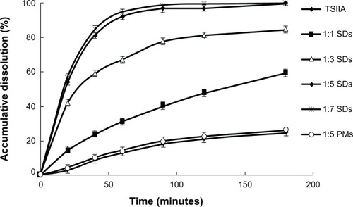
The dissolution profiles of TSIIA from the PMs and SDs are shown in . Pure TSIIA had a poor dissolution rate, and less than 30% of the drug dissolved after 120 minutes. The PMs showed a slight improvement in drug dissolution, which was most likely attributed to an increase in the wetting and local solubilization by the excipients in the diffusion layer,Citation51 which prevented drug aggregation,Citation52 and weak hydrogen bond interaction between the drug and carriers.Citation38 Nevertheless, the SDs with silica nanoparticles had a remarkably higher dissolution rate of TSIIA than pure TSIIA and corresponding PMs (). In addition, the dissolution rate of TSIIA increased with increasing ratios of TSIIA/silica nanoparticles. At a TSIIA/silica nanoparticles ratio of 1:5, more than 90% of the TSIIA was dissolved within 60 minutes, which was approximately a 7-fold increase compared to pure TSIIA. However, the dissolutions of TSIIA (1-hour) prepared at ratios of 1:5 and 1:7 were very similar, which were 92.1% and 94.3%, respectively. This improvement in the dissolution of the drug by the SDs may be attributed to several factors, including a significant reduction in particle size during the formation of a SD, increased surface area, the presence of an amorphous form of the drugCitation53 (as confirmed by DSC and XRD studies), and absorption of the TSIIA onto a large surface area of carrier nanoparticles to enhance drug dissolution, which has been previously described for silica based excipients since the early 1970s.Citation36
Figure 1 The dissolution profiles of TSIIA and SDs and PMs prepared using different ratios of TSIIA/silica nanoparticles (1:1, 1:3, 1:5, and 1:7 SDs and 1:5 PMs).
Note: Each point represents the mean ± standard deviation (n = 3).
Abbreviations: PMs, physical mixtures; SDs, solid dispersions; TSIIA, tanshinone IIA.
SEM
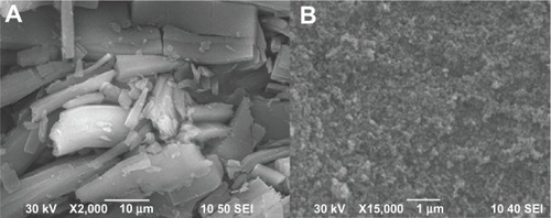
The SEM images of TSIIA and SDs prepared at a 1:5 ratio of TSIIA/silica nanoparticles are shown in . Pure TSIIA () consisted of a mixture of some large crystals with microparticles, which exhibited flat broken needles of different sizes. However, the SDs appeared as irregular-shaped particles, which were smaller than pure TSIIA. Moreover, the original morphology of TSIIA was not visible in the photomicrographs, which suggested that TSIIA dispersed uniformly into the carrier. Thus, the reduced particle size, increased surface area, and close contact between the silica nanoparticles and TSIIA may be responsible for the enhanced drug dissolution of the SDs.
Figure 2 Scanning electron microscopy photomicrographs of (A) TSIIA and (B) SDs prepared using a TSIIA/silica nanoparticles ratio of 1:5.
Abbreviations: SDs, solid dispersions; TSIIA, tanshinone IIA.
DSC
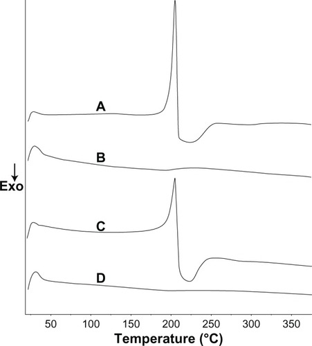
The DSC results of TSIIA, silica nanoparticles, PMs, and SDs consisting of TSIIA/silica nanoparticles at a ratio of 1:5 are shown in . Pure TSIIA () showed a sharp melting peak at an onset temperature of 205.6°C, which indicated its crystalline nature. Observations similar to ours have been reported by Li et al.Citation54 The DSC spectra of PMs () showed that the endothermic peak of the drug was found at 205.6°C, which indicated no interaction between TSIIA and silica nanoparticles in the PMs and that TSIIA existed in a virgin form in the system. DSC of the SDs prepared at a TSIIA/silica nanoparticle ratio of 1:5 () showed that the characteristic peak of TSIIA had completely vanished, which suggested that the drug may have been present in the SDs in an amorphous state, and thus was responsible for increasing the dissolution of the drug.
Figure 3 Differential scanning calorimetry curves of pure TSIIA (A), silica nanoparticles (B), and PMs (C) and SDs prepared at a TSIIA/silica nanoparticles ratio of 1:5 (D).
Abbreviations: Exo, exothermic direction; PMs, physical mixtures; SDs, solid dispersions; TSIIA, tanshi-none IIA.
XRPD
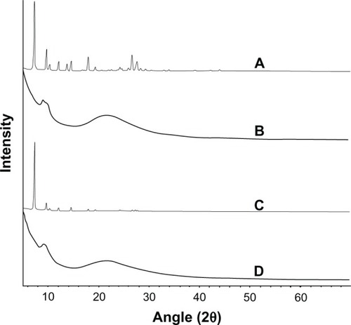
The results of XRPD studies of TSIIA, silica nanoparticles, PMs, and SDs prepared at a TSIIA/silica nanoparticle ratio of 1:5 are shown in . Pure TSIIA was characterized by prominent diffraction peaks in the 2θ angle range of 5°–30° in XRPD (). A broad peak centered at the 2θ angle of 22° was observed within the silica nanoparticle spectra due to its amorphous nature ().Citation55 All the major characteristic crystalline peaks of TSIIA were clearly observed in the PMs diffractograms. However, for the SDs, the discriminatory peaks of TSIIA were clearly abolished which indicated that TSIIA might exist in an amorphous state, which is consistent with the DSC results.
Figure 4 The X-ray powder diffractograms of TSIIA (A), silica nanoparticles (B) and PMs (C) and SDs with a TSIIA/silica nanoparticle ratio of 1:5 (w/w) (D).
Abbreviations: PMs, physical mixtures; SDs, solid dispersions; TSIIA, tanshinone IIA; w/w, weight by weight.
FTIR
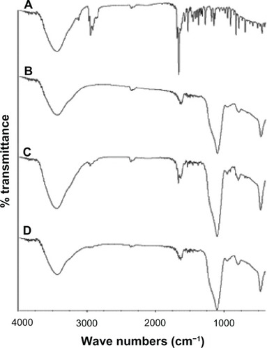
Interactions between TSIIA and silica nanoparticles in the SDs were evaluated with FTIR. The spectra of TSIIA, silica nanoparticles, PMs, and SDs are shown in . No differences were found between spectra of the PMs and SDs. It can be postulated that only weak van der Waals or hydrogen forces are involved in binding TSIIA onto silica nanoparticles.Citation56
Figure 5 The IR spectra of TSIIA (A), silica nanoparticles (B) and PMs (C) and SDs with a TSIIA/silica nanoparticle ratio of 1:5 (w/w) (D).
Abbreviations: IR, infrared; PMs, physical mixtures; SDs, solid dispersions; TSIIA, tanshinone IIA; w/w, weight by weight.
Stability test
It is well established that amorphous drugs formulated in the form of SDs tend to recrystallize upon storage. Thus, in the present study, the stability of the SDs was estimated by examining the drug content and in vitro dissolution studies under storage conditions of 75% RH and 40°C for 6 months. The drug content in SDs prepared at a TSIIA/silica nanoparticle ratio of 1:5 was remarkably unchanged (). The dissolution (60 minutes) of TSIIA from SDs stored for 6 months was slightly reduced compared to that of TSIIA from freshly prepared samples. These results confirmed that silica nanoparticles exhibited a strong stabilization effect on amorphous TSIIA in SDs under severe conditions. This may be because of an interaction between the drug and carriers, and the adsorption on the surface of the amorphous silica nanoparticles, which have been extensively described in the literature.Citation39
Table 1 Stability test of SDs
Plasma concentration-time curve and relative bioavailability
Under the analytical conditions, no endogenous peaks that interfered with TSIIA or loratadine (internal standard) were observed. In addition, the TSIIA peaks could be well distinguished from the loratadine peaks without any interference. Linearity was observed in concentrations of TSIIA within the 8–800 ng/mL range with correlation coefficients of over 0.99 (a typical calibration curve: y = 8.4706x + 5.7152, [r = 0.9976, n = 3]). The limit of quantification was 10 ng/mL. Intra- and interday precisions were below 7%, and the extraction recovery of TSIIA from rat plasma (n = 9) was 80.96% ± 3.82%. The plasma concentration-time curves of TSIIA after oral administration are shown in .
Figure 6 The mean plasma concentration-time curve of TSIIA in rats after oral administration of SDs, PMs, and TSIIA equivalent to 60 mg kg−1 of TSIIA (n = 6).
Notes: The values are represented as the mean ± standard deviation (n = 6/group/time point).
Abbreviations: PMs, physical mixtures; SDs, solid dispersions; TSIIA tanshi-none IIA.
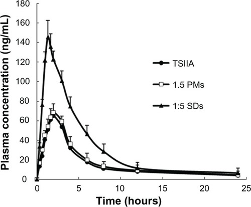
The pharmacokinetic parameters were calculated and are shown in . The AUC from 0 to 24 hours (AUC0–24h) of pure TSIIA and SDs was 379.50 and 862.47, respectively, which yielded a relative bioavailability of 227.3%. The Cmax of the SDs was 152.34 ng/mL, which was a 2.1-fold greater than that of TSIIA. Compared to TSIIA, the SDs had a significantly lower Tmax (P < 0.01), where SDs < TSIIA. This result shows that the TSIIA in the SDs exhibited a faster absorption rate and higher bioavailability, which may be attributed to several characteristics, including that SDs with silica nanoparticles as carriers can strongly potentiate their dissolution and increase the contact area with the gastrointestinal tract, resulting in an accelerated absorption rate and improved bioavailability.Citation57,Citation58 In addition, coexistence of both the carrier and TSIIA in the PMs by physical mixing showed a slightly faster absorption rate and a higher bioavailability, which is potentially a result of the slight improvement in drug release.
Table 2 The main pharmacokinetic parameters of TSIIA after oral administration of TSIIA, PMs or SDs in rats (n = 6) at a dose of 60 mg/kg
In this study, silica nanoparticles, which were used as drug carriers in SDs, showed a high specific surface area and hydrophilic properties, which impart unique advantages that improve their bioavailability. Silica is abundantly distributed in nature, it has good compatibility, and is deemed “Generally Recognized as Safe” (GRAS) by the US Food and Drug Administration (FDA).Citation59 Importantly, the fabrication of silica nanoparticles is simple, scalable, cost-effective, and controllable, and silica nanoparticles have been previously produced on an industrial scale as additives for cosmetics, drugs, printer toners, varnishes, and food. Furthermore, silica nanoparticles are currently being developed for a host of biomedical and biotechnological applications such as DNA transfection, drug delivery, and enzyme immobilization.Citation60–Citation62
Conclusion
In this study, we successfully prepared SDs consisting of TSIIA and silica nanoparticles using the spray-drying method. Both the drug dissolution behavior and oral bioavailability of TSIIA were largely improved by formulating the drug as a SD. In addition, the SDs of TSIIA had excellent stability. Thus, we conclude that silica nanoparticles have practical value as a novel and promising carrier for SDs.
Acknowledgments
This work was supported by the Open Fund of Key Laboratory of New Drug Delivery System of Chinese Materia Medica (2011NDDCM01001).
Disclosure
The authors report no conflicts of interest in this work.
References
- WangPWuXBaoYTSIIA prevents cardiac remodeling through attenuating NAD (P)H oxidase-derived reactive oxygen species production in hypertensive ratsPharmazie201166751752421812327
- SunDDWangHCWangXBTanshinone IIA: a new activator of human cardiac KCNQ1/KCNE1 (I(Ks)) potassium channelsEur J Pharmacol20085901–331732118573250
- GaoJYangGPiRTanshinone IIA protects neonatal rat cardiomyocytes from adriamycin-induced apoptosisTransl Res20081512798718201675
- FuJHuangHLiuJPiRChenJLiuPTSIIA protects cardiac myocytes against oxidative stress-triggered damage and apoptosisEur J Pharmacol20075681–321322117537428
- HaoHWangGCuiNLiJXieLDingZPharmacokinetics, absorption and tissue distribution of tanshinone IIA solid dispersionPlanta Med200672141311131717024606
- LiJWangGLiPHaoHSimultaneous determination of tanshinone IIA and cryptotanshinone in rat plasma by liquid chromatography-electrospray ionization-mass spectrometryJ Chromatogr B Analyt Technol Biomed Life Sci20058261–22630
- YuHSubediRKNepalPREnhancement of solubility and dissolution rate of cryptotanshinone, tanshinone I and tanshinone IIA extracted from Salvia miltiorrhizaArch Pharm Res20123581457146422941489
- YuXYLinSGZhouZWRole of P-glycoprotein in the intestinal absorption of tanshinone IIA, a major active ingredient in the root of Salvia miltiorrhiza BungeCurr Drug Metab20078432534017504222
- MaoSJHouSXLiangZIon-pair reversed-phase HPLC: assay validation of sodium IIA sulfonate in mouse plasmaJ Chromatogr B Analyt Technol Biomed Life Sci20068311–2163168
- WangLJiangXXuWLiCComplexation of tanshinone IIA with 2- hydroxypropyl-beta-cyclodextrin: effect on aqueous solubility, dissolution rate, and intestinal absorption behavior in ratsInt J Pharm20073411–2586717482783
- VasconcelosTSarmentoBCostaPSolid dispersions as strategy to improve oral bioavailability of poor water soluble drugsDrug Discov Today20071223–241068107518061887
- LennernäsHAbrahamssonBThe use of biopharmaceutic classification of drugs in drug discovery and development: current status and future extensionJ Pharm Pharmacol200557327328515807982
- LeunerCDressmanJImproving drug solubility for oral delivery using solid dispersionsEur J Pharm Biopharm2000501476010840192
- StreubelASiepmannJBodmeierRDrug delivery to the upper small intestine window using gastroretentive technologiesCurr Opin Pharmacol20066550150816890020
- TanakaNImaiKOkimotoKDevelopment of novel sustained-release system, disintegration-controlled matrix tablet (DCMT) with solid dispersion granules of nilvadipine (II): in vivo evaluationJ Control Release20061121515616545477
- ShiFZhaoJHLiuYWangZZhangYTFengNPPreparation and characterization of solid lipid nanoparticles loaded with frankincense and myrrh oilInt J Nanomedicine201272033204322619540
- KashanianSAzandaryaniAHDerakhshandehKNew surface-modified solid lipid nanoparticles using N-glutaryl phosphatidylethanolamine as the outer shellInt J Nanomedicine201162393240122114489
- LiJLiuPLiuJPZhangWLYangJKFanYQNovel Tanshinone IIA ternary solid dispersion pellets prepared by a single-step technique: in vitro and in vivo evaluationEur J Pharm Biopharm201280242643222119664
- Parmar KomalRSatapara VijayPShah SunnyRSheth NavinRImprovement of dissolution properties of lamotrigine by inclusion complexation and solid dispersion techniquePharmazie201166211912321434574
- KimMSKimJSParkHJChoWKChaKHHwangSJEnhanced bioavailability of sirolimus via preparation of solid dispersion nanoparticles using a supercritical antisolvent processInt J Nanomedicine201162997300922162657
- LöbenbergRAmidonGLModern bioavailability, bioequivalence and biopharmaceutics classification system: New scientific approaches to international regulatory standardsEur J Pharm Biopharm200050131210840189
- JunghannsJUMüllerRHNanocrystal technology, drug delivery and clinical applicationsInt J Nanomedicine20083329530918990939
- BikiarisDPapageorgiouGZStergiouAPhysicochemical studies on solid dispersions of poorly water-soluble drugs: evaluation of capabilities and limitations of thermal analysis techniquesThermochim Acta20054391–25867
- HeXQPeiLXTongHHZhengYComparison of spray freeze drying and the solvent evaporation method for preparing solid dispersions of baicalein with Pluronic F68 to improve dissolution and oral bioavailabilityAAPS Pharm Sci Tech2011121104113
- KaravasEKtistisGXenakisAGeorgarakisEEffect of hydrogen bonding interactions on the release mechanism of felodipine from nanodispersions with polyvinylpyrrolidoneEur J Pharm Biopharm200663210311416675209
- PokharkarVBMandpeLPPadamwarMNAmbikeAAMahadikKRParadkarADevelopment, characterization and stabilization of amorphous form of a low Tg drugPowder Technol200616712025
- YangCXuXWangJAnZUse of the co-grinding method to enhance the dissolution behavior of a poorly water-soluble drug: generation of solvent-free drug-polymer solid dispersionsChem Pharm Bull (Tokyo)201260783784522790815
- KolašinacNKachrimanisKHomšekIGrujićBDurićZIbrićSSolubility enhancement of desloratadine by solid dispersion in poloxamersInt J Pharm20124361–216117022772487
- BleyHFussneggerBBodmeierRCharacterization and stability of solid dispersions based on PEG/polymer blendsInt J Pharm2010390216517320132875
- ZhaoXLiuXGanLZhouCMoJPreparation and physicochemical characterizations of tanshinone IIA solid dispersionArch Pharm Res201134694995921725816
- JondhaleSBhiseSPoreYPhysicochemical investigations and stability studies of amorphous gliclazideAAPS Pharm Sci Tech2012132448459
- MellaertsRMolsRJammaerJAIncreasing the oral bioavailability of the poorly water soluble drug itraconazole with ordered mesoporous silicaEur J Pharm Biopharm200869122323018164930
- TakeuchiHNagiraSYamamotoHKawashimaYSolid dispersion particles of amorphous indomethacin with fine porous silica particles by using spray-drying methodInt J Pharm20052931–215516415778053
- TakeuchiHNagiraSTanimuraSYamamotoHKawashimaYTabletting of solid dispersion particles consisting of indomethacin and porous silica particlesChem Pharm Bull (Tokyo)200553548749115863917
- TakeuchiHHandaTKawashimaYSpherical solid dispersion containing amorphous tolbutamide embedded in enteric coating polymers or colloidal silica prepared by spray-drying techniqueChem Pharm Bull (Tokyo)1987359380038062830037
- MonkhouseDCLachJLUse of adsorbents in enhancement of drug dissolution. IIJ Pharm Sci1972619143514414341625
- ChauhanBShimpiSParadkarAPreparation and evaluation of glibenclamide-polyglycolized glycerides solid dispersions with silicon dioxide by spray drying techniqueEur J Pharm Sci200526221923016087324
- ChenBWangZQuanGIn vitro and in vivo evaluation of ordered mesoporous silica as a novel adsorbent in liquisolid formulationInt J Nanomedicine2012719920922275835
- DuXHeJSpherical silica micro/nanomaterials with hierarchical structures: synthesis and applicationsNanoscale20113103984400221918775
- CaoXDengWWFuMIn vitro release and in vitro-in vivo correlation for silybin meglumine incorporated into hollow-type mesoporous silica nanoparticlesInt J Nanomedicine2012775376222393284
- GalagudzaMMKorolevDVSoninDLTargeted drug delivery into reversibly injured myocardium with silica nanoparticles: surface functionalization, natural biodistribution, and acute toxicityInt J Nanomedicine2010523123720463939
- CorbalanJJMedinaCJacobyAMalinskiTRadomskiMWAmorphous silica nanoparticles aggregate human platelets: potential implications for vascular homeostasisInt J Nanomedicine2012763163922334785
- GalagudzaMKorolevDPostnovVPassive targeting of ischemic-reperfused myocardium with adenosine-loaded silica nanoparticlesInt J Nanomedicine201271671167822619519
- ThoILiepoldBRosenbergJMaegerleinMBrandlMFrickerGFormation of nano/micro-dispersions with improved dissolution properties upon dispersion of ritonavir melt extrudate in aqueous mediaEur J Pharm Sci2010401253220172027
- WangLCuiFDSunadaHPreparation and evaluation of solid dispersions of nitrendipine prepared with fine silica particles using the melt-mixing methodChem Pharm Bull (Tokyo)2006541374316394546
- YassinAEAlanaziFKEl-BadryMAlsarraIABarakatNSAlanaziFKPreparation and characterization of spironolactone-loaded gelucire microparticles using spray-drying techniqueDrug Dev Ind Pharm200935329730418821154
- Van EerdenbrughBVan SpeybroeckMMolsRItraconazole/TPGS/Aerosil200 solid dispersions: characterization, physical stability and in vivo performanceEur J Pharm Sci200938327027819686846
- KimMSKimJSParkHJChoWKChaKHHwangSJEnhanced bioavailability of sirolimus via preparation of solid dispersion nanoparticles using a supercritical antisolvent processInt J Nanomedicine201162997300922162657
- GuedesFLde OliveiraBGHernandesMZSolid dispersions of imidazolidinedione by PEG and PVP polymers with potential antischistosomal activitiesAAPS Pharm Sci Tech2011121401410
- JondhaleSBhiseSPoreYPhysicochemical investigations and stability studies of amorphous gliclazideAAPS Pharm Sci Tech2012132448459
- VippaguntaSRMaulKATallavajhalaSGrantDJSolid state characterization of nifedipine solid dispersionsInt J Pharm20022361–211112311891075
- KawabataYYamamotoKDebariKOnoueSYamadaSNovel crystalline solid dispersion of tranilast with high photostability and improved oral bioavailabilityEur J Pharm Sci201039425626220038453
- JunghannsJUMüllerRHNanocrystal technology, drug delivery and clinical applicationsInt J Nanomedicine20083329530918990939
- LiJLiuPLiuJPZhangWLYangJKFanYQNovel Tanshinone IIA ternary solid dispersion pellets prepared by a single-step technique: in vitro and in vivo evaluationEur J Pharm Biopharm201280242643222119664
- WangLLuAWangCZhengXZhaoDLiuRNano-fibriform production of silica from natural chrysotileJ Colloid Interface Sci2006295243643916212972
- PlaninšekOKovačičBVrečerFCarvedilol dissolution improvement by preparation of solid dispersions with porous silicaInt J Pharm20114061–2414821219991
- LeunerCDressmanJImproving drug solubility for oral delivery using solid dispersionsEur J Pharm Biopharm2000501476010840192
- HuKLCaoSHuFQFengJFEnhanced oral bioavailability of docetaxel by lecithin nanoparticles: preparation, in vitro, and in vivo evaluationInt J Nanomedicine201273537354522848177
- TangFLiLChenDMesoporous silica nanoparticles: synthesis, biocompatibility and drug deliveryAdv Mater201224121504153422378538
- FalahatiMSabouryAAMa’maniLShafieeARafieepourHAThe effect of functionalization of mesoporous silica nanoparticles on the interaction and stability of confined enzymeInt J Biol Macromol20125041048105422421216
- GalagudzaMKorolevDPostnovVPassive targeting of ischemic-reperfused myocardium with adenosine-loaded silica nanoparticlesInt J Nanomedicine201271671167822619519
- CheangTYTangBXuAWPromising plasmid DNA vector based on APTES-modified silica nanoparticlesInt J Nanomedicine201271061106722403488