 ?Mathematical formulae have been encoded as MathML and are displayed in this HTML version using MathJax in order to improve their display. Uncheck the box to turn MathJax off. This feature requires Javascript. Click on a formula to zoom.
Abstract
The aim of this research was to synthesize and develop a new method for the preparation of iron oxide (Fe3O4) nanoparticles on talc layers using an environmentally friendly process. The Fe3O4 magnetic nanoparticles were synthesized using the chemical co-precipitation method on the exterior surface layer of talc mineral as a solid substrate. Ferric chloride, ferrous chloride, and sodium hydroxide were used as the Fe3O4 precursor and reducing agent in talc. The talc was suspended in deionized water, and then ferrous and ferric ions were added to this solution and stirred. After the absorption of ions on the exterior surface of talc layers, the ions were reduced with sodium hydroxide. The reaction was carried out under a nonoxidizing oxygen-free environment. There were not many changes in the interlamellar space limits (d-spacing = 0.94−0.93 nm); therefore, Fe3O4 nanoparticles formed on the exterior surface of talc, with an average size of 1.95–2.59 nm in diameter. Nanoparticles were characterized using different methods, including powder X-ray diffraction, transmission electron microscopy, emission scanning electron microscopy, energy dispersive X-ray spectroscopy, and Fourier transform infrared spectroscopy. These talc/Fe3O4 nanocomposites may have potential applications in the chemical and biological industries.
Introduction
In the last two decades, inorganic materials with a layered structure have been used to improve the properties of polymer materials with the polymer molecular chain intercalating galleries of adjacent inorganic layers to form nanocomposites, which consist of novel metals and have important applications in medical identification, catalysis, sensors, optics, and electronics due to their shape and size.Citation1–Citation8 Moreover, magnetite (Fe3O4) nanoparticles have attracted increasing research attention in the field of environmental remediation.Citation9
The application of Fe3O4 nanoparticles in the environmental field is mainly due to their much better adsorption and reduction activities than their traditional macro- or microparticle counterparts.Citation10 In addition, Fe3O4 can be easily separated and collected by an external magnetic field. This extraordinary advantage is especially useful for the recovery or reuse of Fe3O4 nanoparticles.Citation11,Citation12 The application of small iron oxide particles in vitro diagnostics has been practiced for nearly 40 years. In the last decade, there has been an increase in the number of investigations using various types of iron oxides including γ-Fe3O4 (maghemite), Fe3O4, and α-Fe3O4 (hematite) particles, which have a core ranging from 5–20 nm in diameter. Of these, Fe3O4 is a very promising candidate0020since its biocompatibility has already been proven.Citation13,Citation14 Fe3O4, is a common magnetic iron oxide that has a cubic inverse spinel structure with oxygen forming a face-centered cubic close packing and iron (Fe) cations occupying interstitial tetrahedral sites and octahedral sites.Citation15
Talc, Mg3Si4O10(OH)2, is a natural compound widely used in the form of a fine powder in several industrial products such as ceramics, putties, paper, paints, flame and ignition retardants, and as an active filler in polymer/silicates composites to improve mechanical characteristics (eg, strength, elasticity, shock resistance) and the nucleation of polymers.Citation16,Citation17 The structure of talc is the well-known 2:1 (T–O–T) layer configuration consisting of a octahedral magnesium (Mg) coordinated sheet (O) sandwiched between two tetrahedral silicon (Si) coordinated sheets (T). The talc structure does not present residual surface charge and the bonds between layers are provided by weak electrostatic and van der Waals forces.Citation18 Talc is known as a two polytypic structure: a two-layer monoclinic 2M structure and a one-layer triclinic 1C1 polytype.Citation19–Citation22
The co-precipitation method is widely used to prepare iron oxide nanoparticles, but the main achievement of this paper was to provide very fine particles (1.95–2.59 nm) with low agglomeration. These magnetic nanoparticles enable easy separation during material preparation and make them suitable for many applications. Moreover, surface decoration of talc with magnetic nanoparticles has led to a new class of nanocomposites materials, which could be also used for environmental purposes as absorbents of metal ions in wastewater treatment.Citation23,Citation24
In this research, talc/Fe3O4 nanocomposites were synthesized at room temperature on the exterior surface of talc layers in aqueous solution using ferric chloride (FeCl3), ferrous chloride (FeCl2), and sodium hydroxide (NaOH) as the iron precursor and reduction agent, respectively. Needless to say, to date, there has not been any research on talc/Fe3O4 nanocomposites using the wet chemical reducing method (ie, lamellar polymeric silicate), which is the subject of this study.
Material and methods
All reagents in this work were of analytical grade and used as received without further purification. Ferric chloride hexahydrate (FeCl3·6H2O) and ferrous chloride tetrahydrate (FeCl2 · 4H2O) of 96% were used as the iron precursor and also, talc powder (<10μ, 3MgO 4SiO2·H2O) were obtained from Sigma-Aldrich (St Louis, MO, USA). NaOH of 99% was obtained from Merck KGaA (Darmstadt, Germany). All these aqueous solutions were used with deionized water.
Synthesis of talc/Fe3O4 nanocomposites
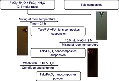
For the synthesis of talc/Fe3O4 nanocomposites, 2.0 g of talc was suspended in 70 mL deionized water, and then the molar ratio in FeCl3 solution was adjusted to 2:1 by adding a measured amount of Fe3+ and Fe2+. The ion solution suspended with talc composites was stirred for 1 hour for impregnation by the external surface of talc layers to prepare talc/Fe3+–Fe2+ composites. The 15 mL freshly prepared NaOH (2.0 M) was added to talc/Fe3+–Fe2+ composites suspension under continuous stirring. The suspensions were finally centrifuged washed twice with ethanol and double distilled water, and kept in a stove at 100°C. All the experiments were conducted at an ambient temperature and under a nonoxidizing oxygen-free environment through the flow of nitrogen gas.
Characterization methods and instruments
The prepared talc/Fe3O4 nanocomposites were characterized by powder X-ray diffraction (PXRD), transmission electron microscopy (TEM), scanning electron microscopy (SEM), energy dispersive X-ray spectroscopy (EDX), and Fourier transform infrared spectroscopy (FT-IR). The structures of the produced Fe3O4 nanoparticles in talc were examined using Philips X’pert PXRD (copper Kα radiation; PANalytical, Almelo, The Netherlands). The changes in the interlamellar spacing of talc and nanoparticles in talc were also studied by using PXRD in the small-angle range of 2θ (5–15 degrees). In addition, the interlamellar space was calculated based on the PXRD peak positions using Bragg’s equation. A wavelength of 0.15418 nm was used for these measurements. The PXRD patterns were recorded at a scan speed of 2 degrees/minute. The TEM observations were carried out using an H-7100 electron microscope (Hitachi Ltd Tokyo, Japan), and the particle size distributions were determined using UTHSCSA ImageTool version 3.00 (The University of Texas Health Science Center, San Antonio, TX, USA). Furthermore, SEM and EDX were performed (XL 30; Philips, Eindhoven, The Netherlands) to study the morphology of talc and talc/Fe3O4 nanocomposites. Moreover, the FT-IR spectra were recorded over the range of 200–4000 cm−1 utilizing the Series 100 FT-IR 1650 spectrophotometer (PerkinElmer, Waltham, MA, USA). After the reactions, the samples were centrifuged by using a high-speed centrifuge (Avanti® J-25; Beckman Coulter, Brea, CA, USA).
Results and discussion
The talc suspension was whitish gray in color, which turned to brown after the addition of FeCl3 · 6H2O and FeCl2 · 4H2O and then to a dark color after the addition of NaOH solution as a reducing agent suspension (–). Conventionally, Fe3O4 nanoparticles are prepared by adding a base to an aqueous mixture of Fe3+and Fe2+ chloride at a 2:1 molar ratio. The chemical reaction of Fe3O4 precipitation is given in EquationEquations 1(1) and Equation2
(2) . The overall reaction may be written as follows:Citation25
Figure 1 Flowchart of the synthesized talc/Fe3O4 nanocomposite powder in (A) talc and (B and C) talc/Fe3+–Fe2+ ion composites.
Abbreviations: EtOH, ethanol; Fe2+, ferrous ion; Fe3+, ferric ion; FeCl2 · 4H2O, ferrous chloride tetrahydrate; FeCl · 6H2O, ferric chloride hexahydrate; Fe3O4, magnetite; H2O, water; NaOH, sodium hydroxide.
The comparison between the PXRD patterns of talc and the prepared Fe3O4/talc nanocomposites under the chemical reduction route fell in the small-angle range of 2θ (9.35–9.50), which indicated the immobilized formation of Fe3O4 nanoparticles on the external surface of the talc layers. The TEM images and size distribution of talc/Fe3O4 nanocomposites showed that the mean diameter of the nanoparticles ranged from about 1.2–3.2 nm. Additionally, the SEM images indicated that there were no structural changes between the initial talc and talc/Fe3O4 nanocomposites. Furthermore, FT-IR spectra showed that there was no chemical interaction between the silicate layers and Fe3O4 nanoparticles in talc/Fe3O4 nanocomposites. The synthesized talc suspensions containing Fe3O4 nanoparticles were found to be unstable over a long period of time, displaying signs of precipitation.
PXRD
As shown in , the original d-spacing (ds) of talc at 2θ 9.35 degrees was 0.94 nm, which gradually decreased to 0.93 nm at 2θ 9.50 degrees by the formation of Fe3O4 nanoparticles on the surface of talc layers. This ds value was direct proof that the Fe3+ ions were bound only on the external surfaces and edges of the talc external layer space. Consequently, the metallic nanoparticles formed only at the exterior layer location, causing a decrease in the basal spacing of talc.Citation15 In this sample, the intensities of reflections and half-widths were constant; therefore, the parallel lamellar structure of the mineral clay was not disrupted by the formation of nanoparticles.
Figure 2 Powder X-ray diffraction of (A,C) talc and (B,D) talc/Fe3O4 nanocomposites with the related peaks.
Abbreviation: Fe3O4, magnetite.
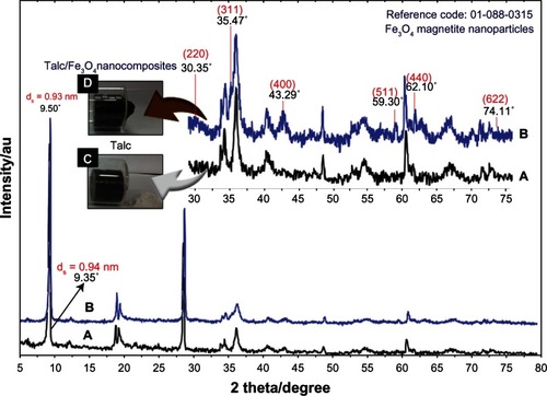
The comparison between the PXRD patterns of the talc and Fe3O4/talc nanocomposites in the small-angle range of 2θ (5–15 degrees) indicated the formation of the intercalated Fe3O4 nanostructure (). As shown in , the PXRD peaks in the wide-angle range of 2θ (30–80 degrees) ascertained that the peaks at 30.35, 35.47, 43.29, 59.30, 62.10, and 74.11 degrees related to the 220, 311, 400, 511, 440, and 622 AU crystalline structures of the Fe3O4 cubic nanocrystal with a spinel structure (reference code: 01-088-0315).Citation26,Citation27
The effect of the magnetic field on the talc and talc/Fe3O4 nanocomposite powder is shown in . These results confirm that there was a significant amount of Fe3O4 nanoparticles on the external layers of talc powder, because the Fe3O4 nanoparticles prepared in talc/Fe3O4 nanocomposites were attracted by magnets.
The average particle size of Fe3O4 nanoparticles in talc substrate can be calculated using the Scherrer Equationequation (3)(3) :
In this equation, K is the Scherrer constant with a value from 0.9–1 (shape factor), λ is the X-ray wavelength (1.5418 Å), β1/2 is the width of the XRD peak at half height, and θ is the Bragg angle. From the Scherrer equation, the average crystallite size of Fe3O4 nanoparticles for Fe3O4/talc nanocomposites is around < 5 nm, which is in line with the TEM and field emission SEM results discussed later.
TEM
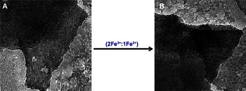
demonstrates TEM images for the size distribution of talc/Fe3O4 nanocomposites containing Fe3O4 nanoparticles. The TEM images and their size distributions revealed that the mean diameter ± standard deviation of Fe3O4 nanoparticles was about 2.27 ± 0.32 nm. Importantly, no morphologic differences were observed between the initial talc and Fe3O4 nanoparticles. The Fe3O4 nanoparticles prepared on the external layer of talc composites are shown by TEM in , which confirm that the structure of talc doesn’t change in FT-IR spectroscopy. The number of Fe3O4 nanoparticles counted in the TEM images was around 374. illustrates the TEM images of pure talc and talc after impregnation with aqueous Fe3+ and Fe2+ ions. demonstrates a typical talc clay image with homogeneously distributed clay flakes, and shows a TEM image for talc/Fe3+–Fe2+ composites without appearance of any nanoparticles.
Figure 3 Transmission electron microscopy image and histogram of particle size distribution for talc/magnetite nanocomposites.
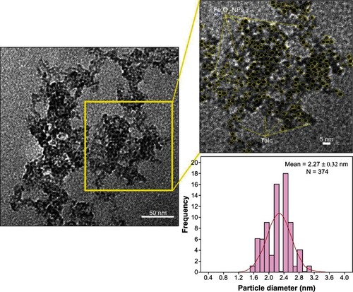
Figure 4 Transmission electron microscopy images of (A) pure talc and (B) talc/Fe3+–Fe2+ ion composites.
Abbreviations: Fe2+, ferrous ion; Fe3+, ferric ion.
SEM
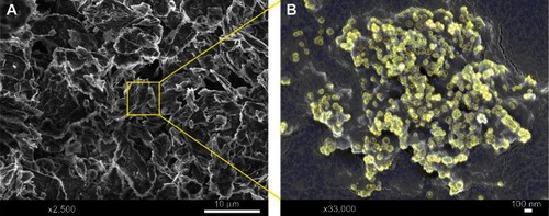
The Fe3O4 nanoparticles prepared on the external layer of talc composites are shown by SEM in , which confirm that the structure of talc doesn’t change in FT-IR spectroscopy. shows the SEM images for the talc/Fe3O4 nanocomposites synthesized with the co-precipitation method. These results confirm that the exterior surface layer of talc as substrate can effectively control the shape and size of the Fe3O4 nanoparticles. The external surface of talc/Fe3O4 nanocomposites with high magnification gradually become shinier due to the presence of small Fe3O4 nanoparticles that aggregate together and create large particles ().
Figure 5 Scanning electron microscopy micrographs of talc/magnetite nanocomposites with (A) low and (B) high magnification.
EDX
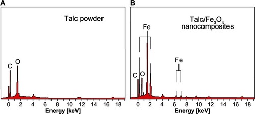
shows the EDX fluorescence spectra for the talc; the peaks around 1.7, 2.4, 2.6, 2.9, 3.6, 4.0, 4.5, 5.0, 6.0, 6.4, and 7.1 keV are related to the binding energies of talc. In , the peaks around 0.2, 0.8, 2.2, 6.4, and 7.0 keV are related to the Fe3O4 nanoparticle elements.Citation28 In addition, the EDX fluorescence spectra for the talc and talc/Fe3O4 nanocomposites confirm the presence of elemental compounds in the talc and Fe3O4 nanoparticles without any impurity peaks. The results indicate that the synthesized Fe3O4 nanoparticles are of high purity.
Figure 6 Energy dispersive X-ray spectroscopy of (A) talc and (B) talc/Fe3O4 nanocomposites.
Abbreviations: C, carbon; Fe, iron; Fe3O4, magnetite; O, oxygen.
FT-IR chemical analysis
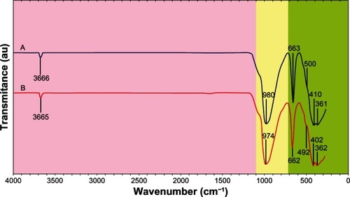
shows the comparison of FT-IR spectra for the silicate host structure of talc and talc/Fe3O4 nanocomposites with NaOH. The positions of vibrational bands in the region 400–1000 cm−1 corresponding to Si–O and other interlayer bonds remained unchanged, and a strong band at 980 cm−1 was associated with the stretching vibration of Si–O. The band at 663 cm−1 was also assigned to the stretching vibration of Si–O, which is usually taken as evidence for a three-dimensional amorphous silica phase. The band at 410–361 cm−1 was assigned to the Si–O–Si bending vibration. The FT-IR spectra indicated the rigidity of silicate layers and nonbonding chemical interaction between the silicate layers and Fe3O4 nanoparticles in talc/Fe3O4 nanocomposites. A broad peak was due to the presence of van der Waals interactions between the hydroxyl groups of H2O with an exterior layer of talc and the partial positive charge on the surface of Fe3O4.Citation15 These peaks, with the enhanced Fe3O4 in the talc/Fe3O4 nanocomposites, shifted to low wave numbers and there was no change in peak intensity.
Figure 7 Fourier transform infrared spectra of (A) talc and (B) talc/magnetite nanocomposites.
Conclusion
The Fe3O4 nanoparticles were successfully prepared by co-precipitation of Fe3+ and Fe2+ in talc as the substrate, with NaOH as the reducing agent. This method was demonstrated to be useful for the preparation of Fe3O4 nanoparticles. The Fe3O4 magnetic nanoparticles with a mean size of 1.95–2.59 nm on the talc layers were synthesized for the first time via a single-step co-precipitation reduction route. This is a cheap, facile, and environmentally friendly method which leads to the formation of Fe3O4 nanoparticles. Also, the Fe3O4 nanoparticles prepared by this route were attracted by magnets. This indicates that the Fe3O4 nanoparticles are formed in the exterior surface of the talc layer.
Acknowledgments
The authors are grateful to the staff of the Department of Chemistry UPM for their help in this research, and Institute of Bioscience (IBS/UPM) for technical assistance.
Disclosure
The authors report no conflicts of interest in this work.
References
- ShameliKAhmadMBJazayeriSDInvestigation of antibacterial properties silver nanoparticles prepared via green methodChem Cent J2012617322839208
- HainfeldJFSlatkinDNFocellaTMSmilowitzHMGold nanoparticles: a new X-ray contrast agentBr J Radiol20067993924825316498039
- SaitoTOhshimaSXuWCAgoHYumuraMIijimaSSize control of metal nanoparticle catalysts for the gas-phase synthesis of single-walled carbon nanotubesJ Phys Chem B200510921106471065216852292
- KellyKLCoronadoEZhaoLLSchatzGCThe optical properties of metal nanoparticles: the influence of size, shape, and dielectric environmentJ Phys Chem B20031073668677
- BallPGarwinLScience at the atomic scaleNature1992355761766
- CavicchiRESilsbeeRHCoulomb suppression of tunnelling rate from small metal particlesPhys Rev Lett1984521614531456
- YuLZhangYPreparation of nano-silver flake by chemical reduction methodRare Metal Mater Eng2010393401404
- ShameliKAhmadBMYunusWZIbrahimNADarroudiMSynthesis and characterization of silver/talc nano composites using the wet chemical reduction methodInt J Nanomedicine2010574375121042420
- FeiCLZhangYYangZSynthesis and magnetic properties of hard magnetic (CoFe2O4)–soft magnetic (Fe3O4) nano-composite ceramics by SPS technologyJ Magn Magn Mater20113231318111816
- ZhouQPramodaKPLeeJMWangKLooLSRole of interface in dispersion and surface energetics of polymer nanocomposites containing hydrophilic POSS and layered silicatesJ Colloid Interface Sci2011355122223021190693
- BookerNAKeirDPriestleyAJRitchieCBSudarmanaDLWoodsMASewage clarification with magnetite particlesWater Sci Technol1991237–917031712
- OrbellJDGodhinoLBiggerSWNguyenTMNgehLNOil spill remediation using magnetic particles: an experiment in environmental technologyJ Chem Educ1997741214461450
- WahajuddinArora SSuperparamagnetic iron oxide nanoparticles: magnetic nanoplatforms as drug carriersInt J Nanomedicine201273445347122848170
- WuWHeQJiangCMagnetic iron oxide nanoparticles: synthesis and surface functionalization strategiesNanoscale Res Lett200831139741521749733
- OliveiraaLRiosRFabrisJDSapagKGargVKLagoRMClay– iron oxide magnetic composites for the adsorption of contaminants in waterAppl Clay Sci2003224169177
- ShameliKAhmadMBAl-MullaEAJShabanzadehPBagheriSAntibacterial effect of silver nanoparticles on talc compositesRes Chem Intermed2013113
- GordonTPerlsteinBHoubaraOFelnerIBaninEMargelSSynthesis and characterization of zinc/iron oxide composite nanoparticles and their antibacterial propertiesColloids Surf A Physiochem Eng Asp201137418
- BrunoMPrencipeMValdreGAb initio quantum-mechanical modeling of pyrophyllite (Al2Si4O10[OH]2) and talc (Mg3Si4O10[OH]2) surfacesPhys Chem Miner20063316371
- PawleyARRedfernSATWoodBJThermal expansivities and compressibilities of hydrous phases in the system MgO–SiO2–H2O: talc, phase A and 10-Å phaseContrib Mineral Petrol1995122301307
- Perez-MaquedaLADuranAPerez-RodriguezJLPreparation of submicron talc particles by sonicationAppl Clay Sci2005281–4245255
- JamilNHPalaniandySAcid medium sonication: a method for the preparation of low density talc nanosheetsPowder Technol20102001–28790
- ZbikMSmartRInfluence of dry grinding on talc and kaolinite morphology: inhibition of nano-bubble formation and improved dispersionMiner Eng2005189969976
- ParkHMLeeWKParkCYChoWJHaCSEnvironmentally friendly polymer hybrids. Part I: mechanical, thermal, and barrier properties of thermoplastic starch/clay nanocompositesJ Mater Sci2003385909915
- YehJMYaoCTHsiehFCPreparation, characterization and electrochemical corrosion studies on environmentally friendly waterborne polyurethane/Na+–MMT clay nanocomposite coatingsEur Polym J2008441030463056
- WuSSunAZhaiFFe3O4 magnetic nanoparticles synthesis from tailings by ultrasonic chemical co-precipitationMater Lett2011651218821884
- YangHDuCJinSTangAPreparation and characterization of SnO2 nanoparticles incorporated into talc porous materials (TPM)Mater Lett2007611737363739
- Pislaru-DanescuLMoregaATelipanGStoicaVNanoparticles of ferrofluid Fe3O4 synthetised by coprecipitation method used in micro-actuation processOptoelectron Adv Mater20104811821186
- ZhaoSYLeeDKKimCWHyunGCYoungHKYoungSKSynthesis of magnetic nanoparticles of Fe3O4 and CoFe2O4 and their surface modification by surfactant adsorptionBull Korean Chem Soc2006272237242