
Yu Y, Zhu T, Li Y, et al. Int J Nanomedicine. 2019;14:7237–7247.
The authors have advised due to an error at the time of figure assembly, Figure 1Ba on page 7240 is incorrect. The correct Figure 1 is as follows.
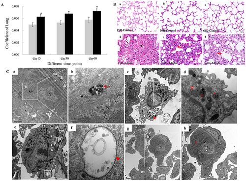
Figure 1 Lung injuries induced by SiNPs through intravenous injection in mice. (A) The coefficients of lungs increased significantly in the SiNP-treated group. Data are expressed as means±SD (n=5). *P<0.05 compared with control. Gray, control; black, admin. (B) Histopathologic changes in mice lungs induced by SiNPs. Black arrow: red blood cells in the area of alveoli; Red arrow: alveoli septum thicken and inflammatory cell infiltration. Scale bar: 20 μm. (C) Ultrastructural observation in mice lungs observed by TEM. (a, b) SiNPs (red arrows) deposited in lungs at 15th day. Scale bar: (a) 1 μm; (b) 200 nm. (c, d) Ultrastructural changes of alveolar macrophages in lungs of SiNP-treated mice at 30th day: extensive vacuolization (red star), mitochondrial fusion (red hollow triangle), mitochondrial cristae disappearance (red asterisk). Scale bar: (c) 0.5 μm; (d) 200 nm. (e, f) Vacuolization (red triangle) in the basophilic granulocyte in lungs of SiNP-treated mice at 30th day. Scale bar: (e) 1 μm; (f) 100 nm. (g, h) Cell cluster consisted of multinucleate cell (hollow star) and type I alveolar epithelial cell (hollow diamond) in lungs of SiNP-treated mice at 60th day. Scale bar: (e) 2 μm; (f) 1 μm.
The authors affirm that this error does not affect the results, discussion, and conclusions of the reported study and apologize for any inconvenience caused to the readers.